


《中国康复理论与实践》 ›› 2025, Vol. 31 ›› Issue (9): 1023-1031.doi: 10.3969/j.issn.1006-9771.2025.09.005
骆丹丹1,2, 沈敏1,3,4( ), 王素娟2, 邱翁歆2, 张宇轩2, 吴蕴2, 王圣虓5
收稿日期:2025-01-17
修回日期:2025-09-07
出版日期:2025-09-25
发布日期:2025-10-10
通讯作者:
沈敏(1971-),女,汉族,江苏南通市人,主任医师,主要研究方向:儿童青少年神经康复,E-mail: 作者简介:骆丹丹(1986-),女,汉族,上海市人,硕士研究生,主要研究方向:学习障碍。
基金资助:
LUO Dandan1,2, SHEN Min1,3,4(), WANG Sujuan2, QIU Wengxin2, ZHANG Yuxuan2, WU Yun2, WANG Shengxiao5
Received:2025-01-17
Revised:2025-09-07
Published:2025-09-25
Online:2025-10-10
Contact:
SHEN Min, E-mail: Supported by:摘要:
目的 采用功能性近红外光谱技术(fNIRS)探究汉语发展性阅读障碍(DD)儿童和典型发育(TD)的健康儿童在静息态下全脑网络的连接特征。
方法 2024年11月至12月,选取复旦大学附属儿科医院6~12岁DD儿童19例(DD组),同时招募年龄和性别匹配的TD儿童18例(TD组),选取额叶皮质(FC)、颞叶皮质(TL)、枕叶皮质(OL)和顶叶皮质(PL)脑区为感兴趣区,采用fNIRS采集两组全脑静息态数据,持续5 min。基于氧合血红蛋白在时间序列上的浓度,计算两组静息态下的功能连接强度,比较两组功能连接强度和脑网络的差异。
结果 DD组全脑功能连接强度高于TD组(t = 2.100, P < 0.05)。DD组右侧OL-右侧FC (t = 2.426, P < 0.05)、右侧OL-左侧FC (t = 2.483, P < 0.05)、右侧TL-右侧FC (t = 2.568, P < 0.05)和右侧TL-左侧FC (t = 2.304, P < 0.05)功能连接强度大于TD组。两组全脑功能连接差异的主要区域位于右侧背外侧前额叶皮质、右侧视觉联合皮质、右侧额叶皮质、左侧额眶区、左侧视觉联合皮质、左侧初级视觉皮质和右侧初级皮质运动区。
结论 DD儿童静息态下右侧枕叶、颞叶及二者与前额叶之间的连接强度较强,提示可能存在右脑代偿左脑阅读网络功能不足的现象。
中图分类号:
骆丹丹, 沈敏, 王素娟, 邱翁歆, 张宇轩, 吴蕴, 王圣虓. 汉语发展性阅读障碍儿童全脑静息态功能连接的特征分析[J]. 《中国康复理论与实践》, 2025, 31(9): 1023-1031.
LUO Dandan, SHEN Min, WANG Sujuan, QIU Wengxin, ZHANG Yuxuan, WU Yun, WANG Shengxiao. Characterisation of whole-brain resting-state functional connectivity in children with Chinese developmental dyslexia[J]. Chinese Journal of Rehabilitation Theory and Practice, 2025, 31(9): 1023-1031.
表1
两组基线资料比较"
| 组别 | n | 性别(男/女)/n | 年龄/岁 | 韦氏IQ |
|---|---|---|---|---|
| TD组 | 18 | 11/7 | 8.50±1.72 | 107.88±11.66 |
| DD组 | 19 | 12/7 | 8.31±1.41 | 100.42±12.43 |
| χ2/t值 | 0.016 | 0.356 | 1.882 | |
| P值 | 0.898 | 0.724 | 0.068 |
表2
ROI与通道对应关系"
| ROI | 通道排布 |
|---|---|
| 左侧FC | CH23、CH22、CH20、CH8、CH10、CH9、CH24 |
| 右侧FC | CH19、CH17、CH5、CH7、CH6、CH21 |
| 左侧TL | CH33、CH32、CH26、CH27、CH25、CH12、CH11 |
| 右侧TL | CH34、CH35、CH2、CH1、CH18、CH4、CH3 |
| 左侧PL | CH44、CH31、CH14、CH43、CH30、CH13 |
| 右侧PL | CH42、CH16、CH29、CH45、CH15、CH28 |
| 左侧OL | CH39、CH40、CH41、CH47、CH48 |
| 右侧OL | CH38、CH37、CH36、CH46 |
图1
48通道在大脑的分布图"
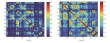
图2
两组基于HbO2全脑功能连接平均强度值图"

表3
两组基于ROI的功能连接强度比较"
| ROI | TD组 | DD组 | t值 | P值 |
|---|---|---|---|---|
| 右侧OL-右侧FC | 0.11 | 0.28 | 2.426 | 0.020 |
| 右侧OL-左侧FC | 0.08 | 0.26 | 2.483 | 0.017 |
| 右侧TL-右侧FC | 0.24 | 0.39 | 2.568 | 0.014 |
| 右侧TL-左侧FC | 0.22 | 0.33 | 2.304 | 0.027 |
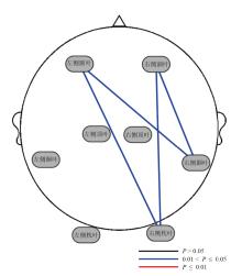
图3
两组基于ROI功能连接强度差异2D图"

表4
TD组与DD组基于通道连接功能连接强度差异"
| 通道 | 连接通道数 | 依据 MNI坐标映射脑区分布 |
|---|---|---|
| CH17 | 18 | 右侧背外侧前额叶皮质 |
| CH46 | 12 | 右侧视觉联合皮质 |
| CH5 | 12 | 右侧额叶皮质 |
| CH8 | 9 | 左侧额眶区 |
| CH47 | 8 | 左侧视觉联合皮质 |
| CH40 | 7 | 左侧初级视觉皮质 |
| CH1 | 7 | 右侧初级皮质运动区 |
| CH37 | 4 | 右侧初级视觉皮质 |
| CH20 | 2 | 左侧额极区 |
| CH30 | 2 | 左侧初级躯体感觉皮质 |
| CH36 | 2 | 右侧视觉联合皮质 |
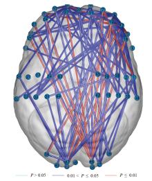
图4
两组基于通道连接功能连接强度差异3D图 注:数字代表相应通道。"

| [1] | World Health Organization. International Classification of Diseases for Mortality And Morbidity Statistics. Eleventh Revision[M]. Geneva: World Health Organization, 2022. |
| [2] | YANG L, LI C, LI X, et al. Prevalence of developmental dyslexia in primary school children: a systematic review and meta-analysis[J]. Brain Sci, 2022, 12(2): 240. |
| [3] |
KALTNER S, JANSEN P. Mental rotation and motor performance in children with developmental dyslexia[J]. Res Dev Disabil, 2014, 35(3): 741-754.
doi: 10.1016/j.ridd.2013.10.003 pmid: 24268351 |
| [4] |
PHILLIPS B A B, ODEGARD T N. Evaluating the impact of dyslexia laws on the identification of specific learning disability and dyslexia[J]. Ann Dyslexia, 2017, 67(3): 356-368.
doi: 10.1007/s11881-017-0148-4 pmid: 29134483 |
| [5] | 李欢, 龙艳林. 近十年国内外汉语阅读障碍干预研究的现状与展望[J]. 中国特殊教育, 2019(7): 47-54. |
| LI H, LONG Y L. Status quo and prospects of intervention research in Chinese dyslexia in the past decade[J]. Chin J Spec Educ, 2019(7): 47-54. | |
| [6] | 王秀红, 姚梦梦, 张蕾, 等. 南京市298例学龄前儿童汉语语音意识发展的影响因素分析[J]. 中华全科医学, 2024, 22(4): 609-613. |
| WANG X H, YAO M M, ZHANG L, et al. Analysis of influencing factors on the development of Chinese phonological awareness among 298 preschool children in Nanjing[J]. Chin J Gener Pract, 2024, 22(4): 609-613. | |
| [7] | HUANG Y, HE M, LI A, et al. Personality, behavior characteristics, and life quality impact of children with dyslexia[J]. Int J Environ Res Public Health, 2020, 17(4): 1415. |
| [8] |
WANG K, LIANG M, WANG L, et al. Altered functional connectivity in early Alzheimer's disease: a resting-state fMRI study[J]. Hum Brain Mapp, 2007, 28(10): 967-978.
doi: 10.1002/hbm.20324 pmid: 17133390 |
| [9] |
CORDES D, HAUGHTON V M, ARFANAKIS K, et al. Mapping functionally related regions of brain with functional connectivity MR imaging[J]. Am J Neuroradiol, 2000, 21(9): 1636-1644.
pmid: 11039342 |
| [10] |
LOWE M J, MOCK B J, SORENSON J A. Functional connectivity in single and multislice echoplanar imaging using resting-state fluctuations[J]. Neuroimage, 1998, 7(2): 119-132.
doi: 10.1006/nimg.1997.0315 pmid: 9558644 |
| [11] |
BISWAL B, YETKIN F Z, HAUGHTON V M, et al. Functional connectivity in the motor cortex of resting human brain using echo-planar MRI[J]. Magn Reson Med, 1995, 34(4): 537-541.
doi: 10.1002/mrm.1910340409 pmid: 8524021 |
| [12] |
HERBET G, DUFFAU H. Revisiting the functional anatomy of the human brain: toward a meta-networking theory of cerebral functions[J]. Physiol Rev, 2020, 100(3): 1181-1228.
doi: 10.1152/physrev.00033.2019 pmid: 32078778 |
| [13] | GALLAGHER A, WALLOIS F, OBRIG H. Functional near-infrared spectroscopy in pediatric clinical research: Different pathophysiologies and promising clinical applications[J]. Neurophotonics, 2023, 10(2): 023517. |
| [14] | VINCENT J L, PATEL G H, FOX M D, et al. Intrinsic functional architecture in the anaesthetized monkey brain[J]. Nature, 2007, 447(7140): 83-86. |
| [15] | NEMMI F, CIGNETTI F, VAUGOYEAU M, et al. Developmental dyslexia, developmental coordination disorder and comorbidity discrimination using multimodal structural and functional neuroimaging[J]. Cortex, 2023, 160: 43-54. |
| [16] | XUE H, WANG Z, TAN Y, et al. Resting-state EEG reveals global network deficiency in dyslexic children[J]. Neuropsychologia, 2020, 138: 107343. |
| [17] |
DIMITRIADIS S I, SIMOS P G, FLETCHER J Μ, et al. Aberrant resting-state functional brain networks in dyslexia: symbolic mutual information analysis of neuromagnetic signals[J]. Int J Psychophysiol, 2018, 126: 20-29.
doi: S0167-8760(17)30117-4 pmid: 29476872 |
| [18] | YU X, FERRADAL S, DUNSTAN J, et al. Patterns of neural functional connectivity in infants at familial risk of developmental dyslexia[J]. JAMA Netw Open, 2022, 5(10): e2236102. |
| [19] | FINN E S, SHEN X, HOLAHAN J M, et al. Disruption of functional networks in dyslexia: a whole-brain, data-driven analysis of connectivity[J]. Biol Psychiatry, 2014, 76(5): 397-404. |
| [20] |
TURKER S, KUHNKE P, JIANG Z, et al. Disrupted network interactions serve as a neural marker of dyslexia[J]. Commun Biol, 2023, 6(1): 1114.
doi: 10.1038/s42003-023-05499-2 pmid: 37923809 |
| [21] | 吴毅. 功能性近红外光谱技术在脑卒中患者康复中的临床应用[J]. 中国康复医学杂志, 2020, 35(11): 1281-1283. |
| [22] | DONIZETE D F D, MARQUES J A P, JOANA B, et al. Task-related brain activity and functional connectivity in upper limb dystonia: a functional magnetic resonance imaging (fMRI) and functional near-infrared spectroscopy (fNIRS) study[J]. Neurophotonics, 2020, 7(4): 045004. |
| [23] | 美国精神医学学会. 精神障碍诊断与统计手册[M]. 北京: 北京大学医学出版社, 2015: 63-64. |
| American Psychiatric Association. The Diagnostic and Statistical Manual of Mental Disorders[M]. Beijing: Peking University Medical Press, 2015: 63-64. | |
| [24] | 王久菊, 孟祥芝, 李虹, 等. 汉语发展性阅读障碍诊断与干预的专家意见[J]. 中国心理卫生杂志, 2023, 37(3): 185-191. |
| WANG J J, MENG X Z, LI H, et al. Expert advice on the diagnosis and intervention of Chinese developmental dyslexia[J]. Chin Ment Health J, 2023, 37(3): 185-191. | |
| [25] | 张亚静, 陈刘昕, 林榕萍, 等. 阅读障碍儿童句法意识、词汇知识和阅读流畅性加工缺陷研究[J]. 中国特殊教育, 2024(8): 62-70. |
| ZHANG Y J, CHEN L X, LIN R P, et al. Research on syntactic awareness,vocabulary knowledge, and reading fluency processing deficits in children with dyslexia[J]. Chin J Spec Educ, 2024(8): 62-70. | |
| [26] | 王孝玲, 陶保平. 小学生识字量测试题库及评价量表[M]. 上海: 上海教育出版社, 1996. |
| WANG X L, TAO B P. Literacy test question bank and evaluation scale for primary school students[M]. Shanghai: Shanghai Education Press, 1996. | |
| [27] | 赵婧, 毕鸿燕, 杨炀. 汉语发展性阅读障碍儿童的快速命名与正字法加工技能[J]. 中国心理卫生杂志, 2012, 26(1): 36-40. |
| ZHAO J, BI H Y, YANG Y. Rapid naming and orthographic processing skill in children with Chinese developmental dyslexia[J]. Chin Ment Health J, 2012, 26(1): 36-40. | |
| [28] |
STRANGMAN G, CULVER P J, THOMPSON H J, et al. A quantitative comparison of simultaneous bold fMRI and NIRS recordings during functional brain activation[J]. Neuroimage, 2002, 17(2): 719-731.
pmid: 12377147 |
| [29] |
SCHURZ M, WIMMER H, RICHLAN F, et al. Resting-state and task-based functional brain connectivity in developmental dyslexia[J]. Cereb Cortex, 2015, 25(10): 3502-3514.
doi: 10.1093/cercor/bhu184 pmid: 25169986 |
| [30] |
KOYAMA M S, KELLY C, SHEHZAD Z, et al. Reading networks at rest[J]. Cereb Cortex, 2010, 20(11): 2549-2559.
doi: 10.1093/cercor/bhq005 pmid: 20139150 |
| [31] | KOYAMA M S, DI MARTINO A, KELLY C, et al. Cortical signatures of dyslexia and remediation: an intrinsic functional connectivity approach[J]. PLoS One, 2013, 8(2): e55454. |
| [32] | CROSS A M, RAMDAJAL R, PETERS L, et al. Resting-state functional connectivity and reading subskills in children[J]. Neuroimage, 2021, 243: 118529. |
| [33] |
QI T, GU B, DING G, et al. More bilateral, more anterior: Alterations of brain organization in the large-scale structural network in Chinese dyslexia[J]. Neuroimage, 2016, 124(Pt A): 63-74.
doi: S1053-8119(15)00808-3 pmid: 26363349 |
| [34] |
PETERSON R L, PENNINGTON B F, OLSON R K. Subtypes of developmental dyslexia: testing the predictions of the dual-route and connectionist frameworks[J]. Cognition, 2013, 126(1): 20-38.
doi: 10.1016/j.cognition.2012.08.007 pmid: 23010562 |
| [35] | 刘丽, 何茵. 汉语发展性阅读障碍的认知神经机制研究及教育启示[J]. 教育发展研究, 2018, 38(24): 64-72. |
| LIU L, HE Y. Neurocognitive basis of Chinese dyslexia and its implications on education[J]. Educ Dev Res, 2018, 38(24): 64-72. | |
| [36] | PANICHELLO M F, BUSCHMAN T J. Shared mechanisms underlie the control of working memory and attention[J]. Nature, 2021, 592(7855): 601-605. |
| [37] | 何红瑶, 高小焱, 刘芳芳, 等. 发展性阅读障碍共患注意缺陷多动障碍儿童视动整合能力特点及相关因素[J]. 中国学校卫生, 2022, 43(5):792-795. |
| HE H Y, GAO X Y, LIU F F, et al. Characteristics and associated factors of visual and motor integration in children with developmental dyslexia and attention deficit hyperactivity disorder[J]. Chin J School Health, 2022, 43(5): 792-795. | |
| [38] | BERMAN S, CICCHINO N, HAJINAZARIAN A, et al. An fMRI study of a dyslexia biomarker[J]. J Young Invest, 2014, 26(1): 1-4. |
| [39] | 钟鑫琪, 比沙拉, 胡晓云, 等. 发育性阅读障碍和注意缺陷多动可疑儿童的情绪行为问题[J]. 中国学校卫生, 2019, 40(10): 1460-1463. |
| ZHONG X Q, BI S L, HU X Y, et al. Emotional and behavioral problems among children with developmental dyslexia and attention deficit hyperactivity disorder[J]. Chin J School Health, 2019, 40(10): 1460-1463. | |
| [40] | 何吴明, 郑剑虹, 戴秀清. 小学生学习障碍检出率和行为特征调查[J]. 岭南师范学院学报, 2022, 43(2): 24-32. |
| HE W M, ZHENG J H, DAI X Q. Investigation of positive rate and learning behavior features of primary school students with learning disabilities in western Guangdong[J]. J Lingnan Normal Univ, 2022, 43(2): 24-32. | |
| [41] | 王鑫洋. 注意缺陷与多动障碍儿童执行功能障碍诊断与康复训练的最新进展[J]. 中国医学创新, 2022, 19(13): 172-175. |
| WANG X Y. Recent advances in executive dysfunction diagnosis and rehabilitation training in children with attention deficit and hyperactivity disorder[J]. Chin Med Innov, 2022, 19(13): 172-175. | |
| [42] |
PATTERSON K, NESTOR P J, ROGERS T T. Where do you know what you know? The representation of semantic knowledge in the human brain[J]. Nat Rev Neurosci, 2007, 8(12): 976-987.
doi: 10.1038/nrn2277 pmid: 18026167 |
| [43] |
HSIEH J K, PRAKASH P R, FLINT R D, et al. Cortical sites critical to language function act as connectors between language subnetworks[J]. Nat Commun, 2024, 15(1): 7897.
doi: 10.1038/s41467-024-51839-z pmid: 39284848 |
| [44] |
MORKEN F, HELLAND T, HUGDAHL K, et al. Reading in dyslexia across literacy development: a longitudinal study of effective connectivity[J]. Neuroimage, 2017, 144(Pt A): 92-100.
doi: S1053-8119(16)30537-7 pmid: 27688204 |
| [45] |
PUGH K R, MENCL W E, SHAYWITZ B A, et al. The angular gyrus in developmental dyslexia: task-specific differences in functional connectivity within posterior cortex[J]. Psychol Sci, 2000, 11(1): 51-56.
pmid: 11228843 |
| [46] |
MENGISIDOU M, MARSHALL C R, STAVRAKAKI S. Semantic fluency difficulties in developmental dyslexia and developmental language disorder (DLD): poor semantic structure of the lexicon or slower retrieval processes?[J]. Int J Lang Commun Disord, 2020, 55(2): 200-215.
doi: 10.1111/1460-6984.12512 pmid: 31697020 |
| [47] | RICHLAN F, STURM D, SCHURZ M, et al. A common left occipito-temporal dysfunction in developmental dyslexia and acquired letter-by-letter reading?[J]. PLoS One, 2010, 5(8): e12073. |
| [48] |
骆丹丹, 沈敏. 基于HT-CHC模式的康复训练对特定学习障碍儿童认知能力的影响[J]. 新医学, 2024, 55(12): 1047-1053.
doi: 10.3969/j.issn.0253-9802.2024.12.011 |
| LUO D D, SHEN M. Effect of rehabilitation training based on HT-CHC mode on cognitive ability of children with specific learning disabilities[J]. New Med, 2024, 55(12): 1047-1053. | |
| [49] | 黄格敏, 申仁洪. 汉语发展性阅读障碍的认知神经机制与干预研究进展[J]. 中国特殊教育, 2022(11): 55-61, 71. |
| HUANG G M, SHEN R H. A study on the cognitive neural mechanism and intervention of Chinese developmental dyslexia[J]. Chin J Spec Educ, 2022(11): 55-61, 71. |
| [1] | 李峤桢, 冯枫, 杜霞, 邵雯, 高咪, 惠琳娜, 袁华, 孙晓龙. 脊髓损伤神经病理性疼痛的脑电信号特征[J]. 《中国康复理论与实践》, 2025, 31(7): 830-837. |
| [2] | 罗红, 徐丽. 重复经颅磁刺激联合重复外周磁刺激对脑出血患者上肢运动功能的效果:基于静息态功能磁共振成像的随机对照试验[J]. 《中国康复理论与实践》, 2024, 30(9): 1060-1068. |
| [3] | 谢丹丹, 陈善佳, 雷蕾, 余果, 余佳慧, 赵嘉培, 何晓阔. 健康人和脑卒中患者视觉反馈步行训练后脑激活特征的功能性近红外光谱技术研究[J]. 《中国康复理论与实践》, 2024, 30(9): 1074-1081. |
| [4] | 刘佳琪, 侯闪闪, 汪鑫煜, 朱崇田, 王孝文. 不同吞咽时期大脑皮质激活特征:基于功能性近红外光谱技术[J]. 《中国康复理论与实践》, 2024, 30(6): 709-718. |
| [5] | 阚超杰, 郭川, 朱仕哲, 眭有昕, 王庆雷, 庄任, 耿阿燕, 王彤. 老年人在认知-平衡双任务下的皮质激活特征[J]. 《中国康复理论与实践》, 2023, 29(10): 1189-1194. |
| [6] | 陆佳敏,闫思念,陈逸浩,陆蓉蓉,吴毅. 脑卒中后认知障碍患者静息态脑网络特征的功能性近红外光谱研究[J]. 《中国康复理论与实践》, 2022, 28(4): 447-452. |
| [7] | 刘绍文,魏聪惠,单新颖,张焱. 下肢截肢患者的脑功能连接[J]. 《中国康复理论与实践》, 2022, 28(1): 90-94. |
| [8] | 英小倩,高轶,廖利民. 健康女性强烈排尿感脑功能网络的小世界特征[J]. 《中国康复理论与实践》, 2021, 27(5): 510-515. |
| [9] | 英小倩,廖利民. 膀胱过度活动症患者大脑功能的连接变化[J]. 《中国康复理论与实践》, 2021, 27(4): 466-471. |
| [10] | 张晓彤,李娜,陈兆聪,梁井凤,喻勇,武惠香,康庄,丘卫红. 右侧小脑对卒中后失语的潜在作用:基于格兰杰因果分析的初步研究[J]. 《中国康复理论与实践》, 2021, 27(12): 1458-1463. |
| [11] | 吴琼,任诗媛,乐赞,葛云祥,马迪,赵红亮,刘刚,王晶,潘钰,窦维蓓. 脑机接口综合康复训练对亚急性期脑卒中疗效的静息态功能磁共振研究[J]. 《中国康复理论与实践》, 2020, 26(1): 77-84. |
| [12] | 石庆丽, 张玉梅, 陈红燕, 白丽君. 脑白质疏松相关认知障碍患者的静息态脑网络及格兰杰因果连接[J]. 《中国康复理论与实践》, 2019, 25(3): 271-278. |
| [13] | 魏娜 a;燕浩;白丽君;姚婧璠 a;李越秀 a;陈红燕 b;张玉梅 . 脑白质疏松患者静息态脑网络磁共振成像的研究[J]. 《中国康复理论与实践》, 2015, 21(07): 793-798. |
| [14] | 赵颖;王强;孙蓉;杜晓霞;宋鲁平;陈灵娟;毕彦超;韩在柱 . 脑损伤静息态低频振幅预测数学认知能力受损情况[J]. 《中国康复理论与实践》, 2015, 21(06): 670-676. |
| [15] | 林志诚;杨珊莉;薛偕华;陶静;陈立典;. 针刺百会穴改善脑卒中患者记忆力的中枢机制[J]. 《中国康复理论与实践》, 2015, 21(02): 184-188. |
| 阅读次数 | ||||||
|
全文 |
|
|||||
|
摘要 |
|
|||||
|
||