


《中国康复理论与实践》 ›› 2021, Vol. 27 ›› Issue (3): 302-309.doi: 10.3969/j.issn.1006-9771.2021.03.009
王琼芬( ),王风波,王科,钟永强,王娇娇
收稿日期:2020-04-01
修回日期:2020-07-07
出版日期:2021-03-25
发布日期:2021-04-02
通讯作者:
王琼芬
E-mail:wqf659@163.com
作者简介:王琼芬(1975-),女,汉族,四川成都市人,硕士,副主任医师,主要研究方向:神经康复。
基金资助:
Qiong-fen WANG(),Feng-bo WANG,Ke WANG,Yong-qiang ZHONG,Jiao-jiao WANG
Received:2020-04-01
Revised:2020-07-07
Published:2021-03-25
Online:2021-04-02
Contact:
Qiong-fen WANG
E-mail:wqf659@163.com
Supported by:摘要: 探讨电针风池穴对急性脑梗死大鼠脑星形胶质细胞和神经元的调节作用。 清洁级Sprague-Dawley雄性大鼠64只,随机数字表法分为假手术组(n = 16)、模型组(n = 16)、非穴组(n = 16)和风池组(n = 16)。后三组采用改良线栓法制备急性脑梗死模型,后两组于造模后分别电针风池旁非穴部位和风池,共7 d。治疗后进行神经功能缺损评分;取脑组织测量脑梗死面积和脑组织含水量;HE染色观察海马组织病理学改变;ELISA法检测海马组织中脑源性神经营养因子(BDNF)、胶质纤维酸性蛋白(GFAP)、神经元特异性烯醇化酶(NSE)的表达水平;采用TUNEL法检测海马神经元细胞凋亡数;采用Western blotting和逆转录实时定量聚合酶链反应(RT-qPCR)检测脑匀浆caspase-3和Bcl-2水平。 与假手术组相比,其他三组神经功能缺损评分、脑梗死面积和含水量升高(P < 0.05),海马组织中GFAP和NSE水平升高(P < 0.05),BDNF降低(P < 0.05),海马神经元凋亡细胞数升高(P < 0.05),caspase-3表达升高(P < 0.05),Bcl-2表达降低(P < 0.05);与模型组和非穴组相比,风池组神经行为学评分、脑梗死面积和含水量降低(P < 0.05),GFAP和NSE水平降低(P < 0.05),BDNF升高(P < 0.05),神经元凋亡细胞数减少(P < 0.05),caspase-3表达降低(P < 0.05),Bcl-2表达增加(P < 0.05)。 电针风池穴可改善急性脑梗死大鼠脑组织损伤,调节脑组织GFAP、NSE和BDNF表达水平,降低神经元凋亡水平。
中图分类号:
王琼芬,王风波,王科,钟永强,王娇娇. 电针风池对急性脑梗死大鼠脑星形胶质细胞和神经元的保护效果[J]. 《中国康复理论与实践》, 2021, 27(3): 302-309.
Qiong-fen WANG,Feng-bo WANG,Ke WANG,Yong-qiang ZHONG,Jiao-jiao WANG. Effect of Electroacupuncture at Fengchi on Astrocytes and Neurons in Rats with Acute Cerebral Infarction[J]. 《Chinese Journal of Rehabilitation Theory and Practice》, 2021, 27(3): 302-309.
表1
各组神经功能缺损评分比较"
| 组别 | n | 评分 |
|---|---|---|
| 假手术组 | 16 | 0 |
| 模型组 | 16 | 2.44±0.51a |
| 非穴组 | 16 | 2.31±0.70a |
| 风池组 | 16 | 1.44±0.63a,b,c |
| H值 | 46.732 | |
| P值 | < 0.001 |
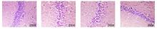
图1
各组海马组织病理学改变(HE染色,×200)假手术组 模型组 非穴组 风池组"
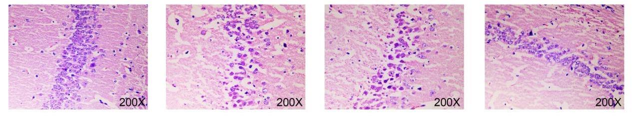
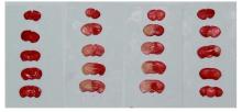
图2
各组脑组织脑梗死面积(TTC染色)假手术组 模型组 非穴组 风池组"
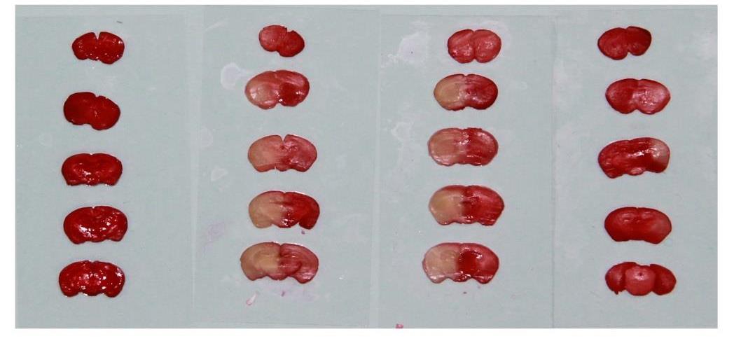
表2
各组大鼠脑梗死面积及脑组织含水量(%)"
| 组别 | n | 梗死面积 | 脑组织含水量 |
|---|---|---|---|
| 假手术组 | 4 | 0 | 68.68±4.64 |
| 模型组 | 4 | 47.51±2.20a | 88.28±6.16a |
| 非穴组 | 4 | 46.84±3.10a | 86.73±5.67a |
| 风池组 | 4 | 24.60±2.12a,b,c | 77.30±4.51a,b,c |
| 统计值 | H = 12.918 | F = 11.851 | |
| P值 | 0.005 | 0.001 |
表3
各组海马组织BDNF、GFAP和NSE水平(pg/ml)"
| 组别 | n | BDNF | GFAP | NSE |
|---|---|---|---|---|
| 假手术组 | 8 | 60.59±4.18 | 293.94±12.28 | 78.88±8.07 |
| 模型组 | 8 | 18.25±1.90a | 414.66±18.32a | 154.11±10.69a |
| 非穴组 | 8 | 18.19±2.18a | 413.36±15.73a | 154.43±9.93a |
| 风池组 | 8 | 41.55±2.75a,b,c | 323.48±14.88a,b,c | 97.31±5.74a,b,c |
| F值 | 402.917 | 128.713 | 135.543 | |
| P值 | < 0.001 | < 0.001 | < 0.001 |
表4
各组海马神经元细胞凋亡数及凋亡相关基因的表达水平"
| 组别 | n | 凋亡细胞数 | caspase-3 mRNA (2-ΔΔCt) | Bcl-2 mRNA (2-ΔΔCt) | caspase-3蛋白 (/GAPDH) | Bcl-2蛋白 (/GAPDH) |
|---|---|---|---|---|---|---|
| 假手术组 | 8 | 12.00±5.89 | 0.96±0.20 | 4.77±0.16 | 0.11±0.09 | 0.80±0.11 |
| 模型组 | 8 | 62.38±12.91a | 7.33±0.98a | 2.46±0.31a | 1.01±0.09a | 0.30±0.10a |
| 非穴组 | 8 | 72.13±10.30a | 7.43±0.74a | 2.91±0.37a | 1.00±0.11a | 0.31±0.10a |
| 风池组 | 8 | 32.63±5.10a,b,c | 4.07±0.67a,b,c | 6.98±0.27a,b,c | 0.37±0.13a,b,c | 2.01±0.10a,b,c |
| F值 | 74.437 | 57.263 | 151.894 | 58.433 | 194.096 | |
| P值 | < 0.001 | < 0.001 | < 0.001 | < 0.001 | < 0.001 |
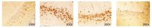
图3
各组大鼠脑海马神经元细胞凋亡数(TUNEL, ×200)假手术组 模型组 非穴组 风池组"
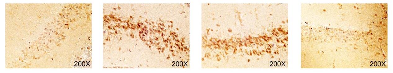

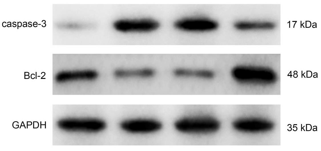
图4
各组caspase-3和Bcl-2蛋白表达水平(Western blotting)假手术组 模型组 非穴组 风池组"
| 1 | 程一升,程霄霄,赵元琛,等. 小续命汤治疗急性脑梗死疗效及对SOD、MCP-1以及BDNF水平的影响[J]. 中华中医药学刊, 2019, 37(2): 181-183. |
| Cheng Y S, Cheng X X, Zhao Y C, et al. Effect of Xiaoxuming decoction on acute cerebral infarction and effect on levels of SOD, MCP-1 and BDNF [J]. Chin J Tradit Chin Med, 2019, 37(2): 181-183. | |
| 2 | 邓希兰. 阿托伐他汀联合氯吡格雷治疗急性脑梗死的临床观察及对血清炎症因子、神经元特异性烯醇化酶的影响[J]. 中国民间疗法, 2018, 26(10): 93-95. |
| Deng X L. Clinical observation of atorvastatin combined with clopidogrel in the treatment of acute cerebral infarction and its effect on serum inflammatory factors and neuron-specific enolase [J]. Chin J Folk Therapeutics, 2018, 26(10): 93-95. | |
| 3 | 陈文华. 脑梗死患者血清S100β和神经元特异性烯醇化酶水平与认知功能障碍的关系研究[J]. 血栓与止血学, 2018, 24(5): 20-22. |
| Chen W H. Relationship between serum S100 and neuron specific enolase levels and cognitive dysfunction in patients with cerebral infarction [J]. Thromb Hemostas, 2018, 24(5): 20-22. | |
| 4 | Li J, Du Y H, Zhang X Z, et al. Effects of electroacupuncture on expression of Ang/Tie-2 mRNA and protein in rats with acute cerebral infarction[J]. J Tradit Chin Med, 2017, 37(5): 659-666. |
| 5 | Ye J, Sun Z, Hu W. Roles of astrocytes in cerebral infarction and related therapeutic strategies [J]. Zhejiang Da Xue Xue Bao Yi Xue Ban, 2018, 47(5): 493-498. |
| 6 | Gorshkov K, Aguisanda F, Thorne N, et al. Astrocytes as targets for drug discovery [J]. Drug Discov Today, 2018, 23(3): 673-680. |
| 7 | 叶涛,朱路文,唐强,等. 电针预处理对脑缺血再灌注大鼠缺血半暗区细胞凋亡及凋亡相关蛋白表达的影响[J]. 中国康复理论与实践, 2018, 24(1): 54-59. |
| Ye T, Zhu L W, Tang Q, et al. Effects of electroacupuncture preconditioning on apoptosis and expression of apoptosis-related proteins in rats after cerebral ischemia-reperfusion [J]. Chin J Rehabil Theory Pract, 2018, 24(1): 54-59. | |
| 8 | Tian R S, Wang S. Electroacupuncture reduced apoptosis of hippocampal neurons in mice with cerebral infarction by regulating the Notch3 signaling pathway [J]. J Mol Neurosci, 2019, 67(3): 456-466. |
| 9 | 秦文熠,荣晓凤,罗勇. 电针通过调节IκB激酶β表达抑制局灶性脑缺血再灌注大鼠脑内炎症损害的机制[J]. 中国康复理论与实践, 2019, 25(4): 407-415. |
| Qin W Y, Rong X F, Luo Y. Effect of electroacupuncture on inflammatory response of rats after focal cerebral ischemia-reperfusion via regulating IκB Kinases β expression [J]. Chin J Rehabil Theory Pract, 2019, 25(4): 407-415. | |
| 10 | Yin T, Chen H P, Wang W, et al. Effect of electroacupuncture preconditioning on neurological deficit and expression of cerebral Cx43 protein in rats with acute cerebral infarction [J]. Zhen Ci Yan Jiu, 2019, 44(7): 497-500. |
| 11 | Zhou P J, Wang A C, Li B, et al. Effect of acupuncture at Fengchi (GB20) on the activity of myosin light chain kinase in the middle meningeal artery of migraine modeled rats [J]. J Tradit Chin Med, 2015, 35(3): 301-305. |
| 12 | Wen Y, Zhang C, Zhao X F, et al. Safety of different acupuncture manipulations for posterior circulation ischemia with vertigo [J]. Neural Regen Res, 2016, 11(8): 1267-1273. |
| 13 | 贾复敏,魏衡,周瑞. TLR4对大鼠急性脑梗死体积与Nrf2、HO-1表达水平的影响[J]. 卒中与神经疾病, 2019, 26(1): 15-18. |
| Jia F M, Wei H, Zhou R. Effects of TLR4 on the volume of acute cerebral infarction and the expression level of Nrf2 and HO-1 in rats [J]. Stroke Nerv Dis, 2019, 26(1): 15-18. | |
| 14 | 裴培,王艳昕,陈怀珍. 电针风池穴对偏头痛大鼠行为学及三叉神经节5-HT7受体表达的影响[J]. 中西医结合心脑血管病杂志, 2017, 15(18): 2247-2251. |
| Pei P, Wang Y X, Chen H Z. Effect of electro-acupuncture at Fengchi on behavior and expression of 5-HT7 receptor in trigeminal ganglion in migraine rats [J]. J Cardiovasc Cerebrovasc Dis, 2017, 15(18): 2247-2251. | |
| 15 | 张潇,张金枝,刘真真. 人参皂苷Rb1对脑梗死大鼠血管新生的作用及机制研究[J]. 解放军医药杂志, 2019, 31(4): 10-15. |
| Zhang X, Zhang J Z, Liu Z Z. Effect and mechanism of ginsenoside Rb1 on angiogenesis in rats with cerebral infarction [J]. PLA J Med, 2019, 31(4): 10-15. | |
| 16 | Li K, Jia J, Wang Z, et al. Elevated serum levels of NSE and S-100β correlate with increased risk of acute cerebral infarction in Asian populations [J]. Med Sci Monit, 2015, 21: 1879-1888. |
| 17 | 杨庆宇,赵锐. 益气活血通络汤对脑梗死患者神经功能缺损及血清胶质纤维酸性蛋白、Ang-Ⅱ、VEGF水平的影响[J]. 中医药信息, 2019, 36(1): 94-98. |
| Yang Q Y, Zhao R. Effect of Yiqi Huoxue Tongluo Decoction on neurological deficit and serum glial fibrillary acidic protein, Ang-Ⅱ, VEGF levels in patients with cerebral infarction [J]. Informat Tradit Chin Med, 2019, 36(1): 94-98. | |
| 18 | Neves J D, Aristimunha D, Vizuete A F, et al. Glial-associated changes in the cerebral cortex after collagenase-induced intracerebral hemorrhage in the rat striatum [J]. Brain Res Bull, 2017, 134: 55-62. |
| 19 | 侯惠莲,田美丽,张冠军,等. SHR-SP脑梗死后微血管形成与星形胶质细胞的关系[J]. 西安交通大学学报(医学版), 2019, 40(1): 84-89. |
| Hou H L, Tian M L, Zhang G J, et al. Relationship between microvessel formation and astrocytes after SHR-SP cerebral infarction [J]. J Xi'an Jiaotong Univ (Med Sci), 2019, 40(1): 84-89. | |
| 20 | Wang Y, Yang J, Du H, et al. Yangyin Tongnao granules enhance neurogenesis in the peri-infarct area and upregulate brain-derived neurotrophic factor and vascular endothelial growth factor after focal cerebral ischemic infarction in rats [J]. Mol Biol Rep, 2019, 46(4): 3817-3826. |
| 21 | Wang Y, Xu S, Pan S, et al. Association of serum neuron-specific enolase and bilirubin levels with cerebral dysfunction and prognosis in large-artery atherosclerotic strokes [J]. J Cell Biochem, 2018, 119(12): 9685-9693. |
| 22 | 陈亚南,王昌铭. 脂蛋白相关性磷脂酶和神经元特异性烯醇化酶在急性脑梗死患者中的动态变化及意义[J]. 中华老年心脑血管病杂志, 2018, 20(3): 290-293. |
| Chen Y N, Wang C M. Change of lipoprotein-associated phospholipase A2 and neuron specific enolase and its significance in patients with acute cerebral infarction [J]. Chin J Geriatr Heart Brain Vess Dis, 2018, 20(3): 290-293. | |
| 23 | 李泽钊,洪震,芮汉臣,等. 小续命汤辅助治疗大面积急性脑梗死的疗效观察[J]. 中国中医急症, 2017, 26(4): 679-681. |
| Li Z Z, Hong Z, Rui H C, et al. J Emerg Tradit Chin Med, 2017, 26(4): 679-681. | |
| 24 | 高剑明,赵军. 电针太阳和风池为主治疗血管性帕金森综合征[J]. 中医药临床杂志, 2017, 29(8): 1303-1305. |
| Gao J M, Zhao J. The Electro-acupuncture of the Taiyang and Fengchi point of 21 cases with vascular Parkinson's syndrome [J]. Chin J Clin Med, 2017, 29(8): 1303-1305. | |
| 25 | 孙柯,梁丽萍,呼小雪,等. 及早经皮穴位电针刺激对高龄老年髋部骨折术后认知的影响[J]. 中国医学物理学杂志, 2018, 35(9): 1075-1079. |
| Sun K, Liang L P, Hu X X, et al. Effect of early percutaneous electroacupuncture stimulation on postoperative cognition of elderly hip fractures [J]. Chin J Med Phys, 2018, 35(9): 1075-1079. | |
| 26 | 韩清,徐鸣曙,张英杰,等. 电针风池穴对脑缺血再灌注大鼠突触素、生长相关蛋白-43的影响[J]. 上海针灸杂志, 2019, 38(6): 674-680. |
| Han Q, Xu M S, Zhang Y J, et al. Effect of electro-acupuncture at Fengchi on synaptophysin and growth-related protein-43 in rats with cerebral ischemia-reperfusion [J]. Shanghai J Acupunct Moxib, 2019, 38(6): 674-680. | |
| 27 | Zheng S M, Zhao F L, Luo Y Y, et al. Clinical effect of electroacupuncture at Baihui and Shuigou points in treatment of brain injury in patients with sepsis-associated encephalopathy [J]. Zhen Ci Yan Jiu, 2020, 45(5): 402-406. |
| 28 | 黄宝丽,李翔翔,张慧,等. 脑梗死患者BDNF水平变化及临床意义[J]. 检验医学与临床, 2016, 13(11): 1525-1527. |
| Huang B L, Li X X, Zhang H, et al. Changes and clinical significance of BDNF in patients with cerebral infarction [J]. Lab Med Clin Med, 2016, 13(11): 1525-1527. | |
| 29 | Shi R X, Ding H T, Li H, et al. Effect of acupuncture stimulation of different acupoint groups on levels of stress hormones and serum brain-derived neurotrophic factor in depression rats [J]. Zhen Ci Yan Jiu, 2015, 40(6): 444-448. |
| 30 | 桂瑶,汤光花,陈永新,等. 溶栓颗粒对急性脑梗死大鼠海马神经元凋亡、脑组织Caspase-3蛋白及外周血中细胞因子水平的影响[J]. 中国中医急症, 2017, 26(10): 79-81. |
| Gui Y, Tang G H, Chen Y X, et al. Effect of Rongshuan Granules on hippocampal neuronal apoptosis, Caspase-3 protein and cytokine levels in peripheral blood of rats with acute cerebral infarction [J]. J Emerg Tradit Chin Med, 2017, 26(10): 79-81. | |
| 31 | 刘明,邓颖,刘杨,等. 蓝布正对大鼠梗死灶周围大脑皮质神经元凋亡和蛋白表达的影响[J]. 中国实验方剂学杂志, 2016, 22(17): 117-121. |
| Liu M, Deng Y, Liu Y, et al. Effects of Gei Herba on neuronal apoptosis and protein expression in peri-infarct cortex of rats with permenant middle cerebral artery occlusion [J]. Chin J Exp Pharmacol, 2016, 22(17): 117-121. | |
| 32 | Xing Y, Yang S D, Wang M M, et al. Electroacupuncture alleviated neuronal apoptosis following ischemic stroke in rats via midkine and ERK/JNK/p38 signaling pathway [J]. J Mol Neurosci, 2018, 66(1): 26-36. |
| 33 | Ma J, Bao L, Xia X, et al. miR-128b promotes cerebral infarction by regulating the expressions of BCL-2 and CAPASE3 [J]. World Neurosurg, 2019, 123: e245-e251. |
| 34 | Che Q Q, Huang T, Zhang Y D, et al. Effect of miR-124 on neuronal apoptosis in rats with cerebral infarction through Wnt/β-catenin signaling pathway [J]. Eur Rev Med Pharmacol Sci, 2019, 23(15): 6657-6664. |
| 35 | 彭静华,金飞,张鸿日,等. 过表达Bcl-2对急性脑梗死大鼠神经功能的影响[J]. 中风与神经疾病, 2017, 34(3): 217-220. |
| Peng J H, Jin F, Zhang H R, et al. Effect of overexpressing Bcl-2 on nerve function in rats with acute cerebral infarction [J]. Stroke Nerv Dis, 2017, 34(3): 217-220. | |
| 36 | Liu F, Jiang Y J, Zhao H J, et al. Electroacupuncture ameliorates cognitive impairment and regulates the expression of apoptosis-related genes Bcl-2 and Bax in rats with cerebral ischaemia-reperfusion injury [J]. Acupunct Med, 2015, 33(6): 478-484. |
| 37 | Hou Z T, Sun Z R, Liu S T, et al. Effects of electroacupuncture intervention on oxygen free radicals and expression of apoptosis-related proteins in rats with ischemic learning and memory disorder [J]. Acupunct Res, 2015, 40(6): 431-438. |
| [1] | 陈怡婷, 王倩, 崔慎红, 李映彩, 张思鈺, 魏衍旭, 任慧, 冷军, 陈斌. 双侧序贯重复经颅磁刺激干预脑卒中患者上肢运动功能的效果[J]. 《中国康复理论与实践》, 2023, 29(8): 926-932. |
| [2] | 李芳, 霍速, 杜巨豹, 刘秀贞, 李小爽, 宋为群. 经颅直流电刺激联合任务导向性康复训练对脊髓损伤大鼠前肢运动障碍的效果[J]. 《中国康复理论与实践》, 2023, 29(7): 777-781. |
| [3] | 崔尧, 丛芳, 黄富表, 曾明, 颜如秀. 不同镜像神经元训练策略下脑与肌肉的活动特征:基于近红外光谱与表面肌电图技术[J]. 《中国康复理论与实践》, 2023, 29(7): 782-790. |
| [4] | 罗兰, 李璐, 金沐. 氙气后处理对脊髓缺血再灌注损伤的效果:Akt信号通路和自噬机制[J]. 《中国康复理论与实践》, 2023, 29(2): 174-181. |
| [5] | 黄志霖, 徐发邵, 施静, 黄淦, 刘梅芳, 张霞辉. 线栓法建立卒中后吞咽障碍的大鼠模型[J]. 《中国康复理论与实践》, 2023, 29(10): 1147-1153. |
| [6] | 秦彦强, 董浩, 孙迎春, 程先宽, 姚海江. 不同针刺方案对卒中后抑郁大鼠神经递质及相关炎性因子的影响[J]. 《中国康复理论与实践》, 2023, 29(1): 30-37. |
| [7] | 张立,吴珊红,赵乐乐,王艳. Netrin-1、Slit2在夹脊电针结合神经松动术调节兔坐骨神经损伤后Rho GTPases失衡的作用[J]. 《中国康复理论与实践》, 2022, 28(8): 914-926. |
| [8] | 缪培,张通,米海霞,张伟东. 不同线栓法复制局灶性脑缺血模型大鼠恢复期学习记忆能力的差异及其机制[J]. 《中国康复理论与实践》, 2022, 28(7): 789-796. |
| [9] | 韩凯月,刘光亮,苏文龙,唐芷晴,张皓. 智能有氧踏车训练对不同病程脑梗死患者的效果[J]. 《中国康复理论与实践》, 2022, 28(7): 822-827. |
| [10] | 周小珏,冯婧,庞日朝,刘捷,张安仁. 隔日限食减轻脊髓损伤大鼠炎症反应的芳香烃受体/细胞因子信号传导抑制因子2/核转录因子-κB信号通路机制[J]. 《中国康复理论与实践》, 2022, 28(5): 544-551. |
| [11] | 宋绍霏,侯园园,王云雷,张通. 异常光周期所致昼夜节律紊乱对大鼠腓肠肌时钟基因和葡萄糖摄取相关基因表达节律的影响[J]. 《中国康复理论与实践》, 2022, 28(5): 552-558. |
| [12] | 李童,方志鹏,邵玉萍,王平. 有氧运动对睡眠剥夺大鼠学习记忆及海马神经元突触可塑性的效果[J]. 《中国康复理论与实践》, 2022, 28(11): 1270-1277. |
| [13] | 丁妍怡,张胜行,刘雨露,余燕,杨敏光,梁胜祥,柳维林,陶静. 电针对血管性认知障碍大鼠脑功能局部一致性的效果[J]. 《中国康复理论与实践》, 2022, 28(1): 55-61. |
| [14] | 戴燕红, 黄嘉英, 罗海龙, 汪睿清, 黄颖安, 陈卓铭, 王红. 脑皮质梗死慢性期非流利性失语症灰质体积的改变[J]. 《中国康复理论与实践》, 2021, 27(7): 785-790. |
| [15] | 王静怡,尹杰,刘建成,庞日朝,王文春. 蜘蛛香环烯醚萜类成分对急性脊髓损伤大鼠神经细胞焦亡的影响[J]. 《中国康复理论与实践》, 2021, 27(6): 653-660. |
| 阅读次数 | ||||||
|
全文 |
|
|||||
|
摘要 |
|
|||||
|
||