


《中国康复理论与实践》 ›› 2021, Vol. 27 ›› Issue (7): 791-796.doi: 10.3969/j.issn.1006-9771.2021.07.009
陈明珍1a,姜凡1a( ),单永1a,洪永锋1b,刘学1a,肖洪波2,陈瑞全2
收稿日期:2020-11-16
修回日期:2021-06-02
出版日期:2021-07-25
发布日期:2021-07-28
通讯作者:
姜凡
E-mail:ahultrasound2005@126.com
作者简介:陈明珍(1994-),女,汉族,安徽淮北市人,硕士研究生,主要研究方向:肌骨超声。|姜凡(1964-),女,汉族,安徽合肥市人,副教授、主任医师,主要研究方向:肌骨超声、妇产超声。
CHEN Ming-zhen1a,JIANG Fan1a(),SHAN Yong1a,HONG Yong-feng1b,LIU Xue1a,XIAO Hong-bo2,CHEN Rui-quan2
Received:2020-11-16
Revised:2021-06-02
Published:2021-07-25
Online:2021-07-28
Contact:
JIANG Fan
E-mail:ahultrasound2005@126.com
摘要:
目的 探讨多模态超声成像在量化评估脑卒中偏瘫患者肌肉痉挛中的应用。
方法 2019年3月至9月,本院脑卒中下肢偏瘫患者44例为试验组,同期健康体检者46例为对照组,对试验组健侧、患侧及对照组右侧腓肠肌内侧头同一位置行二维灰阶超声、剪切波弹性成像和超微血管成像超声检查,记录剪切波速度(SWV)、血流信号值、羽状角(PA)、肌肉厚度(MT)、肌纤维长度(FL)。
结果 试验组患侧SWV明显高于健侧和对照组(t > 6.346, P < 0.01)。静息状态下,试验组患侧FL较健侧和对照组明显缩短(|t| > 6.235, P < 0.01),MT和PA与健侧有显著性差异(|t| > 2.198, P < 0.05);血流信号值低于对照组(t = 2.604, P < 0.05);最大等长收缩状态下,FL缩短较健侧和对照组显著减小(Z > 6.703, P < 0.001)。
结论 超声成像技术能从肌肉结构、血流和硬度等方面定量评价脑卒中患者腓肠肌变化,可为痉挛的诊断和随访提供客观资料。
中图分类号:
陈明珍,姜凡,单永,洪永锋,刘学,肖洪波,陈瑞全. 多模态超声成像在脑卒中患者腓肠肌痉挛评估中的应用[J]. 《中国康复理论与实践》, 2021, 27(7): 791-796.
CHEN Ming-zhen,JIANG Fan,SHAN Yong,HONG Yong-feng,LIU Xue,XIAO Hong-bo,CHEN Rui-quan. Measurement of Structure and Stiffness of Gastrocnemius Muscle for Stroke Patients with Multimodal Ultrasound Imaging[J]. 《Chinese Journal of Rehabilitation Theory and Practice》, 2021, 27(7): 791-796.
表1
两组一般资料比较"
| 组别 | n | 性别(男/女, n) | 年龄(岁) | 类型(出血/梗死, n) | 偏瘫侧(左/右,n) |
|---|---|---|---|---|---|
| 对照组 | 46 | 32/14 | 53.83±10.25 | ||
| 试验组 | 44 | 33/11 | 53.43±13.54 | 29/15 | 25/19 |
| χ2/t值 | 0.156 | 0.331 | |||
| P值 | 0.876 | 0.565 |
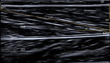
图1
腓肠肌内侧头矢状面声像图 注: GM, gastrocnemius, 腓肠肌; SO, soleus, 比目鱼肌; APO, aponeurosis, 腱膜; Lm, 可视范围内肌纤维长度"
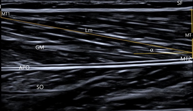
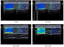
图2
SWE图像"

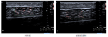
图3
SMI图像"
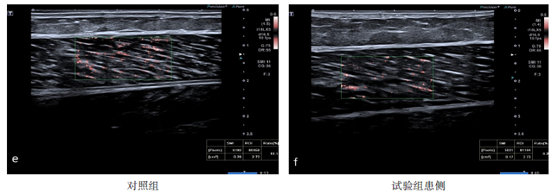
表2
两组静息状态下腓肠肌超声参数比较"
| 组别 | n | FL(mm) | MT(mm) | PA(°) | SWV(m/s) | SMI(%) |
|---|---|---|---|---|---|---|
| 对照组 | 46 | 48.05±2.15 | 13.75±2.68 | 13.07±3.23 | 2.69±0.40 | 7.49±2.11 |
| 试验组健侧 | 44 | 47.69±3.31 | 11.14±2.55 | 12.02±2.93 | 2.72±0.56 | |
| 试验组患侧 | 44 | 43.22±3.40 | 12.25±2.21 | 14.64±3.18 | 3.70±0.85 | 6.28±2.31 |
| t1值 | -6.235 | 2.198 | 4.006 | 6.346 | ||
| P1值 | < 0.001 | 0.031 | <0.001 | < 0.001 | ||
| t2值 | -8.073 | -2.881 | 2.326 | 7.119 | 2.604 | |
| P2值 | < 0.001 | 0.005 | 0.022 | < 0.001 | 0.011 |
表3
两组静息与最大等长收缩状态下腓肠肌FL差值比较(mm)"
| 组别 | n | 差值 |
|---|---|---|
| 对照组 | 46 | 10.91±4.23 |
| 试验组健侧 | 44 | 9.10±4.06 |
| 试验组患侧 | 44 | 3.52±1.94 |
| Z1值 | 2.055 | |
| P1值 | 0.040 | |
| Z2值 | 6.703 | |
| P2值 | < 0.001 | |
| Z3值 | 7.318 | |
| P3值 | < 0.001 |
| [1] |
FEIGIN V L, FOROUZANFAR M H, KRISHNAMURTHI R, et al. Global and regional burden of stroke during 1990-2010: findings from the global burden of disease study 2010[J]. Lancet, 2014, 383(9913):245-254.
doi: 10.1016/S0140-6736(13)61953-4 |
| [2] |
GAO J, RUBIN J M, CHEN J, et al. Ultrasound elastography to assess Botulinum toxin A treatment for post-stroke spasticity: a feasibility study[J]. Ultrasound Med Biol, 2019, 45(5):1094-1102.
doi: 10.1016/j.ultrasmedbio.2018.10.034 |
| [3] |
EBY S, ZHAO H, SONG P, et al. Quantitative evaluation of passive muscle stiffness in chronic stroke[J]. Am J Phys Med Rehabil, 2016, 95(12):899-910.
doi: 10.1097/PHM.0000000000000516 |
| [4] |
PRADINES M, GHEDIRA M, PORTERO R, et al. Ultrasound structural changes in triceps surae after a 1-year daily self-stretch program: a prospective randomized controlled trial in chronic hemiparesis[J]. Neurorehabil Neural Repair, 2019, 33(4):245-259.
doi: 10.1177/1545968319829455 |
| [5] |
GHASEMI E, KHADEMI-KALANTARI K, KHALKHALI-ZAVIEH M, et al. The effect of functional stretching exercises on neural and mechanical properties of the spastic medial gastrocnemius muscle in patients with chronic stroke: a randomized controlled trial[J]. J Stroke Cerebrovasc Dis, 2018, 27(7):1733-1742.
doi: 10.1016/j.jstrokecerebrovasdis.2018.01.024 |
| [6] | 秦鹍, 冯亚男, 李亚鹏, 等. 剪切波弹性成像技术量化评估肌腱肌肉弹性模量的信度[J]. 中国康复理论与实践, 2018, 24(10):1201-1205. |
| QIN K, FENG Y N, LI Y P, et al. Intra- and inter-rater reliability of shear wave elastic imaging technique for elastic modulus measurements of muscle and tendon[J]. Chin Rehabil Theory Pract, 2018, 24(10):1201-1205. | |
| [7] |
LEHOUX M C, SOBCZAK S, CLOUTIER F, et al. Shear wave elastography potential to characterize spastic muscles in stroke survivors: literature review[J]. Clin Biomech (Bristol, Avon), 2020, 72:84-93.
doi: 10.1016/j.clinbiomech.2019.11.025 |
| [8] | 中华医学会神经病学分会, 中华医学会神经病学分会脑血管病学组. 中国各类主要脑血管病诊断要点2019[J]. 中华神经科杂志, 2019, 52(9):710-715. |
| Chinese Society of Neurology, Chinese Stroke Society. Diagnosis of Cerebrovascular Diseases in China (version 2019)[J]. Chin J Neurol, 2019, 52(9):710-715. | |
| [9] |
IKEZOE T, MORI N, NAKAMURA M, et al. Atrophy of the lower limbs in elderly women: is it related to walking ability?[J]. Eur J Appl Physiol, 2011, 111(6):989-995.
doi: 10.1007/s00421-010-1728-8 |
| [10] | 甄希成, 陈新, 张辉. 持续被动运动治疗脑卒中患者下肢肌痉挛的效果[J]. 中国老年学杂志, 2017, 37(4):880-882. |
| ZHEN X C, CHEN X, ZHANG H. Effect of continuous passive exercise on treatment of lower extremity muscle spasm in patients with stroke[J]. Chin J Gerontol, 2017, 37(4):880-882. | |
| [11] |
KUO C, HU G. Post-stroke spasticity: a review of epidemiology, pathophysiology, and treatments[J]. Int J Gerontol, 2018, 12(4):280-284.
doi: 10.1016/j.ijge.2018.05.005 |
| [12] |
JAKUBOWSKI K L, TERMAN A, SANTANA R V C, et al. Passive material properties of stroke-impaired plantarflexor and dorsiflexor muscles[J]. Clin Biomech (Bristol, Avon), 2017, 49:48-55.
doi: 10.1016/j.clinbiomech.2017.08.009 |
| [13] | 樊留博, 韩文胜, 江莹莹, 等. 超声弹性成像评价脑卒中偏瘫肢体硬度对痉挛性偏瘫患者预后的影响[J]. 中华全科医学, 2017, 15(8):1323-1325. |
| FAN L B, HAN W S, JIANG Y Y, et al. Role of elastosonography in evaluating hemiplegic limb hardness and improving the prognosis of spastic hemiplegia patients[J]. Chin General Pract, 2017, 15(8):1323-1325. | |
| [14] |
HONG M J, PARK J B, LEE Y J, et al. Quantitative evaluation of post-stroke spasticity using neurophysiological and radiological tools: a pilot study[J]. Ann Rehabil Med, 2018, 42(3):384-395.
doi: 10.5535/arm.2018.42.3.384 |
| [15] | 邓思宇, 卢茜, 郄淑燕, 等. 等速测试指标与改良Ashworth量表用于踝痉挛评定的相关性研究[J]. 中国康复理论与实践, 2016, 22(2):178-183. |
| DENG S Y, LU Q, QIE S Y, et al. Correlation of isokinetic parameter and Modified Ashworth Scale applied in evaluation of ankle spasticity[J]. Chin Rehabil Theory Pract, 2016, 22(2):178-183. | |
| [16] |
DOS SANTOS A N, ROCHA N A C F. Immediate effect of kinesio taping on knee extensor torque of children with cerebral palsy: three case reports[J]. NeuroRehabilitation, 2018, 43(4):519-523.
doi: 10.3233/NRE-161921 |
| [17] |
MATHEVON L, MICHEL F, AUBRY S, et al. Two-dimensional and shear wave elastography ultrasound: a reliable method to analyse spastic muscles?[J]. Muscle Nerve, 2018, 57(2):222-228.
doi: 10.1002/mus.25716 |
| [18] |
DIAS C P, FREIRE B, GOULART N B, et al. Muscle architecture and torque production in stroke survivors: an observational study[J]. Top Stroke Rehabil, 2017, 24(3):206-213.
doi: 10.1080/10749357.2016.1210873 |
| [19] |
SON J, RYMER W Z, LEE S S M. Limited fascicle shortening and fascicle rotation may be associated with impaired voluntary force-generating capacity in pennate muscles of chronic stroke survivors[J]. Clin Biomech (Bristol, Avon), 2020, 75:105007.
doi: 10.1016/j.clinbiomech.2020.105007 |
| [20] | YANG Y B, ZHANG J, LENG Z P, et al. Evaluation of spasticity after stroke by using ultrasound to measure the muscle architecture parameters: a clinical study[J]. Int J Clin Exp Med, 2014, 7(9):2712-2717. |
| [21] |
D'SOUZA A, BOLSTERLEE B, HERBERT R D. Architecture of the medial gastrocnemius muscle in people who have had a stroke: a diffusion tensor imaging investigation[J]. Clin Biomech (Bristol, Avon), 2020, 74:27-33.
doi: 10.1016/j.clinbiomech.2020.02.004 |
| [22] |
THIELMAN G, YOUREY L. Ultrasound imaging of upper extremity spastic muscle post-stroke and the correlation with function: a pilot study[J]. NeuroRehabilitation, 2019, 45(2):213-220.
doi: 10.3233/NRE-192742 |
| [23] | 刘美快, 徐乐义, 李海燕, 等. 脑卒中患者小腿肌肉形态结构变化的定量超声研究[J]. 中国康复医学杂志, 2018, 33(10):1183-1187. |
| LIU M K, XU L Y, LI H Y, et al. A study on quantitative ultrasound on evaluating the architectural parameters of lower leg muscles in stroke survivors[J]. Chin J Rehabil Med, 2018, 33(10):1183-1187. | |
| [24] |
LEE S S M, SPEAR S, RYMER W Z. Quantifying changes in material properties of stroke-impaired muscle[J]. Clin Biomech (Bristol, Avon), 2015, 30(3):269-275.
doi: 10.1016/j.clinbiomech.2015.01.004 |
| [25] |
LECHARTE T, GROSS R, NORDEZ A, et al. Effect of chronic stretching interventions on the mechanical properties of muscles in patients with stroke: a systematic review[J]. Ann Phys Rehabil Med, 2020, 63(3):222-229.
doi: 10.1016/j.rehab.2019.12.003 |
| [26] |
LIEBER R L, ROBERTS T J, BLEMKER S S, et al. Skeletal muscle mechanics, energetics and plasticity[J]. J Neuroeng Rehabil, 2017, 14(1):108.
doi: 10.1186/s12984-017-0318-y |
| [27] |
FRIDÉN J, LIEBER R L. Spastic muscle cells are shorter and stiffer than normal cells[J]. Muscle Nerve, 2003, 27(2):157-164.
doi: 10.1002/mus.v27:2 |
| [28] |
LEE S S M, JAKUBOWSKI K L, SPEAR S C, et al. Muscle material properties in passive and active stroke-impaired muscle[J]. J Biomech, 2019, 83:197-204.
doi: 10.1016/j.jbiomech.2018.11.043 |
| [29] | LIU J, PAN H, BAO Y, et al. The value of real-time shear wave elastography before and after rehabilitation of upper limb spasm in stroke patients[J]. Biomed Res Int, 2020, 2020:1-7. |
| [30] |
VOLA E A, ALBANO M, DI LUISE C, et al. Use of ultrasound shear wave to measure muscle stiffness in children with cerebral palsy[J]. J Ultrasound, 2018, 21(3):241-247.
doi: 10.1007/s40477-018-0313-6 |
| [31] |
COSGROVE D, PISCAGLIA F, BAMBER J, et al. EFSUMB Guidelines and Recommendations on the Clinical Use of Ultrasound Elastography. Part 2: Clinical Applications[J]. Ultraschall Med, 2013, 34(3):238-253.
doi: 10.1055/s-00000089 |
| [32] |
LENG Y, WANG Z, BIAN R, et al. Alterations of elastic property of spastic muscle with its joint resistance evaluated from shear wave elastography and biomechanical model[J]. Front Neurol, 2019, 10:736.
doi: 10.3389/fneur.2019.00736 |
| [33] |
WU C, HO Y, HSIAO M Y, et al. Evaluation of post-stroke spastic muscle stiffness using shear wave ultrasound elastography[J]. Ultrasound Med Biol, 2017, 43(6):1105-1111.
doi: 10.1016/j.ultrasmedbio.2016.12.008 |
| [34] |
EBY S F, ZHAO H, SONG P, et al. Quantifying spasticity in individual muscles using shear wave elastography[J]. Radiol Case Rep, 2017, 12(2):348-352.
doi: 10.1016/j.radcr.2017.01.004 |
| [35] |
KWON D R, PARK G Y, LEE S U, et al. Spastic cerebral palsy in children: dynamic sonoelastographic findings of medial gastrocnemius[J]. Radiology, 2012, 263(3):794-801.
doi: 10.1148/radiol.12102478 |
| [36] |
LE SANT G, NORDEZ A, HUG F, et al. Effects of stroke injury on the shear modulus of the lower leg muscle during passive dorsiflexion[J]. J Appl Physiol (1985), 2019, 126(1):11-22.
doi: 10.1152/japplphysiol.00968.2017 |
| [37] |
ASKIN A, KALAYCI O T, BAYRAM K B, et al. Strain sonoelastographic evaluation of biceps muscle intrinsic stiffness after botulinum toxin-A injection[J]. Top Stroke Rehabil, 2017, 24(1):12-17.
doi: 10.1080/10749357.2016.1183865 |
| [38] |
HUANG M, MILLER T, YING M, et al. Whole-body vibration modulates leg muscle reflex and blood perfusion among people with chronic stroke: a randomized controlled crossover trial[J]. Sci Rep, 2020, 10(1):1473.
doi: 10.1038/s41598-020-58479-5 |
| [39] | 蓝晓锋, 姜凡, 彭梅, 等. SMI技术对血管性阴茎勃起功能障碍的诊断价值[J]. 中国超声医学杂志, 2018, 34(11):1028-1031. |
| LAN X F, JIANG F, PENG M, et al. The diagnosis value of SMI technology in vascular penile erectile dysfunction[J]. Chin J Ultrasound Med, 2018, 34(11):1028-1031. | |
| [40] |
JIANG Z Z, HUANG Y H, SHEN H L, et al. Clinical applications of superb microvascular imaging in the liver, breast, thyroid, skeletal muscle, and carotid plaques[J]. J Ultrasound Med, 2019, 38(11):2811-2820.
doi: 10.1002/jum.v38.11 |
| [41] |
CALISKAN E, AKKOC O, BAYRAMOGLU Z, et al. Effects of static stretching duration on muscle stiffness and blood flow in the rectus femoris in adolescents[J]. Med Ultrason, 2019, 21(2):136-143.
doi: 10.11152/mu-1859 |
| [1] | 林娜, 高菡璐, 卢惠苹, 陈燕清, 郑军凡, 陈述荣. 虚拟现实技术对脑卒中上肢功能影响的弥散张量成像研究[J]. 《中国康复理论与实践》, 2024, 30(1): 61-67. |
| [2] | 王昊懿, 史亚伟, 鲁俊, 许光旭. 主观垂直感知障碍对脑卒中患者功能影响的回顾性研究[J]. 《中国康复理论与实践》, 2024, 30(1): 68-73. |
| [3] | 陈珺雯, 陈谦, 陈程, 李淑月, 刘玲玲, 吴存书, 龚翔, 鲁俊, 许光旭. 改良八段锦身体活动对脑卒中患者心肺功能、运动功能和日常生活活动能力的效果[J]. 《中国康复理论与实践》, 2024, 30(1): 74-80. |
| [4] | 胡永林, 马颖, 窦超, 陆安民, 江小鸽, 宋新建, 肖玉华. 肩部控制训练联合神经松动术对脑卒中偏瘫患者肩痛及上肢功能的效果[J]. 《中国康复理论与实践》, 2024, 30(1): 81-86. |
| [5] | 王贺, 韩靓, 阚梦凡, 于少泓. 电刺激治疗脑卒中后肩手综合征有效性的系统评价与Meta分析[J]. 《中国康复理论与实践》, 2023, 29(9): 1048-1056. |
| [6] | 胡晓诗, 张琦, 岳青, 梁艳华, 李晓松, 冯啊美, 张燕庆. 矫形弹力绷带对痉挛性偏瘫脑性瘫痪患儿步态对称性和步行能力的效果[J]. 《中国康复理论与实践》, 2023, 29(9): 1083-1089. |
| [7] | 沈星星, 陈伟健, 李聪聪, 李俊毅, 王帅, 叶子璇, 向瑞安, 许学猛. 单侧腰椎间盘突出症患者椎旁肌功能特征[J]. 《中国康复理论与实践》, 2023, 29(9): 1098-1103. |
| [8] | 孙藤方, 任梦婷, 杨琳, 王耀霆, 王红雨, 闫兴洲. 高压氧治疗联合重复外周磁刺激干预脑卒中患者踝运动功能和平衡能力的效果[J]. 《中国康复理论与实践》, 2023, 29(8): 875-881. |
| [9] | 王亚楠, 刘西花. 脑卒中偏瘫患者主观和客观平衡功能测量的相关性及预测效能[J]. 《中国康复理论与实践》, 2023, 29(8): 890-895. |
| [10] | 王海云, 王寅, 周信杰, 何爱群. 基于“中枢-外周-中枢”理论的经颅直流电刺激结合针刺干预脑卒中患者中枢及上肢功能的效果[J]. 《中国康复理论与实践》, 2023, 29(8): 919-925. |
| [11] | 陈怡婷, 王倩, 崔慎红, 李映彩, 张思鈺, 魏衍旭, 任慧, 冷军, 陈斌. 双侧序贯重复经颅磁刺激干预脑卒中患者上肢运动功能的效果[J]. 《中国康复理论与实践》, 2023, 29(8): 926-932. |
| [12] | 李振亚, 孙洁, 郭鹏飞, 王光明. 脑卒中患者口期和咽期吞咽功能改变与误吸的相关性:基于电视透视吞咽检查[J]. 《中国康复理论与实践》, 2023, 29(8): 933-939. |
| [13] | 华玲, 张一楠, 郑玉, 孙俏仪, 房辉, 宋达. 手控节律音乐治疗对脑卒中后单侧空间忽略的效果[J]. 《中国康复理论与实践》, 2023, 29(7): 833-838. |
| [14] | 蒋孝翠, 刘臻, 苏清伦, 赵秦, 夏晓昧, 陆飞. 间歇性Theta节律经颅磁刺激对脑卒中后非流利性失语的影响[J]. 《中国康复理论与实践》, 2023, 29(7): 839-843. |
| [15] | 许苗苗, 李楠, 应颖, 杨凯翔, 杨婧瑞, 李杰, 邱彦群. 重复外周磁刺激对左右颈7神经交叉移位术后脑卒中患者上肢运动功能的效果[J]. 《中国康复理论与实践》, 2023, 29(6): 686-690. |
| 阅读次数 | ||||||
|
全文 |
|
|||||
|
摘要 |
|
|||||
|
||