



《中国康复理论与实践》 ›› 2022, Vol. 28 ›› Issue (4): 429-438.doi: 10.3969/j.issn.1006-9771.2022.04.009
蔡童欣1,雷敏聪1,周逸钧1,孟殿怀1,2( )
收稿日期:2021-12-14
修回日期:2022-03-29
出版日期:2022-04-25
发布日期:2022-05-05
通讯作者:
孟殿怀
E-mail:dhdream@126.com
作者简介:蔡童欣(1998-),女,汉族,山西运城市人,硕士研究生,主要研究方向:异位骨化、神经康复。
基金资助:
CAI Tongxin1,LEI Mincong1,ZHOU Yijun1,MENG Dianhuai1,2()
Received:2021-12-14
Revised:2022-03-29
Published:2022-04-25
Online:2022-05-05
Contact:
MENG Dianhuai
E-mail:dhdream@126.com
Supported by:摘要:
目的 系统综述获得性异位骨化(AHO)相关动物模型,为研究疾病防治方案提供可靠造模方法。方法 检索PubMed、Web of Science、中国知网、万方数据库自建库至2021年11月AHO动物模型相关文献,筛选并提取文献关键内容,采用文献归纳法对各型AHO动物模型进行分析与评价。结果 最终共纳入20篇动物实验研究相关文献,可归纳为创伤后和神经源性两大类异位骨化动物模型,用于模拟AHO的发生发展过程。创伤后异位骨化动物模型主要有肌肉损伤、跟腱切断、肌肉损伤联合关节制动、髋关节损伤、异位植入、爆炸伤和烧伤7种。神经源性异位骨化动物模型主要包括脊髓损伤和脑外伤。目前实验室中常用跟腱切断法和成骨因子植入法造模,此类方法可靠、易行、成功率高,但无法准确解释临床中复杂条件下异位骨化的发病机制。基于爆炸伤、烧伤、神经损伤等条件下造模方法正在完善,为临床研究某些特殊病因所致异位骨化的分子生物学机制及防治方案提供了支持。结论 目前存在的几种造模方法各有利弊,但均无法完整复制人类异位骨化的全部特征,因此,临床上在判定模型的选择方面尚无统一标准。针对疾病的不同病因,选用合适的动物模型,对不同类型AHO在早期制定有效干预措施至关重要。
蔡童欣,雷敏聪,周逸钧,孟殿怀. 获得性异位骨化动物模型的系统综述[J]. 《中国康复理论与实践》, 2022, 28(4): 429-438.
CAI Tongxin,LEI Mincong,ZHOU Yijun,MENG Dianhuai. Animal models of acquired heterotopic ossification: a systematic review[J]. 《Chinese Journal of Rehabilitation Theory and Practice》, 2022, 28(4): 429-438.

图1
文献筛选流程及结果"

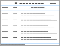
表1
纳入文献的基本特征"
| 纳入文献 | 国家 | 动物种属 | n | 模型类别 | 模型设计 | 造模周期 | 成功率 | 主要评估指标 |
|---|---|---|---|---|---|---|---|---|
| Walton等[ (1983) | 美国 | 绵羊 | 42 | 肌肉损伤模型 | 反复钝力作用于羊大腿深层肌肉致钝挫伤诱导异位骨化。 | 3~12周 | 16.6% | 组织学分析 |
| Moore-Lotridge等[ | 美国 | ABCC6缺陷小鼠 | 101 | 心脏毒素注射到双侧小腿后室肌诱导肌肉损伤,并抑制巨噬细胞介导的纳米羟基磷灰石消退。 | 4周 | 80% | X线 显微CT 组织学分析 能量色散 X 射线 | |
| Yu等[ | 中国 | 雄性SD大鼠 | 80 | 跟腱切断模型 | 第1组:右后肢跟腱切断。 第2组:仅同类皮肤切口。 | 10周 | 第1组:100%。 第2组:0%。 | X线 组织学分析 实时定量PCR |
| Tsailas等[ | 希腊 | 雄性新西兰白兔 | 18 | 肌肉损伤联合关节制动模型 | 石膏固定右膝和踝关节于过伸位+强制暴力活动按摩膝关节,每次5 min,每周6 d,持续5周。 | 10周 | 100% | X 线 |
| Anthonissen等[ | 德国 | 雄性Wistar大鼠 | 20 | 髋关节损伤模型 | 右侧髋关节大转子尖端内侧钻通髓腔,股骨管扩孔至2 mm,钳夹臀大肌、臀中肌3 min。 | 12周 | 90% | 显微CT 改良Brooker评分 |
| Anthonissen等[ | 德国 | 雄性Wistar大鼠 | 50 | 第1组:右髋关节手术+额外的关节周围肌肉损伤。 第2组:右髋关节手术+保护关节周围肌肉。 | 12周 | 第1组:96%,且程度更重。 第2组:86.9%。 | 显微CT 改良Brooker评分 | |
| Li等[ | 美国 | 雄性C57BL/6 J小鼠 | 4 | 异位植入模型 | 双小腿肌注含BMP-2的水凝胶,右侧小腿肌注射心脏毒素。 | 2周 | 左侧:100%。 右侧:100%,且更明显。 | 显微CT 组织学分析 实时定量PCR 蛋白质印迹 |
| Zotz等[ | 巴西 | 雄性Wistar大鼠 | 12 | 双侧髂嵴收集自体骨髓0.35 mL分别注入其双侧股四头肌中。 | 5周 | 100% | 血清碱性磷酸酶 组织学分析 火焰光度法评估股四头肌中钙含量 | |
| Polfer等[ | 美国 | 雄性SD大鼠 | 41 | 爆炸伤模型 | 第1组:单纯暴露于爆炸超压 (120±7) kPa中。 第2组:股骨骨折和周围肌肉挤压伤后,损伤区股骨截肢。 第3组:1+2组干预措施。 | 2~4周 | 第1组:0%。 第2组:66.6%。 第3组:100%。 | X线 显微CT 组织学分析 |
| Epperson等[ | 美国 | 雌性兰布莱绵羊 | 8 | 利用AID模拟军事场景爆炸,同时模拟战场中创伤因素(如骨损伤、止血带、细菌、负压创伤治疗)。 | 24周 | 63% | X线 显微CT 组织学分析 电子背散射分析 | |
| Pavey等[ | 美国 | 雄性SD大鼠 | 48 | 爆炸伤模型+截肢部位接种1×10⁶个细菌菌落。 第1组:接种耐甲氧西林金黄色葡萄球菌。 第2组:接种鲍曼不动杆菌。 第3组:接种不含细菌的载体。 | 12周 | 第1组:100%,且HO程度重。 第2组:100%。 第3组:100%。 | 显微CT 组织学分析 | |
| Tannous等[ | 美国 | 雄性SD大鼠 | 12 | 局部暴露于水下烈性炸药所致高能爆炸中,行指定部位(7只前肢,5只后肢)爆炸截肢。 | 4~24周 | 100% | X线 | |
| Peterson等[ | 美国 | C57BL/6 小鼠 | 50 | 烧伤模型 | 60 ℃铝块置于小鼠背部17秒,制造30%体表面积皮肤烫伤,后行左下肢跟腱切断术。 | 9~15周 | 100% | 显微CT |
| Debaud等[ | 法国 | 雌性C57B16小鼠 | 21 | 脊髓损伤模型 | 第1组:脊髓损伤手术+右后肢外周神经切断+术中双侧腘绳肌心脏毒素注射。 第2组:相同的手术程序+术后第10天双侧心脏毒素注射。 第3组 :假脊髓损伤手术+右后肢外周神经切断+术中双侧腘绳肌心脏毒素注射。 | 4周 | 第1组:100%,且失神经支配侧异位骨化体积显著增加; 第2组:0%; 第3组:50 %。 | 显微CT 组织学分析 实时定量PCR |
| Genêt等[ | 美国 | 雌性C57BL/6小鼠 | 113 | T7~T8间脊髓横断,且右侧腘绳肌内注射心脏毒素。 | 2周 | 99.12%,仅发生在注射心脏毒素侧肢体。 | X线 显微CT 组织学分析 实时定量PCR | |
| Kang等[ | 美国 | 雄性C57/BL6小鼠 | 24 | 实验组:T10节段脊髓完全性损伤,后肢四头肌注射不同剂量(0 g、0.25 g、0.5 g) BMP-2。 对照组:假脊髓损伤+相应剂量BMP-2肌注。 | 2周 | 0.5 µg BMP-2可显著增加四头肌异位骨化含量。 0.25 µg BMP-2仅使实验组产生异位骨化。 0 µg BMP-2不能诱发异位骨化。 | 显微CT 组织学分析 实时定量PCR | |
| Shi等[ | 中国 | 雄性 SD大鼠 | 66 | 脑损伤模型 | 重物撞击顶叶区制造轻度创伤性脑损伤结合右后肢跟腱切断术。 | 10周 | 100% | X线 组织学分析 |
| Ju等[ | 中国 | 雄性SD大鼠 | 44 | 联合脑外伤+烧伤+跟腱切断术。 | 10周 | 100% | X线 组织学分析 实时定量PCR Western Blotting检测 | |
| Brady等[ | 澳大利亚 | 雄性SD大鼠 | 36 | 第1组:三重损伤(脑外伤+股骨骨折+肌肉损伤)。 第2组:单纯外周损伤(假脑外伤+股骨骨折+肌肉损伤)。 第3组:假损伤对照组(假脑外伤+假股骨骨折+假肌肉损伤)。 | 6周 | 第1组:70%。 第2组:20%。 第3组;0%。 | 显微CT 组织学分析 实时定量PCR Western blotting检测 | |
| Anthonissen等[ | 德国 | 雄性Wistar大鼠 | 40 | 第1组:单纯行髋关节手术。 第2组:髋关节手术+中度脑外伤。 第3组:髋关节手术+重度脑外伤。 第4组:仅重度脑外伤。 | 12周 | 第1组:90%。 第2、3组:100%,且3组比2组骨化体积增加。 第4组:0%。 | 显微CT 组织学分析 |
| [1] |
ŁĘGOSZ P, DRELA K, PULIK Ł, et al. Challenges of heterotopic ossification: molecular background and current treatment strategies[J]. Clin Exp Pharmacol Physiol, 2018, 45(12):1229-1235.
doi: 10.1111/1440-1681.13025 |
| [2] |
DEY D, WHEATLEY B M, CHOLOK D, et al. The traumatic bone: trauma-induced heterotopic ossification[J]. Transl Res, 2017, 186:95-111.
doi: 10.1016/j.trsl.2017.06.004 |
| [3] |
MUJTABA B, TAHER A, FIALA M J, et al. Heterotopic ossification: radiological and pathological review[J]. Radiol Oncol, 2019, 53(3):275-284.
doi: 10.2478/raon-2019-0039 |
| [4] | CARPENTIER V T, SALGA M, GATIN L, et al. Early diagnosis of heterotopic ossification among patients admitted to a neurological Post-Intensive Care Rehabilitation Unit[J]. Eur J Phys Rehabil Med, 2021, 57(4):527-534. |
| [5] |
CAPPATO S, GAMBERALE R, BOCCIARDI R, et al. Genetic and acquired heterotopic ossification: a translational tale of mice and men[J]. Biomedicines, 2020, 8(12):611.
doi: 10.3390/biomedicines8120611 |
| [6] |
ALEXANDER K A, TSENG H W, SALGA M, et al. When the nervous system turns skeletal muscles into bones: how to solve the conundrum of neurogenic heterotopic ossification[J]. Curr Osteoporos Rep, 2020, 18(6):666-676.
doi: 10.1007/s11914-020-00636-w |
| [7] | 曾林如, 罗淦, 朱芳兵, 等. 异位骨化预防与治疗的研究进展[J]. 中国骨伤, 2020, 33(3):283-287. |
| ZENG L R, LUO G, ZHU F B, et al. Progress on prevention and treatment of heterotopic ossification[J]. Chin J Orthop Traumatol, 2020, 33(3):283-287. | |
| [8] |
LI L, TUAN R S. Mechanism of traumatic heterotopic ossification: in search of injury-induced osteogenic factors[J]. J Cell Mol Med, 2020, 24(19):11046-11055.
doi: 10.1111/jcmm.15735 |
| [9] | EISENSTEIN N, STAPLEY S, GROVER L. Post-traumatic heterotopic ossification: an old problem in need of new solutions[J]. J Orthop Res, 2018, 36(4):1061-1068. |
| [10] | KAN L, KESSLER J A. Animal models of typical heterotopic ossification[J]. J Biomed Biotechnol, 2011, 2011:309287. |
| [11] |
LI L, JIANG Y, LIN H, et al. Muscle injury promotes heterotopic ossification by stimulating local bone morphogenetic protein-7 production[J]. J Orthop Translat, 2019, 18:142-153.
doi: 10.1016/j.jot.2019.06.001 |
| [12] | WALTON M, ROTHWELL A G. Reactions of thigh tissues of sheep to blunt trauma[J]. Clin Orthop Relat Res, 1983(176):273-281. |
| [13] |
MOORE-LOTRIDGE S N, LI Q, GIBSON B H Y, et al. Trauma-induced nanohydroxyapatite deposition in skeletal muscle is sufficient to drive heterotopic ossification[J]. Calcif Tissue Int, 2019, 104(4):411-425.
doi: 10.1007/s00223-018-0502-5 |
| [14] | ZHANG Q, ZHANG Y, YAN M, et al. βig-h3 enhances chondrogenesis via promoting mesenchymal condensation in rat Achilles tendon heterotopic ossification model[J]. Aging (Albany NY), 2020, 12(8):7030-7041. |
| [15] |
YU D, JU J, XUE F, et al. Expression and significance of related genes in the early stage of post-traumatic heterotopic ossification in a rat model of Achilles tenotomy[J]. Acta Orthop Traumatol Turc, 2021, 55(2):94-101.
doi: 10.5152/j.aott.2021.18480 |
| [16] |
TSAILAS P G, BABIS G C, NIKOLOPOULOS K, et al. The effectiveness of two COX-2 inhibitors in the prophylaxis against heterotopic new bone formation: an experimental study in rabbits[J]. J Surg Res, 2009, 151(1):108-114.
doi: 10.1016/j.jss.2007.12.804 |
| [17] | 叶勇光, 向孝兵, 熊平, 等. 两种异位骨化造模方法的比较研究[J]. 中国中医骨伤科杂志, 2010, 18(2):9-11. |
| YE Y G, XIANG X B, XIONG P, et al. Comparative research on two modeling methods of heterotopic ossification[J]. Chin J Tradit Med Traumatol Orthop, 2010, 18(2):9-11. | |
| [18] |
HÜRLIMANN M, SCHIAPPARELLI F F, ROTIGLIANO N, et al. Influence of surgical approach on heterotopic ossification after total hip arthroplasty: is minimal invasive better? A case control study[J]. BMC Musculoskelet Disord, 2017, 18(1):1-9.
doi: 10.1186/s12891-016-1361-8 |
| [19] | ANTHONISSEN J, OSSENDORF C, HOCK J L, et al. A new small-animal model for the study of acquired heterotopic ossification after hip surgery[J]. Acta Orthop Traumatol Turc, 2015, 49(2):197-202. |
| [20] |
ANTHONISSEN J, OSSENDORF C, HOCK J L, et al. The role of muscular trauma in the development of heterotopic ossification after hip surgery: an animal-model study in rats[J]. Injury, 2016, 47(3):613-616.
doi: 10.1016/j.injury.2015.11.019 |
| [21] |
HAYASHI C, HASEGAWA U, SAITA Y, et al. Osteoblastic bone formation is induced by using nanogel-crosslinking hydrogel as novel scaffold for bone growth factor[J]. J Cell Physiol, 2009, 220(1):1-7.
doi: 10.1002/jcp.21760 |
| [22] |
LEBLANC E, TRENSZ F, HAROUN S, et al. BMP-9-induced muscle heterotopic ossification requires changes to the skeletal muscle microenvironment[J]. J Bone Miner Res, 2011, 26(6):1166-1177.
doi: 10.1002/jbmr.311 |
| [23] |
ZOTZ T G, PAULA J B, MOSER A D. Experimental model of heterotopic ossification in Wistar rats[J]. Braz J Med Biol Res, 2012, 45(6):497-501.
doi: 10.1590/S0100-879X2012007500049 |
| [24] |
JAFFE D E, YOO D, BLEVINS J, et al. Does blast medium affect heterotopic ossification in a blast-amputation model?[J]. Clin Orthop Relat Res, 2015, 473(8):2680-2687.
doi: 10.1007/s11999-015-4320-z |
| [25] | EPPERSON R T, ISAACSON B M, ROTHBERG D L, et al. Developing a combat-relevant translatable large animal model of heterotopic ossification[J]. Bone Rep, 2021, 15:101127. |
| [26] |
POLFER E M, HOPE D N, ELSTER E A, et al. The development of a rat model to investigate the formation of blast-related post-traumatic heterotopic ossification[J]. Bone Joint J, 2015, 97-B(4):572-576.
doi: 10.1302/0301-620X.97B4.34866 |
| [27] | QIN M Y, ATWOOD R E, KETCHUM W A, et al. Characterization of serum levels of testosterone and corticosterone in a blast and amputation rat model of heterotopic ossification[J]. Mol Cell Endocrinol, 2020, 509:110799. |
| [28] |
PAVEY G J, QURESHI A T, HOPE D N, et al. Bioburden increases heterotopic ossification formation in an established rat model[J]. Clin Orthop Relat Res, 2015, 473(9):2840-2847.
doi: 10.1007/s11999-015-4272-3 |
| [29] |
TANNOUS O, GRIFFITH C, O'TOOLE R V, et al. Heterotopic ossification after extremity blast amputation in a Sprague-Dawley rat animal model[J]. J Orthop Trauma, 2011, 25(8):506-510.
doi: 10.1097/BOT.0b013e31821f6265 |
| [30] |
ROBERTSON A D, CHIARAMONTI A M, NGUYEN T P, et al. Failure of indomethacin and radiation to prevent blast-induced heterotopic ossification in a Sprague-Dawley rat model[J]. Clin Orthop Relat Res, 2019, 477(3):644-654.
doi: 10.1097/CORR.0000000000000594 |
| [31] | WILLIAMS D L, EPPERSON R T, TAYLOR N B, et al. System setup to deliver air impact forces to a sheep limb: preparation for model development of blast-related heterotopic ossification[J]. JMIR Res Protoc, 2019, 8(2):e12107. |
| [32] |
RANGANATHAN K, LODER S, AGARWAL S, et al. Heterotopic ossification: basic-science principles and clinical correlates[J]. J Bone Joint Surg Am, 2015, 97(13):1101-1111.
doi: 10.2106/JBJS.N.01056 |
| [33] | PETERSON J R, AGARWAL S, BROWNLEY R C, et al. Direct mouse trauma/burn model of heterotopic ossification[J]. J Vis Exp, 2015(102):e52880. |
| [34] | STURBOIS-NACHEF N, GATIN L, SALGA M, et al. Neurogenic heterotopic ossification in the upper limb[J]. Hand Surg Rehabil, 2022, 41S:S167-S174. |
| [35] | DEBAUD C, SALGA M, BEGOT L, et al. Peripheral denervation participates in heterotopic ossification in a spinal cord injury model[J]. PLoS One, 2017, 12(8):e0182454. |
| [36] |
ZAKRASEK E C, YURKIEWICZ S M, DIRLIKOV B, et al. Use of nonsteroidal anti-inflammatory drugs to prevent heterotopic ossification after spinal cord injury: a retrospective chart review[J]. Spinal Cord, 2019, 57(3):214-220.
doi: 10.1038/s41393-018-0199-3 |
| [37] |
GENÊT F, KULINA I, VAQUETTE C, et al. Neurological heterotopic ossification following spinal cord injury is triggered by macrophage-mediated inflammation in muscle[J]. J Pathol, 2015, 236(2):229-240.
doi: 10.1002/path.4519 |
| [38] |
KANG H, DANG A B, JOSHI S K, et al. Novel mouse model of spinal cord injury-induced heterotopic ossification[J]. J Rehabil Res Dev, 2014, 51(7):1109-1118.
doi: 10.1682/JRRD.2014.01.0019 |
| [39] |
DANG A B, TAY B K, KIM H T, et al. Inhibition of MMP2/MMP9 after spinal cord trauma reduces apoptosis[J]. Spine (Phila Pa 1976), 2008, 33(17):E576-E579.
doi: 10.1097/BRS.0b013e31817ecc87 |
| [40] |
SALGA M, TSENG H W, ALEXANDER K A, et al. Blocking neuromuscular junctions with Botulinum toxin A injection enhances neurological heterotopic ossification development after spinal cord injury in mice[J]. Ann Phys Rehabil Med, 2019, 62(3):189-192.
doi: 10.1016/j.rehab.2019.01.005 |
| [41] | ANTHONISSEN J, STEFFEN C T, HOFMANN A, et al. The pathogenesis of heterotopic ossification after traumatic brain injury. A review of current literature[J]. Acta Orthop Belg, 2020, 86(3):369-377. |
| [42] |
SHI W Z, JU J Y, XIAO H J, et al. Dynamics of MMP-9, MMP-2 and TIMP-1 in a rat model of brain injury combined with traumatic heterotopic ossification[J]. Mol Med Rep, 2017, 15(4):2129-2135.
doi: 10.3892/mmr.2017.6275 |
| [43] |
JU J, YU D, XUE F, et al. Inhibition of NF-ҝB prevents trauma-induced heterotopic ossification in rat model[J]. Connect Tissue Res, 2019, 60(3):304-310.
doi: 10.1080/03008207.2018.1530771 |
| [44] | BRADY R D, ZHAO M Z, WONG K R, et al. A novel rat model of heterotopic ossification after polytrauma with traumatic brain injury[J]. Bone, 2020, 133:115263. |
| [45] |
BRADY R D, CASILLAS-ESPINOSA P M, AGOSTON D V, et al. Modelling traumatic brain injury and posttraumatic epilepsy in rodents[J]. Neurobiol Dis, 2019, 123:8-19.
doi: 10.1016/j.nbd.2018.08.007 |
| [46] |
ANTHONISSEN J, STEFFEN C T, ALESSANDRI B, et al. Traumatic brain injury enhances the formation of heterotopic ossification around the hip: an animal model study[J]. Arch Orthop Trauma Surg, 2020, 140(8):1029-1035.
doi: 10.1007/s00402-019-03326-0 |
| [47] | AMPADIOTAKI M M, EVANGELOPOULOS D S, PALLIS D, et al. New strategies in neurogenic heterotopic ossification[J]. Cureus, 2021, 13(4):e14709. |
| [48] |
PACIFICI M. Acquired and congenital forms of heterotopic ossification: new pathogenic insights and therapeutic opportunities[J]. Curr Opin Pharmacol, 2018, 40:51-58.
doi: 10.1016/j.coph.2018.03.007 |
| [1] | 杨同念, 任杰, 肖辉. 儿童青少年身体活动对焦虑的效果:系统综述的系统综述[J]. 《中国康复理论与实践》, 2025, 31(8): 883-888. |
| [2] | 王晓锋, 胡梦巧, 汪嫣, 魏坤, 徐文竹, 任丹, 马晔. 外骨骼机器人辅助步态训练对脑卒中和脊髓损伤下肢功能康复效果的系统综述[J]. 《中国康复理论与实践》, 2025, 31(8): 914-921. |
| [3] | 顾逸青, 程舒芃, 李勇强, 毛二莉, 励建安. 成年气管切开患者拔管的直接策略与堵管策略对比的系统综述[J]. 《中国康复理论与实践》, 2025, 31(6): 666-673. |
| [4] | 王梅, 朱文平. 身体活动干预2型糖尿病患者的健康效益:系统综述的系统综述[J]. 《中国康复理论与实践》, 2025, 31(5): 553-560. |
| [5] | 叶绿, 常晶, 凌海润. 网络行为成瘾的运动干预效果:系统综述的系统综述[J]. 《中国康复理论与实践》, 2025, 31(4): 431-440. |
| [6] | 朱志强, 徐巧玲, 邵雪云. 肥胖和超重儿童参与身体活动的健康效益:系统综述的系统综述[J]. 《中国康复理论与实践》, 2025, 31(3): 296-305. |
| [7] | 蔡倩, 张溪, 苏海荣, 刘娜, 黄颖, 李际强, 夏金根, 郑德采. 吸气肌训练对脱机失败者脱机疗效的系统综述[J]. 《中国康复理论与实践》, 2025, 31(3): 306-313. |
| [8] | 徐晓红, 钟晓珂, 孙思怡, 张祁, 程怀春. 轻度认知障碍老年人参与艺术治疗心理健康效益的系统综述[J]. 《中国康复理论与实践》, 2025, 31(2): 165-171. |
| [9] | 段富文, 张倍齐, 李丽. 运动干预对运动相关性脑震荡预防作用的系统综述[J]. 《中国康复理论与实践》, 2025, 31(12): 1385-1397. |
| [10] | 杨文睿, 崔思栋, 曾莉. 虚拟与增强现实对孤独症谱系障碍儿童青少年认知、情绪和适应性行为干预效果的系统综述[J]. 《中国康复理论与实践》, 2024, 30(9): 1026-1033. |
| [11] | 周静, 张晓晓, 丁忠冰, 陈建超, 魏星星, 林姝琦. 智力与发展性残疾儿童言语与语言障碍康复的系统综述[J]. 《中国康复理论与实践》, 2024, 30(8): 894-902. |
| [12] | 杨家坤, 张晓侠. 适应性体育活动对智力与发展性残疾儿童功能、活动和参与效益的系统综述[J]. 《中国康复理论与实践》, 2024, 30(7): 768-777. |
| [13] | 唐乐天, 黄兆欣, 刘超, 肖晓飞. 前交叉韧带重建术后患者高血流限制训练康复效果的系统综述[J]. 《中国康复理论与实践》, 2024, 30(7): 789-796. |
| [14] | 魏晓微. 数字赋能技术促进智力与发展性残疾儿童身体活动和健康的系统综述[J]. 《中国康复理论与实践》, 2024, 30(5): 497-504. |
| [15] | 王红志, 杨剑. 虚拟现实技术在脑性瘫痪儿童青少年身体活动和健康中的应用:系统综述的系统综述[J]. 《中国康复理论与实践》, 2024, 30(5): 505-512. |
| 阅读次数 | ||||||
|
全文 |
|
|||||
|
摘要 |
|
|||||
|
||
