



《中国康复理论与实践》 ›› 2022, Vol. 28 ›› Issue (9): 1065-1073.doi: 10.3969/j.issn.1006-9771.2022.09.009
封盼盼1,孙武东2,曲艺2,宋鹏飞2,马明2( )
收稿日期:2022-03-16
修回日期:2022-04-15
出版日期:2022-09-25
发布日期:2022-10-08
通讯作者:
马明
E-mail:nj9868@163.com
作者简介:封盼盼(1997-),女,汉族,江苏连云港市人,硕士研究生,主要研究方向:产后康复。|马明(1980-),男,回族,副主任治疗师,主要研究方向:运动损伤康复。
基金资助:
FENG Panpan1,SUN Wudong2,QU Yi2,SONG Pengfei2,MA Ming2()
Received:2022-03-16
Revised:2022-04-15
Published:2022-09-25
Online:2022-10-08
Contact:
MA Ming
E-mail:nj9868@163.com
Supported by:摘要:
腹直肌分离是产后女性常见疾病,会严重影响患者的生理和心理健康。康复治疗是腹直肌分离的主要干预方式。神经肌肉电刺激和运动疗法均能有效治疗产后腹直肌分离,但最佳治疗方案尚未明确。后续还需开展多中心、大样本研究,对治疗参数进行比较。
中图分类号:
封盼盼,孙武东,曲艺,宋鹏飞,马明. 产后腹直肌分离康复治疗研究进展[J]. 《中国康复理论与实践》, 2022, 28(9): 1065-1073.
FENG Panpan,SUN Wudong,QU Yi,SONG Pengfei,MA Ming. Advance in rehabilitation therapy with diastasis recti abdominis in postpartum women (review)[J]. 《Chinese Journal of Rehabilitation Theory and Practice》, 2022, 28(9): 1065-1073.
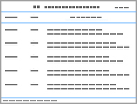
表1
NMES治疗产后DRA"
| 作者 | n | 分组 | 干预参数 | 评估指标 | 研究结论 | |
|---|---|---|---|---|---|---|
| Kamel等[ | 60 | NMES组(n = 30):4片电极置于双侧腹直肌上下,频率、脉宽、通断比分别为80 Hz/0.1~0.5 ms/5 s:10 s/30 min NMES+运动组(n = 30):NMES同上,运动包括卷腹、俄罗斯转体、俯卧抬腿、仰卧抬腿训练 | 介入时间:产后8周 治疗时间:每次30 min,每周3次,共8周 | IRD BMI 腰臀比 腹肌力量 | 两组所有指标均显著改善,NMES+运动组除了BMI外,其他指标改善程度均优于NMES组 | |
| 牛蕾蕾等[ | 117 | 电刺激组(n = 60):4片电极置于双侧腹直肌上下,设定频率,脉宽、通断比为20 Hz/300 μs /5 s:10 s,30 min,再将电极移至双侧的腹内、外斜肌处,以同样参数治疗30 min 对照组(n = 57):空白对照 | 介入时间:产后第1天 治疗时间:每天60 min,共5 d | IRD POP-Q 阴道动态压力 | 两组POP-Q评分、阴道动态压力在产后6~8周均无显著差异;NMES组IRD明显减小 | |
| 华永萍等[ | 84 | 运动组(n = 21):站姿和跪姿收腹、仰卧抬腿、仰卧蹬腿、平板支撑 运动+电刺激组(n = 21):将电极片置于双侧腹直肌、腹横肌和腹斜肌,持续时间为75 Hz/400 μs/7 s,30 Hz/200 μs/8 s,4 Hz/300 μs/11 s,3 Hz/150 μs/5 s,20 min;运动同上 运动+肌贴组(n = 21):一字贴垂直于腹直肌,拉力为0,X贴布垂直于腹直肌,拉力80%;运动同上 运动+电刺激+肌贴组(n = 21):同上 | 介入时间:产后6~24周 治疗时间:每天20 min,共2周 | IRD VAS 腰围 | 四组所有指标均得到改善,其中NMES联合肌贴治疗效果最佳 | |
| 邹燕齐等[ | 90 | NMES组(n = 30):8片电极分别置于双侧腹直肌、腹横肌和腹内、外斜肌上,采用75 Hz/400 μs/7 s, 30 Hz/200 μs/8 s, 4 Hz/300 μs/11 s, 3 Hz/150 μs/5 s进行电刺激 悬吊组(n = 30):进行中立位控制训练 NMES+悬吊组(n = 30):同上 | 介入时间:产后6~24周 治疗时间:每次30 min,共10 d | IRD | 三组IRD均显著降低,NMES组和悬吊组无显著差异,联合组疗效显著优于其他两组 | |
表2
深层核心稳定训练对IRD的作用"
| 作者 | n | 分组 | 测量方式 | 研究结论 |
|---|---|---|---|---|
| Mota等[ (2015) | 84 | 妊娠35~41周:收腹,卷腹 产后6~8周:收腹,卷腹 产后12~14周:收腹,卷腹 产后24~26周:收腹,卷腹 | 超声检查(脐上2 cm,脐上5 cm,脐下2 cm) | 四组患者进行卷腹时IRD均减小;收腹运动则使脐下IRD增加 |
| Theodorsen等[ | 38 | 盆底肌收缩 盆底肌收缩+收腹 | 超声检查(脐上2 cm,脐下2 cm) | 两种训练均使IRD增加,联合收缩时增幅最明显 |
| Gluppe等[ | 38 | 抬头运动 卷腹运动 收腹运动 盆底肌收缩 盆底肌收缩+卷腹 盆底肌收缩+收腹 骨盆的钟摆运动 俄罗斯转体 | 超声检查(脐上2 cm,脐下2 cm) | 抬头和躯干扭转运动减少IRD,卷腹运动减少脐上IRD 盆底肌收缩、收腹、盆底肌收缩联合收腹运动均增加脐下IRD |
| Sancho等[ | 38 | 收腹 卷腹 | 超声检查 (脐上2 cm,脐下2 cm) | 卷腹运动期间脐上IRD减少,收腹和联合卷腹练习时,脐上IRD没有显著差异 |
| Carrera等[ | 98 | 收腹 卷腹 降压运动 | 超声检查 (脐中、脐和剑突之间) | 腹横肌预激活(收腹)增加IRD,但卷腹和降压运动对IRD没有影响 |
| Lee等[ (2016) | 43 | 产后组(n = 26):卷腹,收腹,联合卷腹(先收腹再卷腹) 非产后组(n = 17):同上 | 超声检查 (脐中、脐和剑突之间) | 卷腹之前进行预激活(收腹)使产后DRA患者的IRD增加 |
表3
深层核心稳定性训练对产后DRA的影响"
| 作者 | n | 分组与干预 | 干预参数 | 评估指标 | 研究结论 |
|---|---|---|---|---|---|
| Tuttle等[ (2018) | 30 | 核心锻炼组(n = 10):腹式呼吸、侧平板、死虫训练、坐姿收腹,每个动作重复10次 肌贴组(n = 8):连续佩戴4~7 d,两次治疗间休息2~4 d 联合组(n = 5):同上 空白对照组(n = 7) | 介入时间:产后6~12周 治疗时间:每次30 min,每周4~5次,共12周 | IRD RDQ PFDI-20 | 核心锻炼组和联合组IRD显著降低 |
| 邹燕齐等[ (2020) | 90 | 悬吊组(n = 30):进行仰卧位、俯卧位、侧卧位、中立位控制训练 电刺激组(n = 30):8个电极片,分别置于双侧腹内、外斜肌、腹横肌和腹直肌上 联合组(n = 30):同上 | 介入时间:产后6~24周 治疗时间: 每次30 min,每周5次,共4周 | IRD | 三组IRD均显著降低,电刺激组和悬吊组疗效相似,联合组疗效最好 |
| Awad等[ (2021) | 50 | 平板运动组(n = 25):俯卧中立位平板支撑,单侧平板支撑,巴氏球上平板支撑(3组,每组20次,每次1 min,组间休息2 min) 对照组(n = 25):束腹带固定 | 介入时间:产后12~24周 治疗时间:每次30 min,每周3~4次,共8周 | IRD | 两组IRD均显著降低,但接受渐进式俯卧平板运动的患者IRD改善更明显 |
| Laframboise等[ | 8 | 核心锻炼组(n = 7):以腹式呼吸为主,重点进行腹部等长收缩训练(臀桥、死虫训练) 对照组(n = 1):空白对照 | 介入时间:妊娠24周 治疗时间:每次30 min,每周3次,共12周 | IRD 体质量 UHBE | 核心锻炼组体质量和UHBE评分无显著改善,但休息和活动时的IRD均显著减小 |
| Keshwani等[ | 32 | 核心锻炼组(n = 8):毛巾支撑腹壁,进行收腹、死虫训练,坐姿收腹,渐进式侧平板运动 束腹带组(n = 8):除洗澡和睡觉外全天佩戴 联合组(n = 8):同上 空白对照组(n = 8) | 介入时间:产后3周 治疗时间:每次30 min,每周4~6次,共12周 | IRD 腹肌力量和耐力 VAS PFDI ODI 身体满意度 | 核心锻炼组IRD无改善,但腹部力量得到增强;联合组腹部力量改善最明显 |
| Kazmi等[ (2021) | 40 | 核心锻炼组(n = 20):收腹、坐姿收腹、收腹下蹲、俯卧撑 空白对照组(n = 20) | 治疗时间:每次30 min,每周5次,共8周 | IRD 腹肌力量 | 核心锻炼能缩小IRD,且能增强腹部肌肉力量 |
表4
腹壁强化训练对产后DRA的影响"
| 作者 | n | 分组与干预 | 干预参数 | 评估指标 | 研究结论 |
|---|---|---|---|---|---|
| Sancho等[ | 38 | 收腹运动 卷腹运动 | 介入时间:产后10~12周 | IRD | 卷腹运动期间脐上IRD显著缩小 |
| Mota等[ (2015) | 84 | 妊娠35~41周:收腹,卷腹 产后6~8周:收腹,卷腹 产后12~14周:收腹,卷腹 产后24~26周:收腹,卷腹 | - | IRD | 四组IRD均减小,收腹运动使脐下IRD增加 |
| Pascoal等[ | 20 | 卷腹 | 介入时间:产后24周之内 | IRD | 患者进行卷腹运动时IRD降低 |
| Lee等[ (2016) | 43 | 产后组(n = 26):卷腹,收腹联合卷腹(先收腹再卷腹) 非产后组(n = 17):同上 | - | IRD | 卷腹前进行腹横肌预激活(收腹)使产后DRA患者的IRD增加,但腹外斜肌畸形指数显著减小 |
| Gluppe等[ | 38 | 抬头运动 卷腹运动 盆底肌收缩 盆底肌收缩+卷腹:盆底肌收缩,卷腹 盆底肌收缩+收腹:盆底肌收缩,收腹 骨盆的钟摆运动 俄罗斯转体 | 介入时间:产后6周及以上 | IRD | 抬头和躯干扭转运动使患者的IRD降低,卷腹运动使患者脐上IRD降低。盆底肌收缩、收腹、盆底肌收缩联合收腹运动均使患者脐下IRD增加 |
| Acharry等[ | 30 | 运动:收腹,卷腹,俄罗斯转体,骨盆钟摆运动,每个动作重复5~7次 | 介入时间:产后4周 治疗时间:每天2次天,共2周 | IRD | 运动显著降低IRD |
| Carrera等[ | 98 | 收腹运动 卷腹运动 降压运动 | - | 腹横肌预激活(收腹)增加IRD,卷腹和降压运动对IRD没有影响 | |
| El-Mekawy等[ | 30 | 腹部锻炼组:收腹、骨盆后倾、仰卧起坐、俄罗斯转体,每个动作保持5 s,休息10 s,重复20次 束腹带组:除洗澡睡觉外全天佩戴 | 介入时间:产后2周 治疗时间:每次30 min,每周3次,共6周 | IRD 腹部力量腰臀比 | 两组腰围无变化,但腹部锻炼降低了腰臀比,与束腹带治疗相比,IRD显著减小,腹部力量显著增加 |
| Thabet等[ (2019) | 40 | 核心锻炼+腹部锻炼组(n = 20):佩戴束腹带,进腹式呼吸、收腹、盆底肌收缩、骨盆后倾、俯卧蹬腿、俄罗斯转体等 腹部锻炼组(n = 20):佩戴束腹带,骨盆后倾、俯卧蹬腿、俄罗斯转体等 | 治疗时间:每次30 min,每周3次,共8周 | IRD PF10 | 核心锻炼联合腹部锻炼使IRD降低,生活质量提高 |
| Kamel等[ (2017) | 60 | 运动组(n = 30):卷腹、俄罗斯转体、俯卧蹬腿、死虫训练,每个动作20次,每周增加4次 运动+电刺激组(n = 30):运动同上,电刺激的四个电极分别置于双侧腹直肌上下进行电刺激,30 min | 介入时间:产后8周 治疗时间:每次30 min,每周3次,共8周 | BMI 腰臀比IRD 腹肌力量 | 两组所有指标均显著改善;联合组除了BMI外,其他指标均优于单独运动组 |
表5
盆底肌训练对产后DRA的影响"
| 作者 | n | 分组与干预 | 干预参数 | 评估指标 | 研究结论 |
|---|---|---|---|---|---|
| Mesquita等[ | 50 | 观察组(n = 25):从产后6 h开始运动,包括腹式呼吸、扭转卷腹、骨盆后倾、髋关节内收等刺激盆底肌收缩的运动,每个动作重复10~20次 对照组(n = 25):产后8 h开始,运动方式同上 | 介入时间:产后第1天 治疗时间:6周 | IRD | 两组IRD均降低,产后6 h开始运动使IRD降低12.5%,产后18 h开始运动使IRD降低5.4% |
| Gluppe等[ | 175 | 盆底肌训练组(n = 87):在5个不同体位进行,每个体位进行3组,8~12次最大收缩,分为6~8 s持续收缩和3~4次快速收缩 对照组(n = 88):不干预 | 介入时间:产后6周 治疗时间: 每次45 min,每周1次,共16周 | IRD | 两组在产后6个月和12个月DRA患病率无显著性差异,且运动组产后1年仍有40%的患病率 |
| Theodorsen等[ (2022) | 38 | 盆底肌收缩 收腹 盆底肌收缩+收腹 | 介入时间:产后24周以内 | IRD 尿失禁问卷 ODI骨盆带症状问卷 | 三种训练均使IRD增加,其中联合收缩时IRD的增幅最明显 |
| Gluppe等[ | 38 | 抬头运动 卷腹运动 收腹运动 盆底肌收缩 盆底肌收缩+卷腹:保持盆底肌收缩再进行卷腹 盆底肌收缩+收腹:保持盆底肌收缩再进行收腹 骨盆的钟摆运动 俄罗斯转体:仰卧位,肩胛骨和头部抬离床面的同时身体向一侧侧旋 | 介入时间:产后6周以上 | IRD | 抬头和躯干扭转运动使患者IRD降低,卷腹运动使患者脐上IRD降低。盆底肌收缩、收腹、盆底肌收缩联合收腹运动均使患者脐下IRD增加 |
表6
束腹带和肌内效贴对产后DRA的影响"
| 作者 | n | 分组与干预 | 干预参数 | 评估指标 | 研究结论 |
|---|---|---|---|---|---|
| Keshwani等[ | 32 | 束腹带组(n = 8) 运动组(n = 8):毛巾固定腹部下进行收腹、卷腹、侧平板和死虫运动等运动,每周4~6次,每次30 min以上 运动+束腹带组(n = 8):同上 对照组(n = 8):不干预 | 介入时间:产后3周 治疗时间:12周 | 国际体育活动问卷; IRD ODI 躯干肌的强度和耐力 | 运动组和束腹带组IRD没有得到改善,运动使IRD有所增加,但显著增强腹部力量;联合组腹部力量的增加可持续到24周 |
| El-Mekawy等[ | 30 | 束腹带组(n = 15) 腹部锻炼组(n = 15):收腹、骨盆后倾、仰卧起坐、俄罗斯转体,每个动作保持5 s,休息10 s,重复20次,30 min | 介入时间:产后第2天 治疗时间:6周 | BMI IRD 腹肌力量 腰臀比 | 两组腰臀比显著降低,腹部锻炼组IRD和腹直肌的力量改善程度优于束腹带组 |
| Tuttle等[ (2018) | 33 | 腹部锻炼组(n = 10):四个动作各重复10次,每周4~5次 肌内效贴组(n = 8) 运动+肌内效贴组(n = 5):同上 对照组(n = 10):不干预 | 介入时间: 产后6~12周 治疗时间:12周 | IRD RDQ PFDI-20 | 肌内效贴组IRD有所降低。联合腹部锻炼组IRD显著降低 |
| Ptaszkowska等[ | 24 | 肌内效贴组(n = 13):宽2.5 cm的“一”字型贴布,以75%~100%的拉力,垂直于腹直肌的方向进行贴扎 假肌贴组(n = 11):假贴布,方法同上 | 介入时间: 产后6~48周 治疗时间:48 h | IRD 腹直肌力量 | 肌内效贴组IRD明显减小,假肌内效贴组无明显变化 |
| Depledge等[ | 32 | 运动组(n = 16):收腹,卷腹,死虫训练,侧平板 肌内效贴组(n = 16):2条宽为10 cm的“一”字形贴布以40%的拉力垂直于腹直肌的方向贴于肚脐上下 | 介入时间:产后3周 治疗时间:14周 | IRD | 运动与肌贴均可减少IRD,但在运动期间,使用肌内效贴不会对IRD产生影响 |
| [1] |
MOTA P, PASCOAL A, SANCHO F, et al. Test-retest and intrarater reliability of 2-dimensional ultrasound measurements of distance between rectus abdominis in women[J]. J Orthop Sports Phys Ther, 2012, 42(11): 940-946.
doi: 10.2519/jospt.2012.4115 pmid: 22810966 |
| [2] |
BENJAMIN D, VAN DE WATER A, PEIRIS C. Effects of exercise on diastasis of the rectus abdominis muscle in the antenatal and postnatal periods: a systematic review[J]. Physiotherapy, 2014, 100(1): 1-8.
doi: 10.1016/j.physio.2013.08.005 pmid: 24268942 |
| [3] |
SPERSTAD J, TENNFJORD M, HILDE G, et al. Diastasis recti abdominis during pregnancy and 12 months after childbirth: prevalence, risk factors and report of lumbopelvic pain[J]. Br J Sports Med, 2016, 50(17): 1092-1096.
doi: 10.1136/bjsports-2016-096065 |
| [4] |
BOISSONNAULT J, BLASCHAK M. Incidence of diastasis recti abdominis during the childbearing year[J]. Phys Ther, 1988, 68(7): 1082-1086.
pmid: 2968609 |
| [5] |
FERNANDES D M P, PASCOAL A G, CARITA A I, et al. Prevalence and risk factors of diastasis recti abdominis from late pregnancy to 6 months postpartum, and relationship with lumbo-pelvic pain[J]. Man Ther, 2015, 20(1): 200-205.
doi: 10.1016/j.math.2014.09.002 |
| [6] |
KESHWANI N, MATHUR S, MCLEAN L. Relationship between interrectus distance and symptom severity in women with diastasis recti abdominis in the early postpartum period[J]. Phys Ther, 2018, 98(3): 182-190.
doi: 10.1093/ptj/pzx117 pmid: 29228344 |
| [7] |
LAFRAMBOISE F C, SCHLAFF R A, BARUTH M. Postpartum exercise intervention targeting diastasis recti abdominis[J]. Int J Exerc Sci, 2021, 14(3): 400-409.
pmid: 34055160 |
| [8] |
EMANUELSSON P, GUNNARSSON U, DAHLSTRAND U, et al. Operative correction of abdominal rectus diastasis (ARD) reduces pain and improves abdominal wall muscle strength: a randomized, prospective trial comparing retromuscular mesh repair to double-row, self-retaining sutures[J]. Surgery, 2016, 160(5): 1367-1375.
doi: 10.1016/j.surg.2016.05.035 |
| [9] |
LIAW L J, HSU M J, LIAO C F, et al. The relationships between inter-recti distance measured by ultrasound imaging and abdominal muscle function in postpartum women: a 6-month follow-up study[J]. J Orthop Sports Phys Ther, 2011, 41(6): 435-443.
doi: 10.2519/jospt.2011.3507 |
| [10] |
GILLEARD W L, BROWN J M. Structure and function of the abdominal muscles in primigravid subjects during pregnancy and the immediate postbirth period[J]. Phys Ther, 1996, 76(7): 750-762.
pmid: 8677279 |
| [11] |
CHIARELLO C M, MCAULEY J A, HARTIGAN E H. Immediate effect of active abdominal contraction on inter-recti distance[J]. J Orthop Sports Phys Ther, 2016, 46(3): 177-183.
doi: 10.2519/jospt.2016.6102 pmid: 26813756 |
| [12] | SPERSTAD J B, TENNFJORD M K, HILDE G, et al. Diastasis recti abdominis during pregnancy and 12 months after childbirth: prevalence, risk factors and report of lumbopelvic pain[J]. Br J Sports Med, 2016, 50(17): 1091-1093. |
| [13] |
AXER H, VON KEYSERLINGK D, PRESCHER A. Collagen fibers in linea alba and rectus sheaths[J]. J Surg Res, 2001, 96(2): 239-245.
pmid: 11266279 |
| [14] | 王青, 于晓杰, 杨欣, 等. 产后腹直肌分离发生的影响因素研究[J]. 现代妇产科进展, 2019, 28(12):913-916. |
| WANG Q, YU X J, YANG X, et al. Risk factors for diastasis recti abdominis after delivery[J]. Prog Obstetr Gynecol, 2019, 28(12): 913-916. | |
| [15] |
SPITZNAGLE T M, LEONG F C, VAN DILLEN L R. Prevalence of diastasis recti abdominis in a urogynecological patient population[J]. Int Urogynec J, 2007, 18(3): 321-328.
doi: 10.1007/s00192-006-0143-5 |
| [16] |
GILLEARD W L, BROWN J M M. Structure and function of the abdominal muscles in primigravid subjects during pregnancy and the immediate postbirth period[J]. Phys Ther, 1996, 76(7): 750-762.
pmid: 8677279 |
| [17] |
CHIARELLO C, MCAULEY J, HARTIGAN E. Immediate effect of active abdominal contraction on inter-recti distance[J]. J Orthop And Sports Phys Ther, 2016, 46(3): 177-183.
doi: 10.2519/jospt.2016.6102 |
| [18] |
MOTA P, PASCOAL A, CARITA A, et al. The immediate effects on inter-rectus distance of abdominal crunch and drawing-in exercises during pregnancy and the postpartum period[J]. J Orthop Sports Phys Ther, 2015, 45(10): 781-788.
doi: 10.2519/jospt.2015.5459 pmid: 26304639 |
| [19] |
PASCOAL A, DIONISIO S, CORDEIRO F, et al. Inter-rectus distance in postpartum women can be reduced by isometric contraction of the abdominal muscles: a preliminary case-control study[J]. Physiotherapy, 2014, 100(4): 344-348.
doi: 10.1016/j.physio.2013.11.006 pmid: 24559692 |
| [20] |
WU L, GU Y, GU Y, et al. Diastasis recti abdominis in adult women based on abdominal computed tomography imaging: Prevalence, risk factors and its impact on life[J]. J Clin Nurs, 2021, 30(3-4): 518-527.
doi: 10.1111/jocn.15568 pmid: 33207011 |
| [21] |
DOUBKOVA L, ANDEL R, PALASCAKOVA-SPRINGROVA I, et al. Diastasis of rectus abdominis muscles in low back pain patients[J]. J Back Musculoskel Rehabil, 2018, 31(1): 107-112.
doi: 10.3233/BMR-169687 |
| [22] |
LEE D, LEE L, MCLAUGHLIN L. Stability, continence and breathing: the role of fascia following pregnancy and delivery[J]. J Bodywork Mov Ther, 2008, 12(4): 333-348.
doi: 10.1016/j.jbmt.2008.05.003 |
| [23] |
COLDRON Y, STOKES M, NEWHAM D, et al. Postpartum characteristics of rectus abdominis on ultrasound imaging[J]. Man Ther, 2008, 13(2): 112-121.
doi: 10.1016/j.math.2006.10.001 |
| [24] |
RATH A, ATTALI P, DUMAS J, et al. The abdominal linea alba: an anatomo-radiologic and biomechanical study[J]. Surg Radiol Anat, 1996, 18(4): 281-288.
pmid: 8983107 |
| [25] |
KESHWANI N, HILLS N, MCLEAN L. Inter-rectus distance measurement using ultrasound imaging: does the rater matter?[J]. Physiother Can, 2016, 68(3): 223-229.
pmid: 27909371 |
| [26] |
EMANUELSSON P, DAHLSTRAND U, STRÖMSTEN U, et al. Analysis of the abdominal musculo-aponeurotic anatomy in rectus diastasis: comparison of CT scanning and preoperative clinical assessment with direct measurement intraoperatively[J]. Hernia, 2014, 18(4): 465-471.
doi: 10.1007/s10029-014-1221-0 pmid: 24488508 |
| [27] |
VAN DE WATER A, BENJAMIN D. Measurement methods to assess diastasis of the rectus abdominis muscle (DRAM): a systematic review of their measurement properties and meta-analytic reliability generalisation[J]. Man Ther, 2016, 21: 41-53.
doi: 10.1016/j.math.2015.09.013 |
| [28] |
MENDES D A, NAHAS F, VEIGA D, et al. Ultrasonography for measuring rectus abdominis muscles diastasis[J]. Acta Cir Bras, 2007, 22(3): 182-186.
pmid: 17546290 |
| [29] |
BARBOSA S, DE SÁ R, COCA VELARDE L. Diastasis of rectus abdominis in the immediate puerperium: correlation between imaging diagnosis and clinical examination[J]. Arch Gynecol Obstet, 2013, 288(2): 299-303.
doi: 10.1007/s00404-013-2725-z pmid: 23435798 |
| [30] |
KESHWANI N, MCLEAN L. Ultrasound imaging in postpartum women with diastasis recti: intrarater between-session reliability[J]. J Orthop Sports Phys Ther, 2015, 45(9): 713-718.
doi: 10.2519/jospt.2015.5879 pmid: 26161625 |
| [31] |
MOTA P, PASCOAL A, SANCHO F, et al. Reliability of the inter-rectus distance measured by palpation. Comparison of palpation and ultrasound measurements[J]. Man Ther, 2013, 18(4): 294-298.
doi: 10.1016/j.math.2012.10.013 |
| [32] |
BURSCH S. Interrater reliability of diastasis recti abdominis measurement[J]. Phys Ther, 1987, 67(7): 1077-1079.
pmid: 2955430 |
| [33] | BENJAMIN D R, FRAWLEY H C, SHIELDS N, et al. Establishing measurement properties in the assessment of inter-recti distance of the abdominal muscles in a postnatal women[J]. Musculoskelet Sci Pract, 2020, 49: 102202. |
| [34] |
BENJAMIN D, FRAWLEY H, SHIELDS N, et al. Relationship between diastasis of the rectus abdominis muscle (DRAM) and musculoskeletal dysfunctions, pain and quality of life: a systematic review[J]. Physiotherapy, 2019, 105(1): 24-34.
doi: S0031-9406(18)30132-9 pmid: 30217494 |
| [35] |
KAMEL D, YOUSIF A. Neuromuscular electrical stimulation and strength recovery of postnatal diastasis recti abdominis muscles[J]. Ann Rehabil Med, 2017, 41(3): 465-474.
doi: 10.5535/arm.2017.41.3.465 pmid: 28758085 |
| [36] |
LEE D, HODGES P W. Behavior of the linea alba during a curl-up task in diastasis rectus abdominis: an observational study[J]. J Orthop Sports Phys Ther, 2016, 46(7): 580-589.
doi: 10.2519/jospt.2016.6536 pmid: 27363572 |
| [37] |
TUTTLE L J, FASCHING J, KELLER A, et al. Noninvasive treatment of postpartum diastasis recti abdominis: a pilot study[J]. J Women's Health Phys Ther, 2018, 42(2): 1.
doi: 10.1097/JWH.0000000000000094 |
| [38] |
KIRK B, ELLIOTT-BURKE T. The effect of visceral manipulation on diastasis recti abdominis (DRA): a case series[J]. J Bodyw Mov Ther, 2021, 26: 471-480.
doi: 10.1016/j.jbmt.2020.06.007 pmid: 33992284 |
| [39] |
GREGORY C, BICKEL C. Recruitment patterns in human skeletal muscle during electrical stimulation[J]. Phys Ther, 2005, 85(4): 358-364.
pmid: 15794706 |
| [40] |
SINACORE D, DELITTO A, KING D, et al. Type II fiber activation with electrical stimulation: a preliminary report[J]. Phys ther, 1990, 70(7): 416-422.
pmid: 2356218 |
| [41] |
HENNEMAN E, SOMJEN G, CARPENTER D. Functional significance of cell size in spinal motoneurons[J]. J Neurophys, 1965, 28: 560-580.
doi: 10.1152/jn.1965.28.3.560 |
| [42] |
TINAZZI M, ZARATTINI S, VALERIANI M, et al. Long-lasting modulation of human motor cortex following prolonged transcutaneous electrical nerve stimulation (TENS) of forearm muscles: evidence of reciprocal inhibition and facilitation[J]. Exp Brain Res, 2005, 161(4): 457-464.
doi: 10.1007/s00221-004-2091-y pmid: 15551083 |
| [43] |
ALON G, MCCOMBE S, KOUTSANTONIS S, et al. Comparison of the effects of electrical stimulation and exercise on abdominal musculature[J]. J Orthop Sports Phys Ther, 1987, 8(12): 567-573.
doi: 10.2519/jospt.1987.8.12.567 |
| [44] | 牛蕾蕾, 徐俊, 蔡西国, 等. 产后早期神经肌肉电刺激治疗腹直肌分离的疗效观察[J]. 中华物理医学与康复杂志, 2020, 42(12): 1078-1080. |
| NIU L L, XU J, CAI X G, et al. Effect of early neuromuscular electrical stimulation on inter-rectus distance in postpartum women[J]. Chin J Phys Med Rehabil, 2020, 42(12): 1078-1080. | |
| [45] | 华永萍, 朱红梅, 胡永林, 等. 运动贴扎结合电刺激治疗产后腹直肌分离的效果观察[J]. 中国康复医学杂志, 2022, 37(2): 253-255. |
| HUA Y P, ZHU H M, HU Y L, et al. Effect of kinetic tapingcombined with electrical stimulation on inter-rectus distance in postpartum women[J]. Chin J Rehabil Med, 2022, 37(2): 253-255. | |
| [46] | 邹燕齐, 张光正, 安礼, 等. 盆底神经肌肉电刺激和悬吊训练治疗产后腹直肌分离的效果[J]. 中国康复理论与实践, 2020, 26(11): 1353-1357. |
| ZOU Y Q, ZHANG G Z, AN L, et al. Effects of pelvic floor neuromuscular electrical stimulation and sling exercise training on diastasis recti abdominis after parturition[J]. Chin J Rehabil Theory Pract, 2020, 26(11): 1353-1357. | |
| [47] | THEODORSEN N, FERSUM K, MOE-NILSSEN R, et al. Effect of a specific exercise programme during pregnancy on diastasis recti abdominis: study protocol for a randomised controlled trial[J]. BMJ Open, 2022, 12(2): e056558. |
| [48] |
GLUPPE S, ENGH M, BØ K. Immediate effect of abdominal and pelvic floor muscle exercises on interrecti distance in women with diastasis recti abdominis who were parous[J]. Phys Ther, 2020, 100(8): 1372-1383.
doi: 10.1093/ptj/pzaa070 pmid: 32302393 |
| [49] |
SANCHO M, PASCOAL A, MOTA P, et al. Abdominal exercises affect inter-rectus distance in postpartum women: a two-dimensional ultrasound study[J]. Physiotherapy, 2015, 101(3): 286-291.
doi: 10.1016/j.physio.2015.04.004 pmid: 26094117 |
| [50] | CARRERA P C, DA CUÑA C I, GONZÁLEZ G Y. Immediate effects of different types of abdominal exercises on the inter-rectus distance[J]. Isokinet Exerc Sci, 2021, 29(1): 31-37. |
| [51] | THABET A, ALSHEHRI M. Efficacy of deep core stability exercise program in postpartum women with diastasis recti abdominis: a randomised controlled trial[J]. J Musculoskel Neuron Interac, 2019, 19(1): 62-68. |
| [52] | AWAD E, MOBARK A, ZIDAN A A, et al. Effect of progressive prone plank exercise program on diastasis of rectus abdominis muscle in postpartum women: a randomized controlled trial[J]. J Hum Sport Exerc, 2021, 16: S395-S403. |
| [53] |
LAFRAMBOISE F C, SCHLAFF R A, BARUTH M. Postpartum exercise intervention targeting diastasis recti abdominis[J]. Int J Exerc Sci, 2021, 14(3): 400-409.
pmid: 34055160 |
| [54] |
WALTON L M, COSTA A, LAVANTURE D, et al. The effects of a 6 week dynamic core stability plank exercise program compared to a traditional supine core stability strengthening program on diastasis recti abdominis closure, pain, oswestry disability index (ODI) and pelvic floor disability index scores (PFDI)[J]. Phys Ther Rehabil, 2016, 3(1): 3.
doi: 10.7243/2055-2386-3-3 |
| [55] |
KESHWANI N, MATHUR S, MCLEAN L. The impact of exercise therapy and abdominal binding in the management of diastasis recti abdominis in the early post-partum period: a pilot randomized controlled trial[J]. Physiother Theory Pract, 2021, 37(9): 1018-1033.
doi: 10.1080/09593985.2019.1675207 |
| [56] | KAZMI K, HASHMI G M, BILAL M, et al. Effect of abdominal physiotherapy in reduction of inter recti distance in postpartum females[J]. Pak J Med Health Sci, 2021, 15(6): 1446-1448. |
| [57] |
MARTÍN-RODRÍGUEZ S, BØ K. Is abdominal hypopressive technique effective in the prevention and treatment of pelvic floor dysfunction? Marketing or evidence from high-quality clinical trials?[J]. Br J Sports Med, 2019, 53(2): 135-136.
doi: 10.1136/bjsports-2017-098046 |
| [58] |
RESENDE A P, TORELLI L, ZANETTI M R, et al. Can Abdominal hypopressive technique change levator hiatus area?: A 3-dimensional ultrasound study[J]. Ultrasound Quart, 2016, 32(2): 175-179.
doi: 10.1097/RUQ.0000000000000181 |
| [59] |
RESENDE A P M, BERNARDES B T, STÜPP L, et al. Pelvic floor muscle training is better than hypopressive exercises in pelvic organ prolapse treatment: an assessor-blinded randomized controlled trial[J]. Neurourol Urodyn, 2019, 38(1): 171-179.
doi: 10.1002/nau.23819 |
| [60] | EL-MEKAWY H S, ELDEEB A M, EL-LYTHY M A, et al. Effect of abdominal exercises versus abdominal supporting belt on post-partum abdominal efficiency and rectus separation[J]. Int J Med Health Sci, 2013, 7(1): 75-79. |
| [61] |
ACHARRY N, KUTTY R K. Abdominal exercise with bracing, a therapeutic efficacy in reducing diastasis-recti among postpartal females[J]. Int J Physiother Res, 2015, 3(2): 999-1005.
doi: 10.16965/ijpr.2015.122 |
| [62] |
PASCOAL A G, DIONISIO S, CORDEIRO F, et al. Inter-rectus distance in postpartum women can be reduced by isometric contraction of the abdominal muscles: a preliminary case-control study[J]. Physiotherapy, 2014, 100(4): 344-348.
doi: 10.1016/j.physio.2013.11.006 pmid: 24559692 |
| [63] |
PEREIRA L, BOTELHO S, MARQUES J, et al. Are transversus abdominis/oblique internal and pelvic floor muscles coactivated during pregnancy and postpartum?[J]. Neurourol Urodyn, 2013, 32(5): 416-419.
doi: 10.1002/nau.22315 |
| [64] |
MADILL S, MCLEAN L. Relationship between abdominal and pelvic floor muscle activation and intravaginal pressure during pelvic floor muscle contractions in healthy continent women[J]. Neurourol Urodyn, 2006, 25(7): 722-730.
doi: 10.1002/nau.20285 |
| [65] |
MESQUITA L A, MACHADO A V, ANDRADE A V. Physiotherapy for reduction of diastasis of the recti abdominis muscles in the postpartum period[J]. Rev Bras Ginecol Obstet, 1999, 21(5): 267-272.
doi: 10.1590/S0100-72031999000500004 |
| [66] |
GLUPPE S, HILDE G, TENNFJORD M, et al. Effect of a postpartum training program on the prevalence of diastasis recti abdominis in postpartum primiparous women: a randomized controlled trial[J]. Phys Ther, 2018, 98(4): 260-268.
doi: 10.1093/ptj/pzy008 pmid: 29351646 |
| [67] |
DE MARCO M, ARBIETO E, DA ROZA T, et al. Effects of visceral manipulation associated with pelvic floor muscles training in women with urinary incontinence: a randomized controlled trial[J]. Neurourol Urodyn, 2022, 41(1): 399-408.
doi: 10.1002/nau.24836 |
| [68] |
BRAUMAN D. Diastasis recti: clinical anatomy[J]. Plast Reconstr Surg, 2008, 122(5): 1564-1569.
doi: 10.1097/PRS.0b013e3181882493 pmid: 18971741 |
| [69] |
PUENTEDURA E J, BUCKINGHAM S J, MORTON D, et al. Immediate changes in resting and contracted thickness of transversus abdominis after dry needling of lumbar multifidus in healthy participants: a randomized controlled crossover trial[J]. J Manipulative Physiol Ther, 2017, 40(8): 615-623.
doi: 10.1016/j.jmpt.2017.06.013 |
| [70] |
CHEN F N, ZHOU J P, WU W M, et al. Study on the therapeutic effect of floating needle therapy combined with pressing acupoint embedding for female stress urinary incontinence after childbirth: a randomized trial[J]. Ann Palliat Med, 2021, 10(7): 7786-7793.
doi: 10.21037/apm-21-1382 pmid: 34353065 |
| [71] |
YAO L, LI F, WANG D, et al. Evaluation of acupuncture treatments of postpartum female pelvic floor dysfunction by four-dimensional transperineal pelvic floor ultrasound[J]. Medicine (Baltimore), 2021, 100(42): e27236.
doi: 10.1097/MD.0000000000027236 |
| [72] |
LIU Y, ZHU Y, JIANG L, et al. Efficacy of acupuncture in post-partum with diastasis recti abdominis: a randomized controlled clinical trial study protocol[J]. Front Public Health, 2021, 9: 722572.
doi: 10.3389/fpubh.2021.722572 |
| [73] |
PTASZKOWSKA L, GORECKA J, PAPROCKA-BOROWICZ M, et al. Immediate effects of kinesio taping on rectus abdominis diastasis in postpartum women-preliminary report[J]. J Clin Med, 2021, 10(21): 5043.
doi: 10.3390/jcm10215043 |
| [74] | DEPLEDGE J, MCNAIR P, ELLIS R. Exercises, tubigrip and taping: can they reduce rectus. abdominis diastasis measured three weeks post-partum?[J]. Musculoskelet Sci Pract, 2021, 53: 102381. |
| [1] | 钱李果, 吴同绚, 张巧云, 邢剑, 杨延砚. 康复治疗师继续教育病例库联合案例学习法的设计与运用效果[J]. 《中国康复理论与实践》, 2026, 32(3): 249-257. |
| [2] | 安乐, 覃小霞, 顾彬, 胡国兵, 田维毅. 职业本科康复治疗专业“双师型”教师胜任力模型:基于康复胜任力架构与多源标准的融合研究[J]. 《中国康复理论与实践》, 2026, 32(3): 258-269. |
| [3] | 刘华, 王丽媛, 马欣, 李静, 王丽. 基于OBE-CDIO模式运动康复训练教程课程改革的效果[J]. 《中国康复理论与实践》, 2026, 32(3): 270-275. |
| [4] | 刘霞, 张元民, 葛淑惠, 刘金美, 孙晨鸣, 王元会, 刘静, 李文福. 新医科背景下地方院校康复治疗人才培养模式的构建与实践[J]. 《中国康复理论与实践》, 2026, 32(3): 276-285. |
| [5] | 朱丽丽, 赵嘉欣, 程雪, 王会娟, 陈小霞. 康复期脑卒中患者疾病体验及需求的Meta整合[J]. 《中国康复理论与实践》, 2026, 32(3): 286-293. |
| [6] | 刘平, 周楠, 薛晓杰, 邵伟波. 脑卒中后吞咽障碍康复治疗技术应用研究的文献计量分析[J]. 《中国康复理论与实践》, 2026, 32(3): 294-303. |
| [7] | 蒲心语, 王靖萱, 王虎军, 修安达, 王颖鹏. 重复外周磁刺激在康复治疗中应用的文献计量分析[J]. 《中国康复理论与实践》, 2026, 32(3): 304-316. |
| [8] | 张文静, 李浩, 袁佳佳, 胡怀宁. 远程监督指导下居家肺部音乐疗法对慢性阻塞性肺疾病的效果[J]. 《中国康复理论与实践》, 2026, 32(3): 337-344. |
| [9] | 吴世彩. 康复伦理管理的理论构建与实践探索[J]. 《中国康复理论与实践》, 2026, 32(2): 125-131. |
| [10] | 何照楠, 张婷婷, 何照珂, 孟丽君. 基于BERTopic模型的我国康复伦理研究主题挖掘与内容分析[J]. 《中国康复理论与实践》, 2026, 32(2): 132-141. |
| [11] | 孟丽君. 残疾儿童康复研究伦理审查的挑战与思考[J]. 《中国康复理论与实践》, 2026, 32(2): 142-150. |
| [12] | 李水艳, 李湘, 李彦慧, 陆翊濛, 李杜娟, 杨小良, 谢秋幼. 气道廓清技术在重症康复应用的文献计量分析[J]. 《中国康复理论与实践》, 2026, 32(2): 162-173. |
| [13] | 夏岩, 徐思维, 祝岩岩, 王凤丽, 梁思捷, 李兢业, 梅光艳, 项洁. 基于数字健康模式下个性化帕金森病居家康复的效果[J]. 《中国康复理论与实践》, 2026, 32(2): 181-190. |
| [14] | 祁莉, 张雷, 漆毅, 冯立军, 刘冬梅, 马慧娜. 心肺康复运动对老年慢性阻塞性肺疾病并发慢性心力衰竭患者的效果[J]. 《中国康复理论与实践》, 2026, 32(2): 191-197. |
| [15] | 黄茹, 邱梓瑜, 蔡东星, 卢讯文, 高潮铨. 智能工作模拟训练联合活动调试技术对头痛型脑损伤患者职业相关身体功能的效果[J]. 《中国康复理论与实践》, 2026, 32(2): 215-221. |
| 阅读次数 | ||||||
|
全文 |
|
|||||
|
摘要 |
|
|||||
|
||
