


《中国康复理论与实践》 ›› 2025, Vol. 31 ›› Issue (10): 1143-1155.doi: 10.3969/j.issn.1006-9771.2025.10.005
陈宇航, 杨瑜爱, 卢轩禹, 王昱航, 张蒙园, 张梓寒, 王荟荧, 常静玲( )
)
收稿日期:2025-05-15
修回日期:2025-09-24
出版日期:2025-10-25
发布日期:2025-11-10
通讯作者:
常静玲(1970-),女,博士,主任医师、教授,博士研究生导师,主要研究方向:中医药防治脑病的临床与神经影像研究、中医综合康复治疗方案优化与评价研究,E-mail: ear6979@163.com
作者简介:陈宇航(1994-),男,汉族,福建古田县人,博士研究生,主要研究方向:中医药防治脑病的临床与神经影像研究。
基金资助:
CHEN Yuhang, YANG Yu'ai, LU Xuanyu, WANG Yuhang, ZHANG Mengyuan, ZHANG Zihan, WANG Huiying, CHANG Jingling()
Received:2025-05-15
Revised:2025-09-24
Published:2025-10-25
Online:2025-11-10
Contact:
CHANG Jingling, E-mail: ear6979@163.com
Supported by:摘要:
目的 分析卒中后失语(PSA)和卒中后抑郁(PSD)自发脑活动各自的异常脑区以及共同的变化脑区,探索潜在的单病以及共病的病理学机制。
方法 系统检索建库至2025年4月19日PubMed、Embase、Web of Science、Cochrane Library、中国知网、万方数据库、维普、中国生物医学文献服务系统等数据库,纳入以PSA或PSD为研究对象,以健康人为对照组,以低频波动幅度(ALFF)、分数低频波动幅度(fALFF)和局部一致性(ReHo)为结局指标的静息态功能磁共振成像研究。提取纳入文献的差异性脑区相关结果,采用SDM-PSI V6.23 beta进行神经影像Meta分析。
结果 PSA共纳入17篇文献,涉及患者339例,对照组351例;PSD共纳入5篇文献,涉及患者102例,对照组149例。PSA左侧小脑9区、右侧颞中回、右侧脑岛的自发神经功能活动高于对照组 (P < 0.05),右侧小脑6区、左侧内侧额上回、左侧额中回、右侧前扣带与旁扣带脑回自发神经功能活动低于对照组(P < 0.05);PSD患者右侧楔叶、右侧枕上回的自发神经功能活动高于对照组(P < 0.05),左侧内侧额上回、左侧顶下小叶的自发神经功能活动低于对照组 (P < 0.05)。PSA和PSD在左侧内侧额上回的自发神经功能活动均低于对照组 (P < 0.05)。
结论 PSA的病理学机制可能涉及语言功能网络右侧代偿性亢进与左侧功能抑制,并伴有小脑的交叉性协同活动;PSD的发生可能与右侧枕叶网络功能亢进与左侧额顶叶网络功能抑制有关;左侧内侧额上回(MNI坐标x = 0, y = 26, z = 44)功能抑制很可能是PSA与PSD的语言-情绪整合通路,介导PSA与PSD共病的发生。
中图分类号:
陈宇航, 杨瑜爱, 卢轩禹, 王昱航, 张蒙园, 张梓寒, 王荟荧, 常静玲. 卒中后失语和卒中后抑郁自发脑活动改变的静息态功能磁共振成像Meta分析[J]. 《中国康复理论与实践》, 2025, 31(10): 1143-1155.
CHEN Yuhang, YANG Yu'ai, LU Xuanyu, WANG Yuhang, ZHANG Mengyuan, ZHANG Zihan, WANG Huiying, CHANG Jingling. Spontaneous brain activity changes in post-stroke aphasia and post-stroke depression: a meta-analysis of resting state functional magnetic resonance imaging[J]. Chinese Journal of Rehabilitation Theory and Practice, 2025, 31(10): 1143-1155.
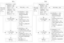
图1
文献筛选流程图"

表1
纳入文献的基本特征和文献质量评价"
| 纳入文献 | 性别(男/ 女)/n | 年龄/岁 | 文化程度/年 | 发病时间 | 病灶大小/cm3 | 诊断 标准 | AHRQ 评分 | |||
|---|---|---|---|---|---|---|---|---|---|---|
| P | C | P | C | P | C | |||||
| PSA | ||||||||||
| Guo等[ | 11/6 | 12/8 | 53.53±14.06 | 54.05±8.43 | 8.71±1.26 | 8.45±1.47 | (9.72±5.30) d | 30.60±42.23 | ABC | 9 |
| Li等[ | 12/3 | 24/6 | 50.60±7.86 | 50.10±7.95 | — | — | (8.25±7.51)个月 | 53.86±34.25 | WAB | 9 |
| Xie等[ | 31/9 | 25/12 | 57.35±11.79 | 55.14±11.39 | 7.58±5.09 | 9.64±4.97 | 2.00(1.00,5.75)周 | 39.29(14.69,79.08) | ABC | 8 |
| Yang等[ | 11/6 | 12/8 | 53.53±14.06 | 54.05±8.43 | 8.71±1.26 | 8.45±1.47 | (9.72±5.30) d | 59.19±58.29 | ABC | 9 |
| Yang等[ | 11/6 | 12/8 | 53.53±14.06 | 54.05±8.43 | 8.71±1.26 | 8.45±1.47 | (9.72±5.30) d | 59.19±58.29 | ABC | 9 |
| Zhang等[ | 27/9 | 17/7 | 58.83±9.76 | 55.17±6.13 | 11.50±3.19 | 12.38±2.45 | (66.19±35.19) d | 3.06±3.62 | WAB | 9 |
| 戴燕红等[ | 9/6 | 10/8 | 56.13±7.01 | 53.83±5.55 | 10.07±2.84 | 9.00±2.30 | (262.80±6.14) d | — | ABC | 9 |
| 李闯等[ | 6/4 | 5/5 | 59.00±7.86 | 60.20±7.63 | 5.60±1.14 | 6.20±0.84 | (5.50±1.02) d | — | ABC | 8 |
| 李春星等[ | 12/8 | 12/8 | 64.50±8.00 | 52.30±7.80 | — | — | — | — | WAB | 8 |
| 李晓琳等[ | 13/2 | 13/2 | 59.87±11.53 | 56.73±10.29 | 12.07±2.87 | 13.40±2.59 | (57.93±33.22) d | — | WAB | 10 |
| 刘会茹等[ | 6/4 | 6/4 | 59.00±17.40 | 50.00±3.80 | 10.50±2.10 | 8.80±4.00 | — | — | ABC | 9 |
| 王顺娟等[ | 8/6 | 11/5 | 65.80±6.40 | 64.40±3.20 | 7.50±4.10 | 6.20±5.60 | — | — | ABC | 10 |
| 许光等[ | 10/7 | 13/6 | 49.61±2.92 | 52.71±3.21 | 10.29±3.05 | 12.48±3.03 | — | — | ABC | 9 |
| 许光等[ | 10/7 | 13/6 | 49.61±2.92 | 52.71±3.21 | 10.29±3.05 | 12.48±3.03 | — | — | ABC | 8 |
| 张晓彤等[ | 15/4 | 12/5 | 48.63±7.50 | 45.76±7.41 | — | — | — | — | WAB | 8 |
| 周克贵等[ | 8/6 | 11/5 | 65.80±6.40 | 64.40±3.20 | 7.50±4.10 | 6.20±5.60 | — | — | ABC | 10 |
| Li等[ | 32/14 | 24/16 | 53.41±12.47 | 48.79±12.87 | 10.83±4.24 | 11.85±2.69 | — | — | WAB | 8 |
| PSD | ||||||||||
| Wu等[ | 21/18 | 43/31 | 60.00±9.41 | 57.43±6.83 | — | — | (9.92±4.92) d | 0.13±0.18 | DSM-V | 10 |
| Yao等[ | 8/7 | 9/12 | 64.13±6.01 | 60.67±6.95 | 9.40±3.38 | 10.29±2.61 | (62.87±15.73) d | 1.65±1.46 | HAMD | 9 |
| Yao等[ | 8/7 | 9/12 | 64.13±6.01 | 60.67±6.95 | 9.40±3.38 | 10.29±2.61 | (62.87±15.73) d | 1.65±1.46 | HAMD | 9 |
| 侯晶晶等[ | 10/3 | 8/5 | 54.00±9.70 | 58.00±12.20 | — | — | (10.8±2.8) d | — | DSM-V | 8 |
| 张慧萍等[ | 14/6 | 15/5 | 54.20±9.50 | 58.40±12.30 | — | — | (10.9±2.8) d | — | — | 8 |
表2
纳入文献的影像特征"
| 纳入文献 | 品牌 | 场强 | TR/ms | TE/ms | 分析工具包 | FWHM/mm | 统计阈值 | 结局指标 |
|---|---|---|---|---|---|---|---|---|
| PSA | ||||||||
| Guo等[ | Siemens | 3.0 T | 2000 | 30 | DPABI, DynamicBC | — | Alphasim(P校正 < 0.05) | ALFF |
| Li等[ | Siemens | 3.0 T | 2000 | 30 | DPARSF, SPM12 | 6 | FWE(P校正 < 0.05) | ReHo, ALFF |
| Xie等[ | GE | 3.0 T | 2400 | 30 | SPM8, DPARSF | 4 | FWE(P校正 < 0.05) | ALFF |
| Yang等[ | Siemens | 3.0 T | 2000 | 30 | SPM8, DPARSF | 8 | FDR(P校正 < 0.05) | ALFF |
| Yang等[ | Siemens | 3.0 T | 2000 | 30 | SPM8, DPARSF | 8 | FDR(P校正 < 0.05) | ReHo |
| Zhang等[ | Siemens | 3.0 T | 2000 | 30 | SPM12, DPABI | 6 | TFCE(P校正 < 0.05) | ALFF |
| 戴燕红等[ | GE | 3.0 T | 2100 | 30 | DPASRSF, REST | — | Alphasim(P校正 < 0.005) | fALFF |
| 李闯等[ | Siemens | 3.0 T | 2000 | 30 | SPM8, DPARSF | — | — | ReHo |
| 李春星等[ | Siemens | 3.0 T | 2330 | 30 | SPM8, DPARSF | 6 | Alphasim(P校正 < 0.005) | ALFF |
| 李晓琳等[ | Siemens | 3.0 T | 2000 | 30 | DPARSF, SPM12 | — | GRF(P校正 < 0.05) | ALFF |
| 刘会茹等[ | GE | 3.0 T | 2000 | 30 | REST | — | Alphasim(P校正 < 0.001) | ALFF |
| 王顺娟等[ | 联影 | 3.0 T | 2500 | 60 | SPM8, DPARSF | 4 | Alphasim(P校正 < 0.05) | ReHo |
| 许光等[ | Philips | 1.5 T | 2000 | 50 | SPM8, REST | — | Alphasim(P校正 < 0.01) | ReHo |
| 许光等[ | Philips | 1.5 T | 2000 | 50 | DPARSF, REST | 4 | Alphasim(P校正 < 0.01) | fALFF |
| 张晓彤等[ | GE | 3.0 T | 2000 | 35 | Restplus | 6 | PNA < 0.05 | ALFF |
| 周克贵等[ | 联影 | 3.0 T | 2500 | 60 | SPM8, DPARSF | — | Alphasim(P校正 < 0.05) | ReHo |
| Li等[ | Siemens | 3.0 T | 1000 | 30 | RESTplus, SPM12 | 6 | FDR(P校正 < 0.05) | ReHo, fALFF |
| PSD | ||||||||
| Wu等[ | GE | 3.0 T | 2000 | 30 | SPM12, DPABI | 6 | FDR(P校正 < 0.05) | ALFF |
| Yao等[ | Siemens | 3.0 T | 2500 | 30 | DPARSF, SPM12 | 6 | FDR(P校正 < 0.05) | ALFF |
| Yao等[ | Siemens | 3.0 T | 2500 | 30 | DPARSF, SPM12 | 6 | GRF(P校正 < 0.05) | ReHo, fALFF |
| 侯晶晶等[ | Siemens | 3.0 T | 2000 | 30 | SPM5, REST | 8 | P不校正 < 0.01 | ReHo |
| 张慧萍等[ | Philips | 3.0 T | — | — | — | — | P不校正 < 0.05 | ReHo |
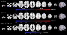
图2
PSA与PSD自发脑活动改变区域"
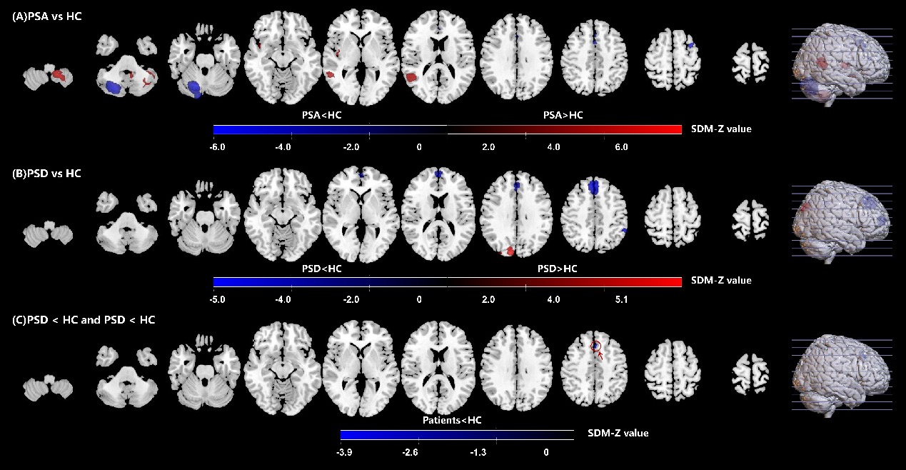
表3
PSA与PSD的自发神经功能活动差异区域"
| MNI坐标(x, y, z) | AAL脑区(Brodmann分区) | 团块大小/voxels | SDM-Z值 | P值 | I2/% |
|---|---|---|---|---|---|
| PSA > 对照组 | |||||
| -18, -50, -48 | 左侧小脑9区(NA) | 1050 | 5.978 | 0.001 | 1.042 |
| 56, -50, 4 | 右侧颞中回(BA21) | 435 | 4.592 | 0.001 | 18.489 |
| 42, -10, 6 | 右侧脑岛(BA48) | 101 | 4.535 | 0.008 | 0.972 |
| 42, 6, -14 | 右侧脑岛(BA48) | 19 | 4.414 | 0.032 | 0.858 |
| PSA < 对照组 | |||||
| 18, -68, -28 | 右侧小脑6区(BA19) | 1822 | -6.002 | 0.001 | 1.554 |
| 2, 24, 40 | 左侧内侧额上回(BA32) | 242 | -4.793 | 0.006 | 5.643 |
| -34, 8, 60 | 左侧额中回(BA8) | 45 | -5.064 | 0.014 | 11.292 |
| 6, 32, 22 | 右侧前扣带与旁扣带脑回(BA32) | 40 | -4.234 | 0.030 | 1.224 |
| PSD > 对照组 | |||||
| 10, -86, 32 | 右侧楔叶(BA19) | 265 | 5.014 | 0.001 | 0.209 |
| 30, -88, 30 | 右侧枕上回(NA) | 13 | 4.051 | 0.038 | 9.236 |
| PSD < 对照组 | |||||
| 2, 38, 42 | 左侧内侧额上回(BA9) | 954 | -5.104 | 0.001 | 4.256 |
| -2, 60, 10 | 左侧内侧额上回(BA10) | 299 | -4.514 | 0.001 | 1.419 |
| -56, -44, 42 | 左侧顶下小叶(BA40) | 119 | -4.428 | 0.005 | 3.653 |
| PSA < 对照组且PSD < 对照组 | |||||
| 0, 26, 44 | 左侧内侧额上回(BA8) | 65 | -3.883 | 0.008 | 48.200 |

图3
PSA与PSD共同差异脑区的头皮投影位置"

| [1] |
WU S M, WU B, LIU M, et al. Stroke in China: advances and challenges in epidemiology, prevention, and management[J]. Lancet Neurol, 2019, 18(4): 394-405.
doi: S1474-4422(18)30500-3 pmid: 30878104 |
| [2] |
FEIGIN V L, BRAININ M, NORRVING B, et al. World Stroke Organization (WSO): Global stroke fact sheet 2022[J]. Int J Stroke, 2022, 17(1): 18-29.
doi: 10.1177/17474930211065917 pmid: 34986727 |
| [3] |
GINEX V, GILARDONE G, VIGANÒ M, et al. Interaction between recovery of motor and language abilities after stroke[J]. Arch Phys Med Rehabil, 2020, 101(8): 1367-1376.
doi: 10.1016/j.apmr.2020.04.010 |
| [4] | TOWFIGHI A, OVBIAGELE B, EL HUSSEINI N, et al. Poststroke depression: a scientific statement for healthcare professionals from the American Heart Association/American Stroke Association[J]. Stroke, 2017, 48(2): e30-e43. |
| [5] |
WILSON S M, ENTRUP J L, SCHNECK S M, et al. Recovery from aphasia in the first year after stroke[J]. Brain, 2023, 146(3): 1021-1039.
doi: 10.1093/brain/awac129 |
| [6] |
CAI W, MUELLER C, LI Y J, et al. Post stroke depression and risk of stroke recurrence and mortality: a systematic review and meta-analysis[J]. Ageing Res Rev, 2019, 50: 102-109.
doi: S1568-1637(18)30315-5 pmid: 30711712 |
| [7] |
LIN H L, SUNG F C, MUO C H, et al. Depression risk in post-stroke aphasia patients: a nationwide population-based cohort study[J]. Neuroepidemiology, 2023, 57(3): 162-169.
doi: 10.1159/000530070 |
| [8] |
SHEHATA G A, EL MISTIKAWI T, RISHA A S K, et al. The effect of aphasia upon personality traits, depression and anxiety among stroke patients[J]. J Affect Disord, 2015, 172: 312-314.
doi: 10.1016/j.jad.2014.10.027 |
| [9] |
ASHAIE S A, HURWITZ R, CHERNEY L R. Depression and subthreshold depression in stroke-related aphasia[J]. Arch Phys Med Rehabil, 2019, 100(7): 1294-1299.
doi: 10.1016/j.apmr.2019.01.024 |
| [10] |
ZANELLA C, LAURES-GORE J, DOTSON V M, et al. Incidence of post-stroke depression symptoms and potential risk factors in adults with aphasia in a comprehensive stroke center[J]. Top Stroke Rehabil, 2022, 30(5): 448-458.
doi: 10.1080/10749357.2022.2070363 |
| [11] |
MEDEIROS G C, ROY D, KONTOS N, et al. Post-stroke depression: a 2020 updated review[J]. Gen Hosp Psychiatry, 2020, 66: 70-80.
doi: 10.1016/j.genhosppsych.2020.06.011 |
| [12] |
EDELKRAUT L, LÓPEZ-BARROSO D, TORRES-PRIORIS M J, et al. Spectrum of neuropsychiatric symptoms in chronic post-stroke aphasia[J]. World J Psychiatry, 2022, 12(3): 450-469.
doi: 10.5498/wjp.v12.i3.450 pmid: 35433325 |
| [13] |
HAN Y, JING Y, LI X, et al. Clinical characteristics of post-stroke basal ganglia aphasia and the study of language-related white matter tracts based on diffusion spectrum imaging[J]. NeuroImage, 2024, 295: 120664.
doi: 10.1016/j.neuroimage.2024.120664 |
| [14] | 苑杰, 姜伟时, 刘颖, 等. 卒中后抑郁与病灶分布关系的研究进展[J]. 世界最新医学信息文摘, 2019, 19(80): 103-104. |
| YUAN J, JIANG W S, LIU Y. A study on the relationship between post-stroke depression and the distribution of lesions[J]. World Latest Med Inf, 2019, 19(80): 103-104. | |
| [15] |
VILLA R F, FERRARI F, MORETTI A. Post-stroke depression: mechanisms and pharmacological treatment[J]. Pharmacol Ther, 2018, 184: 131-144.
doi: 10.1016/j.pharmthera.2017.11.005 |
| [16] |
WU X M, WANG L Y, JIANG H B, et al. Frequency-dependent and time-variant alterations of neural activity in post-stroke depression: a resting-state fMRI study[J]. Neuroimage Clin, 2023, 38: 103445.
doi: 10.1016/j.nicl.2023.103445 |
| [17] |
TANG X Y, GUO Z X, CHEN G M, et al. A multimodal meta-analytical evidence of functional and structural brain abnormalities across Alzheimer's disease spectrum[J]. Ageing Res Rev, 2024, 95: 102240.
doi: 10.1016/j.arr.2024.102240 |
| [18] |
ZHANG X Y, WANG W, BAI X, et al. Alterations in regional homogeneity and multiple frequency amplitudes of low-frequency fluctuation in patients with new daily persistent headache: a resting-state functional magnetic resonance imaging study[J]. J Headache Pain, 2023, 24(1): 14.
doi: 10.1186/s10194-023-01543-y pmid: 36814220 |
| [19] | ALBAJES-EIZAGIRRE A, SOLANES A, FULLANA M A, et al. Meta-analysis of voxel-based neuroimaging studies using seed-based d mapping with permutation of subject images (SDM-PSI)[J]. J Vis Exp, 2019(153): e59841. |
| [20] | PAGE M J, MOHER D, BOSSUYT P M, et al. PRISMA 2020 explanation and elaboration: updated guidance and exemplars for reporting systematic reviews[J]. BMJ, 2021, 372: n160. |
| [21] |
ALBAJES-EIZAGIRRE A, SOLANES A, VIETA E, et al. Voxel-based meta-analysis via permutation of subject images (PSI): theory and implementation for SDM[J]. NeuroImage, 2019, 186: 174-184.
doi: 10.1016/j.neuroimage.2018.10.077 |
| [22] |
RADUA J, RUBIA K, CANALES-RODRÍGUEZ E J, et al. Anisotropic kernels for coordinate-based meta-analyses of neuroimaging studies[J]. Front Psychiatry, 2014, 5: 13.
doi: 10.3389/fpsyt.2014.00013 pmid: 24575054 |
| [23] |
LIM L, RADUA J, RUBIA K. Gray matter abnormalities in childhood maltreatment: a voxel-wise meta-analysis[J]. Am J Psychiatry, 2014, 171(8): 854-863.
doi: 10.1176/appi.ajp.2014.13101427 |
| [24] |
GUO J, BISWAL B B, HAN S, et al. Altered dynamics of brain segregation and integration in poststroke aphasia[J]. Hum Brain Mapp, 2019, 40(11): 3398-3409.
doi: 10.1002/hbm.24605 pmid: 31016854 |
| [25] |
LI H Z, ZHANG H, XU S, et al. Altered spontaneous brain activity in poststroke aphasia: a resting-state fMRI study[J]. Brain Sci, 2023, 13(2): 300.
doi: 10.3390/brainsci13020300 |
| [26] |
XIE X H, ZHANG T, BAI T J, et al. Resting-state neural-activity alterations in subacute aphasia after stroke[J]. Brain Sci, 2022, 12(5): 678.
doi: 10.3390/brainsci12050678 |
| [27] |
YANG M, LI J, LI Y B, et al. Altered intrinsic regional activity and interregional functional connectivity in post-stroke aphasia[J]. Sci Rep, 2016, 6: 24803.
doi: 10.1038/srep24803 pmid: 27091494 |
| [28] |
YANG M, LI J, YAO D Z, et al. Disrupted intrinsic local synchronization in poststroke aphasia[J]. Medicine (Baltimore), 2016, 95(11): e3101.
doi: 10.1097/MD.0000000000003101 |
| [29] |
ZHANG B L, CHANG J L, PARK J, et al. Uncinate fasciculus and its cortical terminals in aphasia after subcortical stroke: a multi-modal MRI study[J]. Neuroimage Clin, 2021, 30: 102597.
doi: 10.1016/j.nicl.2021.102597 |
| [30] | 戴燕红, 王红, 罗海龙, 等. 弱活性与强连接:脑梗死后慢性期失语症的静息态脑功能研究[J]. 中国康复医学杂志, 2021, 36(12): 1496-1504. |
| DAI Y H, WANG H, LUO H L, et al. Weak activity and strong connectivity: resting state brain function in chronic aphasia after cerebral infarction[J]. Chin J Rehabil Med, 2021, 36(12): 1496-1504. | |
| [31] | 李闯, 李爱琴, 曹爱华, 等. 脑梗死后运动性失语的发生机制:基于局部一致性的静息态功能MRl研究[J]. 神经病学与神经康复学杂志, 2022, 18(1): 15-21. |
| LI C, LI A Q, CAO A H, et al. Application of ReHo in motor aphasia in patients with stroke: a BOLD-fMRl study[J]. J Neurol Neurorehabil, 2022, 18(1): 15-21. | |
| [32] | 李春星, 李华, 卓兵芝, 等. 卒中后失语患者脑功能成像与失语商的相关性[J]. 中华行为医学与脑科学杂志, 2013, 22(6): 517-519. |
| LI C X, LI H, ZHUO B Z, et al. A correlativity study of amplitude of low frequency fluctuation change of resting-state brain activity and aphasia quotient in aphasia patients after stroke[J]. Chin J Behav Med Brain Sci, 2013, 22(6): 517-519. | |
| [33] |
李晓琳, 徐敏杰, 曹云, 等. 卒中后失语静息态脑功能成像亚频段低频振幅脑活动研究[J]. 中国康复理论与实践, 2021, 27(5): 497-503.
doi: 10.3969/j.issn.1006-9771.2021.05.001 |
| LI X L, XU M J, CAO Y, et al. Frequency-dependent alterations in amplitude of low-frequency fluctuations in resting-state functional magnetic resonance imaging of post stroke aphasia[J]. Chin J Rehabil Theory Pract, 2021, 27(5): 497-503. | |
| [34] | 刘会茹, 王维卓, 王欣, 等. 脑梗死运动性失语患者的静息态脑功能研究[J]. 医学影像学杂志, 2014, 24(10): 1674-1679. |
| LIU H R, WANG W Z, WANG X, et al. Study of resting-state functional MRI in motor aphasia after ischemic stroke[J]. J Med Imaging, 2014, 24(10): 1674-1679. | |
| [35] | 王顺娟, 夏进东, 周克贵. 脑梗死后运动性失语的脑功能活动局部一致性研究[J]. 临床放射学杂志, 2020, 39(9): 1719-1723. |
| WANG S J, XIA J D, ZHOU K G. Study on the homogeneity of cerebral regional functional area of motor aphasia after cerebral infarction[J]. J Clin Radiol, 2020, 39(9): 1719-1723. | |
| [36] | 许光, 马晓芬, 江桂华, 等. 缺血性卒中后非流利型失语患者局部一致性变化:静息态功能性磁共振成像研究[J]. 国际脑血管病杂志, 2013, 21(10): 764-768. |
| XU G, MA X F, JIANG G H, et al. Regional homogeneity changes in patients with non-fluent aphasia after ischemic stroke: a resting-state functional magnetic resonance imaging study[J]. Int J Cerebrovasc Dis, 2013, 21(10): 764-768. | |
| [37] | 许光, 马晓芬, 江桂华, 等. 缺血性卒中后非流利型失语患者静息态功能磁共振比率低波振幅的研究[J]. 实用医学杂志, 2014(7): 1016-1020. |
| XU G, MA X F, JIANG G H, et al. Resting-state fMRI fALFF analysis in patients with non-fluent aphasia after ischemic stroke[J]. J Pract Med, 2014(7): 1016-1020. | |
| [38] |
张晓彤, 李娜, 陈兆聪, 等. 右侧小脑对卒中后失语的潜在作用:基于格兰杰因果分析的初步研究[J]. 中国康复理论与实践, 2021, 27(12): 1458-1463.
doi: 10.3969/j.issn.1006-9771.2021.12.012 |
| ZHANG X T, LI N, CHEN Z C, et al. Potential role of right cerebellum in post-stroke aphasia: a preliminary study based on Granger causality analysis[J]. Chin J Rehabil Theory Pract, 2021, 27(12): 1458-1463. | |
| [39] | 周克贵, 王顺娟, 杨柳, 等. 脑梗死后运动性失语的局部脑功能活动强度研究[J]. 中华老年心脑血管病杂志, 2020, 22(2): 123-126. |
| ZHOU K G, WANG S J, YANG L, et al. Intensity of local cerebral functional activity after motor aphasia due to cerebral infarction[J]. Chin J Geriatr Heart Brain Vessel Dis, 2020, 22(2): 123-126. | |
| [40] |
LI S Q, YUAN Z N, LI Y X, et al. Abnormal resting-state neural activities of language and non-language cognitive function impairments in stroke patients with aphasia: a cross-sectional study[J]. Clin Neurol Neurosurg, 2025, 251: 108849.
doi: 10.1016/j.clineuro.2025.108849 |
| [41] |
YAO G Q, LI J, WANG J J, et al. Improved resting-state functional dynamics in post-stroke depressive patients after Shugan Jieyu Capsule treatment[J]. Front Neurosci, 2020, 14: 297.
doi: 10.3389/fnins.2020.00297 |
| [42] |
YAO G Q, ZHANG X Q, LI J, et al. Improving depressive symptoms of post-stroke depression using the Shugan Jieyu Capsule: a resting-state functional magnetic resonance imaging study[J]. Front Neurol, 2022, 13: 860290.
doi: 10.3389/fneur.2022.860290 |
| [43] | 侯晶晶, 王春雪, 张宁, 等. 急性缺血性脑卒中伴发抑郁障碍的静息态脑功能磁共振成像研究[J]. 中国神经免疫学和神经病学杂志, 2011, 18(4): 264-268. |
| HOU J J, WANG C X, ZHANG N, et al. Study on resting-state functional magnetic resonance imaging in acute ischemic stroke patients with depressive disorder[J]. Chin J Neuroimmunol Neurol, 2011, 18(4): 264-268. | |
| [44] | 张慧萍, 陆强彬, 陆梦茹, 等. 急性缺血性卒中伴发抑郁障碍患者的脑功能核磁共振成像研究[J]. 国际精神病学杂志, 2018, 45(1): 121-123. |
| ZHANG H P, LU Q B, LU M R, et al. Brain functional magnetic resonance imaging of patients with acute ischemic stroke complicated with depressive disorder[J]. J Int Psychiatry, 2018, 45(1): 121-123. | |
| [45] |
HICKOK G. The dual stream model of speech and language processing[J]. Handb Clin Neurol, 2022, 185: 57-69.
doi: 10.1016/B978-0-12-823384-9.00003-7 pmid: 35078610 |
| [46] |
CHANG E F, RAYGOR K P, BERGER M S. Contemporary model of language organization: an overview for neurosurgeons[J]. J Neurosurg, 2015, 122(2): 250-261.
doi: 10.3171/2014.10.JNS132647 pmid: 25423277 |
| [47] |
樊瑞文, 李晓琳, 黄幸, 等. 基于语言双流模型的卒中后失语右脑功能网络研究[J]. 中国康复理论与实践, 2020, 26(5): 572-578.
doi: 10.3969/j.issn.1006-9771.2020.05.016 |
| FAN R W, LI X L, HUANG X, et al. Functional connectivities in right hemisphere for poststroke aphasia: based on dual stream model[J]. Chin J Rehabil Theory Pract, 2020, 26(5): 572-578. | |
| [48] |
樊瑞文, 黄幸, 李晓琳, 等. 卒中后失语非损伤侧脑电功率谱网络特征研究[J]. 中国康复理论与实践, 2020, 26(6): 692-696.
doi: 10.3969/j.issn.1006-9771.2020.06.013 |
| FAN R W, HUANG X, LI X L, et al. Characteristics of electroencephalogram power spectrum network on ininjured side of post-stroke aphasia[J]. Chin J Rehabil Theory Pract, 2020, 26(6): 692-696. | |
| [49] | 吴建满, 李银官, 范秋玲, 等. 失语症患者静息态fMRI成像研究[J]. 医学影像学杂志, 2019, 29(5): 731-735, 743. |
| WU J M, LI Y G, FAN Q L, et al. Study on resting-state functional MRI in aphasia[J]. J Med Imaging, 2019, 29(5): 731-735, 743. | |
| [50] |
VAN HEES S, MCMAHON K, ANGWIN A, et al. A functional MRI study of the relationship between naming treatment outcomes and resting state functional connectivity in post-stroke aphasia[J]. Hum Brain Mapp, 2014, 35(8): 3919-3931.
doi: 10.1002/hbm.22448 pmid: 24453137 |
| [51] |
YUAN Q M, LI H H, DU B Q, et al. The cerebellum and cognition: further evidence for its role in language control[J]. Cereb Cortex, 2022, 33(1): 35-49.
doi: 10.1093/cercor/bhac051 pmid: 35226917 |
| [52] |
JOBSON K R, HOFFMAN L J, METOKI A, et al. Language and the cerebellum: structural connectivity to the eloquent brain[J]. Neurobiol Lang, 2024, 5(3): 652-675.
doi: 10.1162/nol_a_00085 |
| [53] | 潘梦洁, 陈峰, 林明方, 等. 基于局部一致性的重度抑郁症患者脑静息态功能磁共振成像研究[J]. 海南医学, 2016, 27(3): 363-367. |
| PAN M J, CHEN F, LIN M F, et al. Study of brain regional homogeneity in patients with major depressive disorder using resting-state functional MRI[J]. Hainan Med J, 2016, 27(3): 363-367. | |
| [54] | 徐康丽, 安兰花, 张金生, 等. 功能磁共振在中医药治疗缺血性脑卒中领域的研究热点与前沿[J]. 中国组织工程研究, 2024, 28(11): 1789-1796. |
| XU K L, AN L H, ZHANG J S, et al. Research hotspots and frontiers of functional magnetic resonance imaging in treatment of ischemic stroke by traditional Chinese medicine[J]. Chin J Tissue Eng Res, 2024, 28(11): 1789-1796. | |
| [55] |
WU L, ZHANG T, ZHANG S. Comparative study of magnetic resonance imaging-based neuroimaging methods in older adults with depression[J]. Psychiatry Res Neuroimaging, 2023, 331: 111637.
doi: 10.1016/j.pscychresns.2023.111637 |
| [56] |
LI K, ZHANG M, ZHANG H S, et al. The spontaneous activity and functional network of the occipital cortex is correlated with state anxiety in healthy adults[J]. Neurosci Lett, 2020, 715: 134596.
doi: 10.1016/j.neulet.2019.134596 |
| [57] |
WU F F, LU Q B, KONG Y, et al. A comprehensive overview of the role of visual cortex malfunction in depressive disorders: opportunities and challenges[J]. Neurosci Bull, 2023, 39(9): 1426-1438.
doi: 10.1007/s12264-023-01052-7 pmid: 36995569 |
| [58] | 傅成伟, 陈霞, 侯小燕, 等. 经皮耳迷走神经刺激治疗帕金森病抑郁的临床疗效及机制研究[J]. 中国中西医结合影像学杂志, 2025, 23(1): 7-11. |
| FU C W, CHEN X, HOU X Y, et al. Clinical efficacy and mechanism of transcutaneous auricular vagus nerve stimulation in treatment of depression in Parkinson disease[J]. Chin Imaging J Integr Tradit West Med, 2025, 23(1): 7-11. | |
| [59] |
LIN S, HUANG L, LUO Z C, et al. The ATP level in the medial prefrontal cortex regulates depressive-like behavior via the medial prefrontal cortex-lateral habenula pathway[J]. Biol Psychiatry, 2022, 92(3): 179-192.
doi: 10.1016/j.biopsych.2022.02.014 |
| [60] |
PRICE R B, DUMAN R. Neuroplasticity in cognitive and psychological mechanisms of depression: an integrative model[J]. Mol Psychiatry, 2020, 25(3): 530-543.
doi: 10.1038/s41380-019-0615-x |
| [61] | 毛星刚, 杨秋子, 姬昂, 等. 融合脑网络的多模态三维可视化技术在脑胶质瘤手术个体化解剖的应用分析[J]. 中国微侵袭神经外科杂志, 2025, 29(1): 12-18. |
| MAO X G, YANG Q Z, JI A, et al. Application and analysis of multimodal 3-D visualization technology integrating brain networks in individualized anatomy of glioma surgery[J]. Chin J Minim Invasive Neurosurg, 2025, 29(1): 12-18. | |
| [62] |
LA CORTE E, ELDAHABY D, GRECO E, et al. The frontal aslant tract: a systematic review for neurosurgical applications[J]. Front Neurol, 2021, 12: 641586.
doi: 10.3389/fneur.2021.641586 |
| [63] |
DICK A S, GARIC D, GRAZIANO P, et al. The frontal aslant tract (FAT) and its role in speech, language and executive function[J]. Cortex, 2019, 111: 148-163.
doi: S0010-9452(18)30348-4 pmid: 30481666 |
| [64] |
ZHONG A J, BALDO J V, DRONKERS N F, et al. The unique role of the frontal aslant tract in speech and language processing[J]. Neuroimage Clin, 2022, 34: 103020.
doi: 10.1016/j.nicl.2022.103020 |
| [65] |
GONG J Y, WANG J J, QIU S J, et al. Common and distinct patterns of intrinsic brain activity alterations in major depression and bipolar disorder: voxel-based meta-analysis[J]. Transl Psychiatry, 2020, 10(1): 353.
doi: 10.1038/s41398-020-01036-5 |
| [66] | 吴俊娥. 电针治疗卒中后抑郁临床研究及其对大鼠mPFC神经元影响[D]. 广州: 广州中医药大学, 2023. |
| WU J E. Clinical study of electroacupuncture treatment of post-stroke depression and its effects on rat mPFC neurons[D]. Guangzhou: Guangzhou University of Chinese Medicine, 2023. | |
| [67] | 赵德福, 赵瑜, 杨孝芳. 督脉取穴针刺联合Schuell语言康复训练对脑卒中后失语症患者言语功能、MoCA评分及语言中枢活动功能的影响[J]. 临床和实验医学杂志, 2021, 20(8): 886-890. |
| ZHAO D F, ZHAO Y, YANG X F. Effect of Du Mai acupoint acupuncture combined with Schuell language rehabilitation training on the speech function, MoCA and the function of language center in aphasia patients after stroke[J]. J Clin Exp Med, 2021, 20(8): 886-890. | |
| [68] | 张震, 何腾, 王旭, 等. 基于"督脉入络脑"探讨针刺督脉经穴治疗脑病的神经传导机制[J]. 山东中医杂志, 2024, 43(8): 795-803. |
| ZHANG Z, HE T, WANG X, et al. Nerve conduction mechanism of treating encephalopathy by acupuncturing acupoints on Du-Meridian based on "Governor Vessel Entering into Collateral-Brain"[J]. Shandong J Tradit Chin Med, 2024, 43(8): 795-803. | |
| [69] | 李棋龙, 田浩梅, 陈楚淘. 神庭穴的古代应用研究[J]. 辽宁中医杂志, 2023, 50(2): 77-80. |
| LI Q L, TIAN H M, CHEN C T. Study on ancient application of Shenting (GV24)[J]. Liaoning J Tradit Chin Med, 2023, 50(2): 77-80. | |
| [70] | 许磊, 何玲, 李慧, 等. 基于数据挖掘的针灸治疗失语症选穴规律研究[J]. 中国针灸, 2023, 43(4): 471-478. |
| XU L, HE L, LI H, et al. The rules of acupoint selection of acupuncture and moxibustion for aphasia based on data mining[J]. Chin Acupunct Moxibustion, 2023, 43(4): 471-478. | |
| [71] | 徐建文, 贾红玲. 基于数据挖掘技术的针刺治疗运动性失语选穴规律探析[J]. 针灸临床杂志, 2023, 39(1): 67-73. |
| XU J W, JIA H L. Analysis on law of points selection in acupuncture treatment for motor aphasia based on data mining technology[J]. J Clin Acupunct Moxibustion, 2023, 39(1): 67-73. | |
| [72] | 赵蕊, 徐敏杰, 李翔宇, 等. 基于形神思维探讨从督论治卒中后失语[J]. 环球中医药, 2024, 17(4): 670-675. |
| [73] | 楚惠, 赖国安, 许创润, 等. 靳三针调神针法治疗帕金森病轻中度抑郁的临床研究[J]. 针灸临床杂志, 2024, 40(5): 10-16. |
| CHU H, LAI G A, XU C R, et al. Clinical study of Jin's Three-Needle regulating shen acupuncture in treatment of mild-to-moderate Parkinson's disease depression[J]. J Clin Acupunct Moxibustion, 2024, 40(5): 10-16. | |
| [74] | 高小梅, 薛建芳, 施晓瑜, 等. 针灸治疗中风后抑郁研究概述[J]. 光明中医, 2025, 40(4): 800-803. |
| GAO X M, XUE J F, SHI X Y, et al. An overview of acupuncture and moxibustion in the treatment of post-stroke depression[J]. Guangming J Chin Med, 2025, 40(4): 800-803. | |
| [75] | 杨嘉誉. 电针神庭、百会穴通过负反馈调控突触可塑性改善MCAO/R大鼠学习记忆的机制探讨[D]. 福州: 福建中医药大学, 2024. |
| YANG J Y. Mechanism of electroacupuncture at Shenting and Baihui points to improve learning memory in MCAO/R rats through negative feedback modulation of synaptic plasticity[D]. Fuzhou: Fujian University of Traditional Chinese Medicine, 2024. | |
| [76] | 杨柳笛, 李红蕾. 电针百会、神庭对脂多糖诱导的抑郁样小鼠行为及海马NOD样受体蛋白3炎症小体的影响[J]. 河北中医, 2023, 45(10): 1693-1697, 1702. |
| YANG L D, LI H L. Efficacy of electroacupuneture at Baihui (GV20) and Shenting (GV24) on lipopolysaccharide-induced depression-like behavior in mice and its effect on NOD-like receptor protein 3 inflammasome in hippocampal region[J]. Hebei J Tradit Chin Med, 2023, 45(10): 1693-1697, 1702. |
| [1] | 张梓寒, 管津智, 黄幸, 周莉, 张雅宣, 张蒙园, 常静玲. 卒中后失语患者汉语词图匹配任务态脑电的时域及时频特征[J]. 《中国康复理论与实践》, 2025, 31(8): 947-957. |
| [2] | 杜雯倩, 张旭, 陈继伟, 王兴, 朱昆. 运动干预对帕金森病冻结步态效果的Meta分析[J]. 《中国康复理论与实践》, 2025, 31(7): 781-789. |
| [3] | 山磊, 刘影, 张欣, 迟茜茜, 朱晓敏. 加速间歇性爆发性θ波刺激治疗卒中后抑郁的效果[J]. 《中国康复理论与实践》, 2025, 31(7): 822-829. |
| [4] | 张勇, 蔡增, 徐凤萍, 刘丹, 常红娟. 音乐治疗对孤独症谱系障碍儿童干预效果的Meta分析[J]. 《中国康复理论与实践》, 2025, 31(4): 423-430. |
| [5] | 杨益成, 王丹丹, 沈群策, 张磊, 吴雪萍. 癌症患者化疗期间运动干预焦虑和抑郁状态效果的Meta分析[J]. 《中国康复理论与实践》, 2025, 31(2): 184-193. |
| [6] | 杨三峡, 刘清栾, 万嘉敏. 音乐干预对阿尔茨海默病患者激活模式区域差异的功能磁共振成像Meta分析[J]. 《中国康复理论与实践》, 2025, 31(10): 1156-1163. |
| [7] | 赵英楠, 郑鑫彤, 刘俊玲, 王紫丹, 吴泓悦, 李冰, 李燕. 阈值吸气肌训练对肺癌术后患者肺康复影响的Meta分析[J]. 《中国康复理论与实践》, 2025, 31(10): 1164-1171. |
| [8] | 温艳飞, 杨露, 班玥, Ykabaru Daniela BERBESI NORIEGA, 张郝琪, 王丽, 刘华. ICF框架下运动疗法对慢性非特异性颈痛效果的Meta分析[J]. 《中国康复理论与实践》, 2024, 30(7): 778-788. |
| [9] | 吕美玲, 王洁, 曾维斯, 温晓婷, 楚鑫. 虚拟现实技术对帕金森病患者认知功能和生活质量影响的Meta分析[J]. 《中国康复理论与实践》, 2024, 30(6): 648-656. |
| [10] | 崔甜甜, 杨钰琳, 崔腾腾, 马丽虹. 不同强化训练对脑性瘫痪儿童上肢运动功能效果的网状Meta分析[J]. 《中国康复理论与实践》, 2024, 30(4): 437-448. |
| [11] | 魏辰, 王子贤, 李淑璠, 王芃, 贾舒祺, 田英. 镜像疗法对脑卒中患者上肢运动功能和日常生活活动能力影响的Meta分析[J]. 《中国康复理论与实践》, 2024, 30(3): 281-291. |
| [12] | 黄幸, 常静玲, 张梓寒, 李颖. 卒中后失语工作记忆的事件相关电位及时频特征[J]. 《中国康复理论与实践》, 2024, 30(3): 316-325. |
| [13] | 王铭琛, 张文宇, 张贤祚, 臧万里. 经颅直流电刺激对帕金森病患者认知功能和生活质量影响的Meta分析[J]. 《中国康复理论与实践》, 2024, 30(2): 183-188. |
| [14] | 李芳, 刘慧珍, 梅利平, 张通, 张豪杰, 李冰洁, 赵军. 脑梗死患者康复科住院期间卒中后抑郁程度的相关因素[J]. 《中国康复理论与实践》, 2024, 30(2): 217-222. |
| [15] | 张明兰, 张玲玲, 王丽莎, 刘莉, 高润, 饶江, 刘婉, 夏子安, 张传文, 程欣欣. 自主神经功能对卒中后抑郁患者运动功能的影响[J]. 《中国康复理论与实践》, 2024, 30(2): 223-231. |
| 阅读次数 | ||||||
|
全文 |
|
|||||
|
摘要 |
|
|||||
|
||