



《中国康复理论与实践》 ›› 2026, Vol. 32 ›› Issue (3): 329-336.doi: 10.3969/j.issn.1006-9771.2026.03.009
杨玲1, 王庆雷2, 王洁1, 徐雯婕1, 王彤2, 郭川2( ), 钱雪1()
收稿日期:2025-11-06
修回日期:2026-01-28
出版日期:2026-03-25
发布日期:2026-04-01
通讯作者:
钱雪(1983-),女,汉族,江苏镇江市人,副主任护师,主要研究方向:康复护理及康复管理,E-mail:qianxuenurse@126.com;郭川(1988-),男,汉族,江苏连云港市人,博士,副主任技师,硕士研究生导师,主要研究方向:神经系统疾病康复治疗,E-mail:guochuan@njmu.edu.cn。
作者简介:杨玲(1988-),女,汉族,江苏常州市人,主管护师,主要研究方向:康复护理及心理护理
基金资助:
YANG Ling1, WANG Qinglei2, WANG Jie1, XU Wenjie1, WANG Tong2, GUO Chuan2(), QIAN Xue1()
Received:2025-11-06
Revised:2026-01-28
Published:2026-03-25
Online:2026-04-01
Contact:
QIAN Xue, E-mail: Supported by:摘要:
目的 运用功能性近红外光谱成像技术,观察卒中后焦虑(PSA)患者前额叶皮质的激活及功能连接特征,探讨其潜在神经机制。
方法 2024年12月至2025年9月,于常州市德安医院招募脑卒中患者120例,根据汉密尔顿焦虑量表(HAMA)将患者分为PSA组(n = 60)和非PSA组(n = 60)。所有患者均佩戴18通道fNIRS采集帽进行检测,观察两组额极区与背外侧前额叶区(DLPFC)在静息态下的功能连接差异,以及任务态下相应脑区激活情况。
结果 静息态功能连接分析显示,两组在额极区和DLPFC间的网络连接均无显著性差异(|t| < 1.301, P > 0.05)。任务态激活结果显示,与非PSA组相比,PSA组健侧额极区激活降低(Z = -2.063, P < 0.05),且该区域激活水平与HAMA评分呈负相关(ρ = -0.201, P = 0.028)。
结论 PSA患者在任务态下健侧额极激活水平降低,且激活水平与焦虑程度呈负相关。
中图分类号:
杨玲, 王庆雷, 王洁, 徐雯婕, 王彤, 郭川, 钱雪. 基于功能性近红外光谱成像的卒中后焦虑患者前额叶皮质激活特征及机制研究[J]. 《中国康复理论与实践》, 2026, 32(3): 329-336.
YANG Ling, WANG Qinglei, WANG Jie, XU Wenjie, WANG Tong, GUO Chuan, QIAN Xue. Activation patterns and mechanism in the prefrontal cortex of post-stroke anxiety patients: a study using functional near-infrared spectroscopy[J]. Chinese Journal of Rehabilitation Theory and Practice, 2026, 32(3): 329-336.
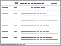
表1
两组一般资料比较"
| 组别 | PSA组(n = 60) | 非PSA组(n = 60) | t/χ²/Z值 | P值 |
|---|---|---|---|---|
| 年龄/岁 | 62.20±11.81 | 62.27±12.35 | -0.030 | 0.976 |
| 性别/n | 43/17 | 44/16 | 0.042 | 0.838 |
| 卒中类型(缺血性/出血性)/n | 50/10 | 42/18 | 2.981 | 0.084 |
| 卒中侧(左/右)/n | 26/34 | 31/29 | 0.835 | 0.361 |
| MoCA评分 | 20.00(16.00, 24.00) | 22.50(18.00, 25.00) | -1.607 | 0.108 |
| HAMA评分 | 12.00(8.00, 15.75) | 2.00(0.00, 3.00) | -9.458 | < 0.001 |
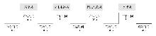
图1
VFT设计"
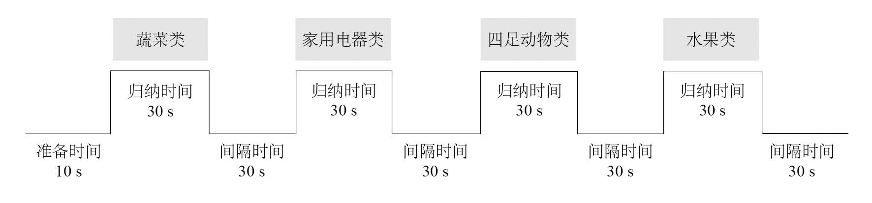
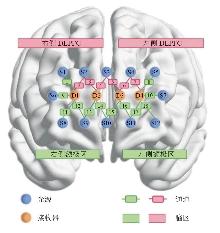
图2
fNIRS光源、接收器及通道排布"
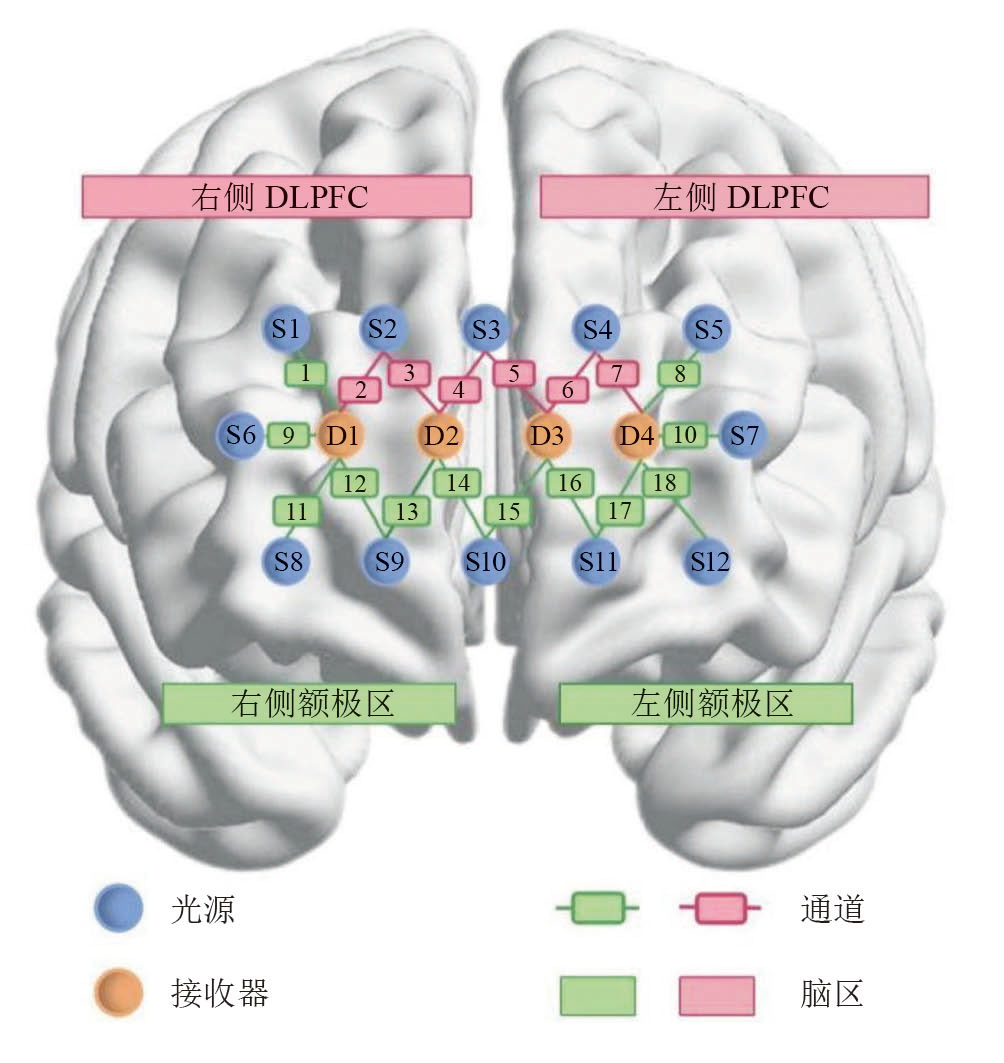
表2
两组静息态功能连接比较(z值)"
| ROI-ROI | PSA组(n = 60) | 非PSA组(n = 60) | t值 | P值 |
|---|---|---|---|---|
| 患侧额极-患侧DLPFC | 1.159±0.547 | 1.129±0.535 | 0.300 | 0.764 |
| 患侧额极-健侧DLPFC | 0.893±0.488 | 0.960±0.461 | -0.769 | 0.443 |
| 患侧额极-健侧额极 | 1.205±0.446 | 1.192±0.430 | 0.160 | 0.873 |
| 患侧DLPFC-健侧DLPFC | 1.091±0.572 | 1.211±0.432 | -1.301 | 0.196 |
| 患侧DLPFC-健侧额极 | 0.904±0.519 | 0.960±0.473 | -0.613 | 0.541 |
| 健侧DLPFC-健侧额极 | 1.162±0.525 | 1.204±0.578 | -0.415 | 0.679 |
表3
两组VFT脑激活比较"
| ROI | PSA组(n = 60) | 非PSA组(n = 60) | Z值 | P值 |
|---|---|---|---|---|
| 患侧额极 | 0.077(-0.029, 0.140) | 0.106(0.005, 0.275) | -1.375 | 0.169 |
| 患侧DLPFC | 0.044(-0.036, 0.142) | 0.100(-0.014, 0.210) | -1.533 | 0.125 |
| 健侧DLPFC | 0.027(-0.031, 0.094) | 0.081(-0.034, 0.211) | -1.470 | 0.142 |
| 健侧额极 | 0.084(-0.027, 0.140) | 0.100(-0.006, 0.251) | -2.063 | 0.039 |
表4
VFT中各ROI与HAMA评分的相关性分析"
| ROI | ρ | P |
|---|---|---|
| 患侧额极 | -0.124 | 0.178 |
| 患侧DLPFC | -0.138 | 0.132 |
| 健侧DLPFC | -0.129 | 0.159 |
| 健侧额极 | -0.201 | 0.028 |
| [1] |
MEDEIROS G C, ROY D, KONTOS N, et al. Post-stroke depression: a 2020 updated review[J]. Gen Hosp Psychiatry, 2020, 66: 70-80.
doi: 10.1016/j.genhosppsych.2020.06.011 |
| [2] | LIU L, MARSHALL I J, PEI R, et al. Natural history of depression up to 18 years after stroke: a population-based South London Stroke Register study[J]. Lancet Reg Health Eur, 2024, 40: 100882. |
| [3] |
RAFSTEN L, DANIELSSON A, SUNNERHAGEN K S. Anxiety after stroke: a systematic review and meta-analysis[J]. J Rehabil Med, 2018, 50(9): 769-778.
doi: 10.2340/16501977-2384 pmid: 30184240 |
| [4] |
XIAO M, HUANG G, FENG L, et al. Impact of sleep quality on post-stroke anxiety in stroke patients[J]. Brain Behav, 2020, 10(12): e01716.
doi: 10.1002/brb3.v10.12 |
| [5] | 吕敏, 李雪冰. 脑卒中后焦虑障碍研究现状[J]. 中国康复医学杂志, 2021, 36(3): 353-359. |
| LÜ M, LI X B. Research status of post-stroke anxiety disorder[J]. Chin J Rehabil Med, 2021, 36(3): 353-359. | |
| [6] | WRIGHT F, WU S, CHUN H Y, et al. Factors associated with poststroke anxiety: a systematic review and meta-analysis[J]. Stroke Res Treat, 2017, 2017: 2124743. |
| [7] |
OESTREICH L K L, LO J W, DI BIASE M A, et al. Network analysis of neuropsychiatric, cognitive, and functional complications of stroke: implications for novel treatment targets[J]. Psychiatry Clin Neurosci, 2024, 78(4): 229-236.
doi: 10.1111/pcn.v78.4 |
| [8] |
MACKENZIE J J, MORENO-GOMEZ V. Poststroke anxiety: the other poststroke mood disorder[J]. Stroke, 2024, 55(11): 2703-2704.
doi: 10.1161/STROKEAHA.124.048771 pmid: 39417252 |
| [9] |
GUO B, ZHANG M, HAO W, et al. Neuroinflammation mechanisms of neuromodulation therapies for anxiety and depression[J]. Transl Psychiatry, 2023, 13(1): 5.
doi: 10.1038/s41398-022-02297-y |
| [10] |
GIL-PATERNA P, FURMARK T. Imaging the cerebellum in post-traumatic stress and anxiety disorders: a mini-review[J]. Front Syst Neurosci, 2023, 17: 1197350.
doi: 10.3389/fnsys.2023.1197350 |
| [11] |
PIRONDINI E, KINANY N, SUEUR C L, et al. Post-stroke reorganization of transient brain activity characterizes deficits and recovery of cognitive functions[J]. Neuroimage, 2022, 255: 119201.
doi: 10.1016/j.neuroimage.2022.119201 |
| [12] |
ZUO L, LAN X, ZHOU Y, et al. Cerebellar-cerebral circuits functional connectivity in patients with cognitive impairment after basal ganglia stroke: a pilot study[J]. Front Aging Neurosci, 2025, 17: 1478891.
doi: 10.3389/fnagi.2025.1478891 |
| [13] |
WU H, LU B, XIANG N, et al. Different activation in dorsolateral prefrontal cortex between anxious depression and non-anxious depression during an autobiographical memory task: a fNIRS study[J]. J Affect Disord, 2024, 362: 585-594.
doi: 10.1016/j.jad.2024.07.031 |
| [14] |
BALDASSO B D, RAZA S Z, ISLAM S S, et al. Disrupted hemodynamic response within dorsolateral prefrontal cortex during cognitive tasks among people with multiple sclerosis-related fatigue[J]. PLoS One, 2024, 19(6): e0303211.
doi: 10.1371/journal.pone.0303211 |
| [15] |
PAN C, LI G, SUN W, et al. Neural substrates of poststroke depression: current opinions and methodology trends[J]. Front Neurosci, 2022, 16: 812410.
doi: 10.3389/fnins.2022.812410 |
| [16] |
PENG Y, ZHENG Y, YUAN Z, et al. The characteristics of brain network in patient with post-stroke depression under cognitive task condition[J]. Front Neurosci, 2023, 17: 1242543.
doi: 10.3389/fnins.2023.1242543 |
| [17] |
ELLIOTT M L, KNODT A R, IRELAND D, et al. What is the test-retest reliability of common task-functional MRI measures? New empirical evidence and a meta-analysis[J]. Psychol Sci, 2020, 31(7): 792-806.
doi: 10.1177/0956797620916786 pmid: 32489141 |
| [18] |
崔威, 李春光, 徐嘉诚, 等. 功能性近红外光谱技术在神经疾病中的应用[J]. 中国康复理论与实践, 2020, 26(7): 771-774.
doi: 10.3969/j.issn.1006-9771.2020.07.006 |
| CUI W, LI C G, XU J C, et al. Advance in functional near-in‐ frared spectroscopy for some neurological diseases (review)[J]. Chin J Rehabil Theory Pract, 2020, 26(7): 771-774. | |
| [19] |
罗启航, 吴羽皙, 张嘉璇, 等. 老年人平衡功能的脑网络变化:基于功能性近红外线光谱研究[J]. 中国康复理论与实践, 2023, 29(2): 238-242.
doi: 10.3969/j.issn.1006-9771.2023.02.013 |
| LUO Q H, WU Y X, ZHANG J X, et al. Changes in brain network of balance function in the elderly: a study based on functional near-infrared spectroscopy[J]. Chin J Rehabil Theory Pract, 2023, 29(2): 238-242. | |
| [20] |
PINTI P, TACHTSIDIS I, HAMILTON A, et al. The present and future use of functional near-infrared spectroscopy (fNIRS) for cognitive neuroscience[J]. Ann N Y Acad Sci, 2020, 1464(1): 5-29.
doi: 10.1111/nyas.v1464.1 |
| [21] |
BALADA F, ALUJA A, GARCIA O, et al. Gender differences in prefrontal cortex response to negative emotional stimuli in drivers[J]. Brain Sci, 2024, 14(9): 884.
doi: 10.3390/brainsci14090884 |
| [22] |
ZHUANG R, ZHU S, SUI Y, et al. Age-related differences in stepping reactions to a balance perturbation: a functional near-infrared spectroscopy and surface electromyography study[J]. Brain Sci, 2022, 12(11): 1479.
doi: 10.3390/brainsci12111479 |
| [23] | 中华医学会神经病学分会, 中华医学会神经病学分会脑血管病学组. 中国脑血管疾病分类2015诊断标准[J]. 中华神经科杂志, 2017, 50(3): 168-171. |
| Chinese Society of Neurology, Chinese Stroke Society. Diagnostic criteria of Chinese classification of cerebrovascular Diseases (2015)[J]. Chin J Neurology, 2017, 50(3): 168-171. | |
| [24] |
WANG J, ZHAO D, LIN M, et al. Post-stroke anxiety analysis via machine learning methods[J]. Front Aging Neurosci, 2021, 13: 657937.
doi: 10.3389/fnagi.2021.657937 |
| [25] |
FAUL F, ERDFELDER E, LANG A G, et al. G*Power 3: a flexible statistical power analysis program for the social, behavioral, and biomedical sciences[J]. Behav Res Methods, 2007, 39(2): 175-191.
doi: 10.3758/bf03193146 pmid: 17695343 |
| [26] |
ZHANG Y, FENG Y, LIU L, et al. Abnormal prefrontal cortical activation during the GO/NOGO and verbal fluency tasks in adult patients with comorbid generalized anxiety disorder and attention-deficit/hyperactivity disorder: an fNIRS study[J]. J Psychiatr Res, 2024, 172: 281-290.
doi: 10.1016/j.jpsychires.2024.02.053 pmid: 38428164 |
| [27] |
REN Y, CUI G, FENG K, et al. A scoping review of utilization of the verbal fluency task in Chinese and Japanese clinical settings with near-infrared spectroscopy[J]. Front Psychiatry, 2024, 15: 1282546.
doi: 10.3389/fpsyt.2024.1282546 |
| [28] |
LIU T T, FALAHPOUR M. Vigilance effects in resting-state fMRI[J]. Front Neurosci, 2020, 14: 321.
doi: 10.3389/fnins.2020.00321 |
| [29] |
MARTIN C G, HE B J, CHANG C. State-related neural influences on fMRI connectivity estimation[J]. NeuroImage, 2021, 244: 118590.
doi: 10.1016/j.neuroimage.2021.118590 |
| [30] |
ZUGMAN A, JETT L, ANTONACCI C, et al. A systematic review and meta-analysis of resting-state fMRI in anxiety disorders: need for data sharing to move the field forward[J]. J Anxiety Disord, 2023, 99: 102773.
doi: 10.1016/j.janxdis.2023.102773 |
| [31] | DIXON M L, CHRISTOFF K. The lateral frontopolar cortex and complex goal-directed cognition[J]. J Neurosci, 2021, 41(8): 1639-1651. |
| [32] |
WANG D, LIN B, HUANG Y, et al. Exploring neural correlates between anxiety and inhibitory ability: evidence from task-based fNIRS[J]. Depress Anxiety, 2024, 2024: 8680134.
doi: 10.1155/da.v2024.1 |
| [33] |
SHEN Y, WU B, YU J, et al. Functional near-infrared spectroscopy (fNIRS) in patients with major depressive disorder, generalized anxiety disorder and their comorbidity: comparison with healthy controls[J]. Asian J Psychiatr, 2025, 105: 104382.
doi: 10.1016/j.ajp.2025.104382 |
| [34] | TSUJIMOTO M, MATSUZAKI Y, YAMAYA N, et al. Brain activation and functional connectivity of reappraisal and acceptance for anxious events[J]. eNeuro, 2023, 10(6): ENEURO.0033-23.2023. |
| [35] | COLE M W, REPŠE S, ANTICEVIC A. The frontopolar cortex and flexible cognitive control[J]. Trends Cogn Sci, 2023, 27(6): 487-500. |
| [36] | BISHOP S J, JENKINS R, LAWRENCE A D. Neural mechanisms of attentional control in anxiety[J]. Nat Neurosci, 2020, 23(7): 909-920. |
| [37] |
LI M Z, SHI Y L, HE X J, et al. Resting-state fMRI and post-ischemic stroke functional recovery: unraveling causality and predicting therapeutic targets[J]. Int J Mol Sci, 2025, 26(8): 3608.
doi: 10.3390/ijms26083608 |
| [38] |
HUAI Y, YANG W, LV Y, et al. Enriched rehabilitation on brain functional connectivity in patients with post-stroke cognitive impairment[J]. Front Neurol, 2024, 15: 1503737.
doi: 10.3389/fneur.2024.1503737 |
| [39] |
LEVY R. The prefrontal cortex: from monkey to man[J]. Brain, 2024, 147(3): 794-815.
doi: 10.1093/brain/awad389 |
| [40] |
DOMIC-SIEDE M, IRANI M, VALDÉS J, et al. Theta activity from frontopolar cortex shows different roles in cognitive planning performance[J]. NeuroImage, 2021, 226: 117557.
doi: 10.1016/j.neuroimage.2020.117557 |
| [41] |
BRAMSON B, MEIJER S, VAN NULAND A, et al. Anxious individuals shift emotion control from lateral frontal pole to dorsolateral prefrontal cortex[J]. Nat Commun, 2023, 14: 4880.
doi: 10.1038/s41467-023-40666-3 pmid: 37573436 |
| [42] |
BOISVERT M, DUGRÉ J R, POTVIN S. Patterns of abnormal activations in severe mental disorders: a transdiagnostic data-driven meta-analysis of task-based fMRI studies[J]. Psychol Med, 2024, 54(13): 1-12.
doi: 10.1017/S0033291723002738 |
| [43] |
PICÓ-PÉREZ M, HALL S J E, TIEMANN L, et al. Neural predictors of cognitive-behavior therapy outcome in anxiety-related disorders: a meta-analysis of task-based fMRI studies[J]. Psychol Med, 2023, 53(8): 3387-3395.
doi: 10.1017/S0033291721005444 |
| [44] |
LANG X, WEN D, LI Q, et al. fNIRS evaluation of frontal and temporal cortex activation by verbal fluency task and high-level cognition task for detecting anxiety and depression[J]. Front Psychiatry, 2021, 12: 690121.
doi: 10.3389/fpsyt.2021.690121 |
| [45] |
PANG X, HUANG L, HE H, et al. Reorganization of dynamic network in stroke patients and its potential for predicting motor recovery[J]. Neural Plast, 2024, 2024: 9932927.
doi: 10.1155/np.v2024.1 |
| [46] |
WU X, XU K, LI T, et al. Abnormal intrinsic functional hubs and connectivity in patients with post-stroke depression[J]. Ann Clin Transl Neurol, 2024, 11(7): 1852-1867.
doi: 10.1002/acn3.v11.7 |
| [1] | 朱丽丽, 赵嘉欣, 程雪, 王会娟, 陈小霞. 康复期脑卒中患者疾病体验及需求的Meta整合[J]. 《中国康复理论与实践》, 2026, 32(3): 286-293. |
| [2] | 刘平, 周楠, 薛晓杰, 邵伟波. 脑卒中后吞咽障碍康复治疗技术应用研究的文献计量分析[J]. 《中国康复理论与实践》, 2026, 32(3): 294-303. |
| [3] | 岳圆, 张通, 刘元旻, 王亚囡. 下肢外骨骼机器人对卒中后小脑性共济失调患者平衡和步行功能的影响[J]. 《中国康复理论与实践》, 2026, 32(1): 23-29. |
| [4] | 吴澄, 章云枫, 王卫宁, 余克威, 张艳正, 沈嘉蓉, 吴毅. 肘腕功能支具对脑卒中偏瘫患者足底压力和平衡功能的效果[J]. 《中国康复理论与实践》, 2026, 32(1): 30-39. |
| [5] | 张军风, 陈昊, 杜宇征, 李琛, 于涛, 杨元庆. 2004年至2024年脑卒中后肢体痉挛非药物疗法的文献计量分析[J]. 《中国康复理论与实践》, 2026, 32(1): 45-58. |
| [6] | 高凯祥, 芦海涛. 2015年至2024年神经康复研究的文献计量分析[J]. 《中国康复理论与实践》, 2026, 32(1): 59-68. |
| [7] | 王潇婧, 魏婧怡, 卫晨, 王冉, 马赛, 刘西花. 针刺同步构音训练对脑卒中后痉挛型构音障碍的效果[J]. 《中国康复理论与实践》, 2025, 31(9): 1009-1016. |
| [8] | 骆丹丹, 沈敏, 王素娟, 邱翁歆, 张宇轩, 吴蕴, 王圣虓. 汉语发展性阅读障碍儿童全脑静息态功能连接的特征分析[J]. 《中国康复理论与实践》, 2025, 31(9): 1023-1031. |
| [9] | 高云汉, 侯闪闪, 汪鑫煜, 朱崇田. 基于功能性近红外光谱探讨脑机接口对脑卒中患者上肢运动功能障碍的效果[J]. 《中国康复理论与实践》, 2025, 31(9): 1066-1073. |
| [10] | 张子昂, 陈静, 沈孟茹, 耿宗晓, 韩雪, 赵旭, 徐磊. 不同运动模式对脑卒中患者步行及平衡功能的效果比较[J]. 《中国康复理论与实践》, 2025, 31(8): 896-905. |
| [11] | 王晓锋, 胡梦巧, 汪嫣, 魏坤, 徐文竹, 任丹, 马晔. 外骨骼机器人辅助步态训练对脑卒中和脊髓损伤下肢功能康复效果的系统综述[J]. 《中国康复理论与实践》, 2025, 31(8): 914-921. |
| [12] | 孙婉婷, 艾丽皮乃·亚森, 龚翔, 肖悦, 甘兆丹, 刘铭洁, 曾兰婷, 马姝玥, 鲁俊, 许光旭. 基于运动序列学习探讨高频重复经颅磁刺激对脑卒中患者上肢功能的效果[J]. 《中国康复理论与实践》, 2025, 31(7): 812-821. |
| [13] | 刘兰群, 李艳丽, 梁家琦, 陈爽, 刘慧林. 头针结合计算机辅助训练对脑卒中后记忆障碍的效果[J]. 《中国康复理论与实践》, 2025, 31(7): 862-868. |
| [14] | 刘璇, 高玲, 褚凤明, 陈杰, 张明. 脑机接口联合上肢康复机器人对脑卒中患者上肢功能的影响[J]. 《中国康复理论与实践》, 2025, 31(6): 703-710. |
| [15] | 付国军, 余秀芳, 吕鑫, 吉璐, 刘华庆. 复合电磁刺激联合下颌抗阻训练对卒中后吞咽障碍的效果[J]. 《中国康复理论与实践》, 2025, 31(6): 721-728. |
| 阅读次数 | ||||||
|
全文 |
|
|||||
|
摘要 |
|
|||||
|
||
