


《中国康复理论与实践》 ›› 2020, Vol. 26 ›› Issue (4): 407-422.doi: 10.3969/j.issn.1006-9771.2020.04.007
张松1,2,黄英如1,2( ),石一峰1,2,刘云霄1,2,冼华1,2
收稿日期:2019-12-17
修回日期:2020-01-08
出版日期:2020-04-25
发布日期:2020-04-27
通讯作者:
黄英如
E-mail:hyr12678@126.com
作者简介:张松(1987-),男,汉族,重庆市人,硕士研究生,主要研究方向:周围神经损伤修复。
基金资助:
ZHANG Song1,2,HUANG Ying-ru1,2(),SHI Yi-feng1,2,LIU Yun-xiao1,2,XIAN Hua1,2
Received:2019-12-17
Revised:2020-01-08
Published:2020-04-25
Online:2020-04-27
Contact:
HUANG Ying-ru
E-mail:hyr12678@126.com
Supported by:摘要:
目的 探讨内源性神经营养因子(ENTFs)对冷冻保存大鼠坐骨神经同种异体移植后神经再生的影响。方法 15 mm雌性Sprague-Dawley大鼠坐骨神经置于DMEM溶液中,37 ℃、5% CO2分别体外预处理1 d、3 d、7 d、14 d和21 d (A组、B组、C组、D组和E组),设置新鲜神经对照组(F组)。Western blotting检测神经的胶质细胞源性神经营养因子(GDNF)、神经生长因子(NGF)、Bcl-2、Bax、Caspase-3、主要组织相容性复合体(MHC)-Ⅰ、MHC-Ⅱ蛋白表达。将上述6组神经置于冷冻保存液中液氮保存4周,Calcein-AM/Propidium Iodide染色、激光共聚焦显微镜观察保存后神经活细胞和死细胞情况。用上述冷冻保存4周的坐骨神经和新鲜坐骨神经(G组),同种异体移植修复雌性Wistar大鼠坐骨神经10 mm缺损(A′组、B′组、C′组、D′组、E′组、F′组和G′组),设置同系移植组(H′组)。移植术后1周,免疫荧光染色观察CD8+T细胞、巨噬细胞入侵移植物情况,ELISA法检测受者血清白细胞介素(IL)-2、干扰素(IFN)-γ、肿瘤坏死因子(TNF)-α水平;移植术后20周,电生理检测肌肉复合动作电位(CMAP)和神经传导速度(NCV),称重计算腓肠肌湿重比,神经丝(NF)200免疫荧光染色、甲苯胺蓝染色和透射电镜观察再生神经组织学。结果 与F组相比,C组、D组和E组GDNF、NGF蛋白表达均增加(P < 0.05);B~E组Bcl-2蛋白表达降低( P < 0.05),Bax和Caspase-3蛋白表达均增加( P < 0.05);A组~E组MHC-Ⅰ、MHC-Ⅱ蛋白表达均降低( P <0.05)。坐骨神经冷冻保存4周后,与F组和G组相比,C组、D组和E组活细胞数量降低。同种异体移植术后1周,与F′组和G′组相比,C′组、D′组和E′组移植物CD8+T细胞、巨噬细胞减少,受者血清IL-2、TNF-α水平降低(P < 0.05)。移植术后20周,C′组、D′组和E′组CMAP、NCV、腓肠肌湿重比、再生有髓神经纤维数及髓鞘厚度均显著优于F′组和G′组( P <0.05),C′组、D′组和E′组移植神经NF200表达高于F′组和G′组。结论 体外预处理大鼠坐骨神经能诱导ENTFs表达,高表达ENTFs的坐骨神经冷冻保存后异体移植能促进受者神经再生和功能恢复。
中图分类号:
张松,黄英如,石一峰,刘云霄,冼华. 内源性神经营养因子促进冷冻保存大鼠坐骨神经异体移植后神经再生的作用[J]. 《中国康复理论与实践》, 2020, 26(4): 407-422.
ZHANG Song,HUANG Ying-ru,SHI Yi-feng,LIU Yun-xiao,XIAN Hua. Effects of Endogenous Neurotrophic Factors on Nerve Regeneration after Cryopreserved Sciatic Nerve Allograft in Rats[J]. 《Chinese Journal of Rehabilitation Theory and Practice》, 2020, 26(4): 407-422.
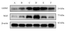
图1
各预处理组坐骨神经GDNF、NGF蛋白表达"

表1
各预处理组坐骨神经GDNF、NGF蛋白表达"
| 组别 | n | GDNF/β-actin | NGF/β-actin |
|---|---|---|---|
| A组 | 6 | 0.186±0.026 | 0.092±0.023 |
| B组 | 6 | 0.454±0.032a | 0.251±0.029a |
| C组 | 6 | 0.937±0.030a,b | 0.864±0.063a,b |
| D组 | 6 | 1.242±0.041a,b,c | 1.212±0.040a,b,c, |
| E组 | 6 | 0.982±0.058a,b,d | 0.458±0.042a,b,c,d |
| F组 | 6 | 0.542±0.025a,c,d,e | 0.265±0.023a,c,d,e |
| F值 | 339.661 | 358.905 | |
| P值 | < 0.001 | < 0.001 |
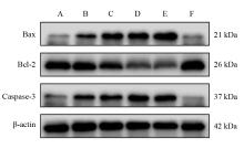
图2
各预处理组坐骨神经Bcl-2、Bax、Caspase-3蛋白表达"
表2
各预处理组坐骨神经Bcl-2、Bax、Caspase-3蛋白表达"
| 组别 | n | Bax/β-actin | Bcl-2/β-actin | Bax/Bcl-2 | Caspase-3/β-actin |
|---|---|---|---|---|---|
| A组 | 6 | 0.203±0.031 | 1.128±0.066 | 0.180±0.030 | 0.255±0.019 |
| B组 | 6 | 0.446±0.026a | 1.061±0.038 | 0.422±0.035 | 0.528±0.031a |
| C组 | 6 | 0.754±0.037a,b | 0.894±0.038a,b | 0.843±0.020a,b, | 0.613±0.028a |
| D组 | 6 | 0.858±0.047a,b | 0.691±0.027a,b,c | 1.244±0.090a,b,c | 0.741±0.035a,b,c |
| E组 | 6 | 1.001±0.064a,b,c,d | 0.629±0.034a,b,c | 1.597±0.177a,b,c,d | 0.750±0.046a,b,c |
| F组 | 6 | 0.189±0.043b,c,d,e | 1.267±0.094b,c,d,e | 0.149±0.028b,c,d,e | 0.167±0.030b,c,d,e |
| F值 | 192.279 | 63.485 | 148.898 | 171.621 | |
| P值 | < 0.001 | < 0.001 | < 0.001 | < 0.001 |
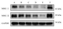
图3
各预处理组坐骨神经MHC-Ⅰ、MHC-Ⅱ蛋白表达"

表3
各预处理组坐骨神经MHC-Ⅰ、MHC-Ⅱ蛋白表达"
| 组别 | n | MHC-Ⅰ/GAPDH | MHC-Ⅱ/GAPDH |
|---|---|---|---|
| A组 | 6 | 0.311±0.021 | 0.639±0.033 |
| B组 | 6 | 0.154±0.014a | 0.576±0.022 |
| C组 | 6 | 0.128±0.010a | 0.467±0.024a,b |
| D组 | 6 | 0.070±0.006a,b,c | 0.311±0.019a,b,c |
| E组 | 6 | 0.045±0.004a,b,c | 0.139±0.015a,b,c,d |
| F组 | 6 | 0.422±0.021a,b,c,d,e | 0.816±0.036a,b,c,d,e |
| F值 | 358.905 | 349.990 | |
| P值 | < 0.001 | < 0.001 |
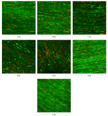
图4
各组坐骨神经比较(Calcein-AM/Propidium Iodide染色,激光共聚焦显微镜,×400,bar=50 µm)"

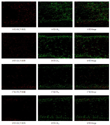
图5
移植术后1周A′~D′组移植神经段CD8+T细胞、巨噬细胞入侵情况(免疫荧光染色,×200) "

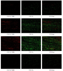
图6
移植术后1周E′~H′组移植神经段CD8+T细胞、巨噬细胞入侵情况(免疫荧光染色,×200) "

表4
移植术后1周,各组血清IL-2、IFN-γ、TNF-α水平(ng/L)"
| 组别 | n | IL-2 | IFN-γ | TNF-α |
|---|---|---|---|---|
| A′组 | 6 | 44.373±2.598 | 238.883±14.146 | 182.373±12.779 |
| B′组 | 6 | 43.786±2.604 | 234.133±19.821 | 164.290±8.081 |
| C′组 | 6 | 39.139±0.780 | 226.973±12.371 | 153.900±6.210a |
| D′组 | 6 | 36.864±2.130a,b | 225.423±11.857 | 136.743±4.721a,b |
| E′组 | 6 | 35.121±1.122a,b | 213.220±15.041 | 128.970±6.730a,b,c |
| F′组 | 6 | 44.848±1.858c,d,e | 257.103±14.482e | 184.997±2.015c,d,e |
| G′组 | 6 | 48.575±1.221c,d,e | 338.757±9.551a,b,c,d,e,f | 190.680±4.430b,c,d,e |
| H′组 | 6 | 35.173±1.721a,b,f,g | 203.947±11.377f,g | 123.757±3.462a,b,c,f,g |
| F值 | 22.218 | 27.632 | 45.485 | |
| P值 | < 0.001 | < 0.001 | < 0.001 |
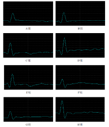
图7
移植术后20周各组CMAP波形 注:刺激电流4 mA"
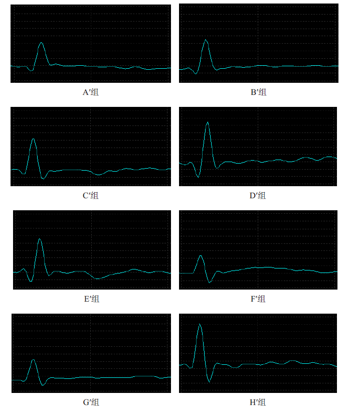
表5
移植术后20周各组CMAP和NCV比较"
| 组别 | n | CMAP(mA) | NCV(m/s) |
|---|---|---|---|
| A′组 | 6 | 0.127±0.031 | 4.983±0.649 |
| B′组 | 6 | 0.202±0.036 | 5.367±0.592 |
| C′组 | 6 | 0.235±0.051a | 6.417±0.436a |
| D′组 | 6 | 0.432±0.050a,b,c | 8.733±0.622a,b,c |
| E′组 | 6 | 0.273±0.044a,d | 7.083±0.739a,b,d |
| F′组 | 6 | 0.108±0.041b,c,d,e | 4.217±0.306b,c,d,e |
| G′组 | 6 | 0.062±0.025b,c,d,e | 4.067±0.592b,c,d,e |
| H′组 | 6 | 0.475±0.043a,b,c,e,f,g | 9.117±0.542a,b,c,e,f,g |
| F值 | 80.228 | 68.753 | |
| P值 | < 0.001 | < 0.001 |
表6
移植术后20周各组腓肠肌湿重比"
| 组别 | n | 腓肠肌湿重比 |
|---|---|---|
| A′组 | 6 | 0.523±0.020 |
| B′组 | 6 | 0.557±0.021 |
| C′组 | 6 | 0.615±0.024a |
| D′组 | 6 | 0.765±0.031a,b,c |
| E′组 | 6 | 0.642±0.026a,b,d |
| F′组 | 6 | 0.484±0.010c,d,e |
| G′组 | 6 | 0.444±0.022a,b,c,d,e |
| H′组 | 6 | 0.782±0.039a,b,c,e,f,g |
| F值 | 71.790 | |
| P值 | < 0.001 |
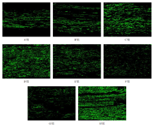
图8
移植术后20周各组移植神经段再生神经NF200(免疫荧光染色,×400)"

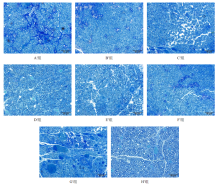
图9
移植术后20周各组移植神经段再生神经(甲苯胺蓝染色,bar = 50 μm)"

表7
移植术后20周各组轴突密度与髓鞘厚度比较"
| 组别 | n | 轴突密度(/mm2) | 髓鞘厚度(µm) |
|---|---|---|---|
| A′组 | 6 | 11776.524±816.723 | 0.604±0.060 |
| B′组 | 6 | 13302.008±1808.003 | 0.712±0.057 |
| C′组 | 6 | 15970.413±427.301a | 0.749±0.074a |
| D′组 | 6 | 21256.583±1009.335a,b,c | 0.989±0.058a,b,c |
| E′组 | 6 | 16877.374±1916.679a,d | 0.764±0.047a,d |
| F′组 | 6 | 11570.388±1277.176c,d,e | 0.564±0.050b,c,d,e |
| G′组 | 6 | 7471.276±689.650a,b,c,d,e,f | 0.421±0.034a,b,c,d,e,f |
| H′组 | 6 | 22674.714±1806.836a,b,c,e,f,g | 1.010±0.096a,b,c,e,f,g |
| F值 | 44.513 | 76.527 | |
| P值 | < 0.001 | < 0.001 |
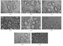
图10
移植术后20周各组移植神经段再生神经(透射电镜,×6000,bar = 2 μm)"

| [1] |
Eggers R, de Winter F, Hoyng S A, et al. Timed GDNF gene therapy using an immune-evasive gene switch promotes long distance axon regeneration[J]. Brain, 2019, 142(2):295-311.
doi: 10.1093/brain/awy340 pmid: 30649249 |
| [2] |
Hsu M N, Liao H T, Truong V A, et al. CRISPR-based activation of endogenous neurotrophic genes in adipose stem cell sheets to stimulate peripheral nerve regeneration[J]. Theranostics, 2019, 9(21):6099-6111.
doi: 10.7150/thno.36790 |
| [3] |
Giusti G, Lee J Y, Kremer T, et al. The influence of vascularization of transplanted processed allograft nerve on return of motor function in rats[J]. Microsurgery, 2016, 36(2):134-143.
doi: 10.1002/micr.22371 |
| [4] |
Han G H, Peng J, Liu P, et al. Therapeutic strategies for peripheral nerve injury: decellularized nerve conduits and Schwann cell transplantation[J]. Neural Regen Res, 2019, 14(8):1343-1351.
doi: 10.4103/1673-5374.253511 |
| [5] |
Lin T, Liu S, Chen S, et al. Hydrogel derived from porcine decellularized nerve tissue as a promising biomaterial for repairing peripheral nerve defects[J]. Acta Biomater, 2018, 73:326-338.
doi: 10.1016/j.actbio.2018.04.001 |
| [6] |
Szynkaruk M, Kemp S W, Wood M D, et al. Experimental and clinical evidence for use of decellularized nerve allografts in peripheral nerve gap reconstruction[J]. Tissue Eng Part B Rev, 2013, 19(1):83-96.
doi: 10.1089/ten.teb.2012.0275 |
| [7] | 李子健, 黄英如, 曾欢欢, 等. 丹参酮Ⅱ_A磺酸钠促进低温保存的大鼠坐骨神经同种异体移植后神经再生[J]. 中国中药杂志, 2018, 43(9):1907-1914. |
| [8] |
Poppler L H, Ee X, Schellhardt L, et al. Axonal growth arrests after an increased accumulation of Schwann cells expressing senescence markers and stromal cells in acellular nerve allografts[J]. Tissue Eng Part A, 2016, 22(13-14):949-961.
doi: 10.1089/ten.tea.2016.0003 |
| [9] |
Saheb-Al-Zamani M, Yan Y, Farber S J, et al. Limited regeneration in long acellular nerve allografts is associated with increased Schwann cell senescence[J]. Exp Neurol, 2013, 247:165-177.
doi: 10.1016/j.expneurol.2013.04.011 pmid: 23644284 |
| [10] |
Jesuraj N J, Santosa K B, Macewan M R, et al. Schwann cells seeded in acellular nerve grafts improve functional recovery[J]. Muscle Nerve, 2014, 49(2):267-276.
doi: 10.1002/mus.23885 pmid: 23625513 |
| [11] | 李子健, 黄英如, 曾欢欢, 等. 冷诱导RNA结合蛋白促进冷冻保存大鼠坐骨神经异体移植后神经再生的作用[J]. 中国康复理论与实践, 2018, 24(4):391-400. |
| [12] | Ogier M, Kron M, Katz D M. Neurotrophic factors in development and regulation of respiratory control[J]. Compr Physiol, 2013, 3(3):1125-1134. |
| [13] |
Arthur-Farraj P J, Latouche M, Wilton D K, et al. c-Jun reprograms Schwann cells of injured nerves to generate a repair cell essential for regeneration[J]. Neuron, 2012, 75(4):633-647.
doi: 10.1016/j.neuron.2012.06.021 pmid: 22920255 |
| [14] |
Gomez-Sanchez J A, Pilch K S, van der Lans M, et al. After nerve injury, lineage tracing shows that myelin and remak schwann cells elongate extensively and branch to form repair Schwann cells, which shorten radically on remyelination[J]. J Neurosci, 2017, 37(37):9086-9099.
doi: 10.1523/JNEUROSCI.1453-17.2017 |
| [15] |
Jessen K R. The repair Schwann cell and its function in regenerating nerves[J]. J Physiol, 2016, 594(13):3521-3531.
doi: 10.1113/JP270874 |
| [16] |
Eggers R, de Winter F, Arkenaar C, et al. Enhanced regeneration and reinnervation following timed GDNF gene therapy in a cervical ventral root avulsion[J]. Exp Neurol, 2019, 321:113037.
doi: S0014-4886(19)30186-4 pmid: 31425689 |
| [17] |
Eggers R, Tannemaat M R, Ehlert E M, et al. A spatio-temporal analysis of motoneuron survival, axonal regeneration and neurotrophic factor expression after lumbar ventral root avulsion and implantation[J]. Exp Neurol, 2010, 223(1):207-220.
doi: 10.1016/j.expneurol.2009.07.021 pmid: 19646436 |
| [18] |
Boyer R B, Sexton K W, Rodriguez-Feo C L, et al. Adjuvant neurotrophic factors in peripheral nerve repair with chondroitin sulfate proteoglycan-reduced acellular nerve allografts[J]. J Surg Res, 2015, 193(2):969-977.
doi: 10.1016/j.jss.2014.09.023 |
| [19] | 石晓伟, 黄亮亮, 夏冰, 等. 控释神经生长因子的阵列微管促进神经再生[J]. 中国矫形外科杂志, 2019, 27(13):1205-1210. |
| [20] |
Santos D, Giudetti G, Micera S, et al. Focal release of neurotrophic factors by biodegradable microspheres enhance motor and sensory axonal regeneration in vitro and in vivo[J]. Brain Res, 2016, 1636:93-106.
doi: 10.1016/j.brainres.2016.01.051 |
| [21] |
Tajdaran K, Gordon T, Wood M D, et al. A glial cell line-derived neurotrophic factor delivery system enhances nerve regeneration across acellular nerve allografts[J]. Acta Biomater, 2016, 29:62-70.
doi: S1742-7061(15)30133-1 pmid: 26441127 |
| [22] | 黄海涛, 刘华蔚, 胡敏. 神经营养因子促周围神经再生的研究进展[J]. 神经解剖学杂志, 2013, 29(5):599-602. |
| [23] |
Allen S J, Watson J J, Shoemark D K, et al. GDNF, NGF and BDNF as therapeutic options for neurodegeneration[J]. Pharmacol Ther, 2013, 138(2):155-175.
doi: 10.1016/j.pharmthera.2013.01.004 |
| [24] | 范红石, 王艳, 陈国平. 周围神经损伤后轴突再生微环境的研究进展[J]. 中国康复理论与实践, 2015, 31(3):288-291. |
| [25] |
Kokai L E, Ghaznavi A M, Marra K G, et al. Incorporation of double-walled microspheres into polymer nerve guides for the sustained delivery of glial cell line-derived neurotrophic factor[J]. Biomaterials, 2010, 31(8):2313-2322.
doi: 10.1016/j.biomaterials.2009.11.075 |
| [26] |
Kemp S W, Webb A A, Dhaliwal S, et al. Dose and duration of nerve growth factor (NGF) administration determine the extent of behavioral recovery following peripheral nerve injury in the rat[J]. Exp Neurol, 2011, 229(2):460-470.
doi: 10.1016/j.expneurol.2011.03.017 |
| [27] | 闫斌, 王靖博, 张宏, 等. 槲皮素对高糖培养海马神经元凋亡及Akt、p-Akt、Bcl-2、Bax蛋白表达的影响[J]. 中国康复理论与实践, 2017, 23(12):1390-1396. |
| [28] | 高玲, 赵建军, 王冰梅, 等. 益髓解毒法对血管性痴呆大鼠海马神经元Caspase-3、Caspase-9 mRNA及蛋白表达的影响[J]. 中华中医药杂志, 2019, 34(12):5656-5660. |
| [29] |
Snigdha S, Smith E D, Prieto G A, et al. Caspase-3 activation as a bifurcation point between plasticity and cell death[J]. Neurosci Bull, 2012, 28(1):14-24.
doi: 10.1007/s12264-012-1057-5 pmid: 22233886 |
| [30] |
Davila E, Byrne G W, Labreche P T, et al. T-cell responses during pig-to-primate xenotransplantation[J]. Xenotransplantation, 2006, 13(1):31-40.
doi: 10.1111/xen.2006.13.issue-1 |
| [31] |
Horne P H, Zimmerer J M, Fisher M G, et al. Critical role of effector macrophages in mediating CD4-dependent alloimmune injury of transplanted liver parenchymal cells[J]. J Immunol, 2008, 181(2):1224-1231.
doi: 10.4049/jimmunol.181.2.1224 |
| [32] |
Zimmerer J M, Horne P H, Fisher M G, et al. Unique CD8+ T cell-mediated immune responses primed in the liver[J]. Transplantation, 2016, 100(9):1907-1915.
doi: 10.1097/TP.0000000000001290 pmid: 27379551 |
| [33] | 刘畅, 王红艳. CD8+T细胞活化与分化的分子机制[J]. 中国免疫学杂志, 2017, 33(4):481-487. |
| [34] |
Gerner M Y, Casey K A, Kastenmuller W, et al. Dendritic cell and antigen dispersal landscapes regulate T cell immunity[J]. J Exp Med, 2017, 214(10):3105-3122.
doi: 10.1084/jem.20170335 |
| [35] |
Matia I, Fellmer P, Splith K, et al. Immunosuppressive protocol with delayed use of low-dose tacrolimus after aortic transplantation suppresses donor-specific anti-MHC class I and class II antibody production in rats[J]. Ann Transplant, 2014, 19:225-232.
doi: 10.12659/AOT.889870 |
| [36] |
Meyer zu Hörste G, Hu W, Hartung H P, et al. The immunocompetence of Schwann cells[J]. Muscle Nerve, 2008, 37(1):3-13.
pmid: 17823955 |
| [37] |
Whitlock E L, Myckatyn T M, Tong A Y, et al. Dynamic quantification of host Schwann cell migration into peripheral nerve allografts[J]. Exp Neurol, 2010, 225(2):310-319.
doi: 10.1016/j.expneurol.2010.07.001 |
| [1] | 李芳, 霍速, 杜巨豹, 刘秀贞, 李小爽, 宋为群. 经颅直流电刺激联合任务导向性康复训练对脊髓损伤大鼠前肢运动障碍的效果[J]. 《中国康复理论与实践》, 2023, 29(7): 777-781. |
| [2] | 罗兰, 李璐, 金沐. 氙气后处理对脊髓缺血再灌注损伤的效果:Akt信号通路和自噬机制[J]. 《中国康复理论与实践》, 2023, 29(2): 174-181. |
| [3] | 黄志霖, 徐发邵, 施静, 黄淦, 刘梅芳, 张霞辉. 线栓法建立卒中后吞咽障碍的大鼠模型[J]. 《中国康复理论与实践》, 2023, 29(10): 1147-1153. |
| [4] | 秦彦强, 董浩, 孙迎春, 程先宽, 姚海江. 不同针刺方案对卒中后抑郁大鼠神经递质及相关炎性因子的影响[J]. 《中国康复理论与实践》, 2023, 29(1): 30-37. |
| [5] | 缪培,张通,米海霞,张伟东. 不同线栓法复制局灶性脑缺血模型大鼠恢复期学习记忆能力的差异及其机制[J]. 《中国康复理论与实践》, 2022, 28(7): 789-796. |
| [6] | 周小珏,冯婧,庞日朝,刘捷,张安仁. 隔日限食减轻脊髓损伤大鼠炎症反应的芳香烃受体/细胞因子信号传导抑制因子2/核转录因子-κB信号通路机制[J]. 《中国康复理论与实践》, 2022, 28(5): 544-551. |
| [7] | 宋绍霏,侯园园,王云雷,张通. 异常光周期所致昼夜节律紊乱对大鼠腓肠肌时钟基因和葡萄糖摄取相关基因表达节律的影响[J]. 《中国康复理论与实践》, 2022, 28(5): 552-558. |
| [8] | 李童,方志鹏,邵玉萍,王平. 有氧运动对睡眠剥夺大鼠学习记忆及海马神经元突触可塑性的效果[J]. 《中国康复理论与实践》, 2022, 28(11): 1270-1277. |
| [9] | 王静怡,尹杰,刘建成,庞日朝,王文春. 蜘蛛香环烯醚萜类成分对急性脊髓损伤大鼠神经细胞焦亡的影响[J]. 《中国康复理论与实践》, 2021, 27(6): 653-660. |
| [10] | 周文美,陶陶,吴霜,王廷龙,杨正奕,张莹. 丰富环境对脑缺血再灌注损伤大鼠神经功能和缺血半暗带区葡萄糖代谢的影响[J]. 《中国康复理论与实践》, 2021, 27(5): 522-529. |
| [11] | 王琼芬,王风波,王科,钟永强,王娇娇. 电针风池对急性脑梗死大鼠脑星形胶质细胞和神经元的保护效果[J]. 《中国康复理论与实践》, 2021, 27(3): 302-309. |
| [12] | 李向哲,丁洁,王庆华,董传明,王彤,吴勤峰. 减重平板训练对脊髓损伤大鼠神经病理性疼痛及脊髓后角谷氨酸脱羧酶-65/67表达的影响[J]. 《中国康复理论与实践》, 2021, 27(2): 131-136. |
| [13] | 艾坤,许明,刘琼,邓石峰,刘继生,祁芳,易细芹,瞿启睿,张泓. 电针对骶上脊髓损伤后逼尿肌亢进大鼠逼尿肌中环腺苷酸和蛋白激酶A含量及肌球蛋白轻链激酶磷酸化的影响[J]. 《中国康复理论与实践》, 2021, 27(2): 137-144. |
| [14] | 凌梦钰,杨一卓,刘帅,叶超群. 运动训练对脑缺血再灌注大鼠认知功能及前额皮层神经元核抗原和突触素1表达的效果[J]. 《中国康复理论与实践》, 2021, 27(11): 1272-1281. |
| [15] | 彭志锋, 张义平, 张继红. 早期运动联合戊四氮对脑缺血再灌注大鼠神经功能的效果及其机制[J]. 《中国康复理论与实践》, 2021, 27(1): 27-30. |
| 阅读次数 | ||||||
|
全文 |
|
|||||
|
摘要 |
|
|||||
|
||