


《中国康复理论与实践》 ›› 2020, Vol. 26 ›› Issue (5): 572-578.doi: 10.3969/j.issn.1006-9771.2020.05.016
樊瑞文,李晓琳,黄幸,徐敏杰,曹云,张丹莉,常静玲( )
)
收稿日期:2019-12-17
修回日期:2020-01-17
出版日期:2020-05-25
发布日期:2020-05-29
通讯作者:
常静玲
E-mail:ear6979@163.com
作者简介:樊瑞文(1991-),女,汉族,江苏镇江市人,博士研究生,主要研究方向:中医药防治脑病的临床与神经影像学实验研究。
基金资助:
FAN Rui-wen,LI Xiao-lin,HUANG Xing,XU Min-jie,CAO Yun,ZHANG Dan-li,CHANG Jing-ling()
Received:2019-12-17
Revised:2020-01-17
Published:2020-05-25
Online:2020-05-29
Contact:
CHANG Jing-ling
E-mail:ear6979@163.com
Supported by:摘要:
目的 探究右脑功能网络在左侧脑损伤卒中后失语(PSA)的效应特点。方法 2018年12月至2019年6月,招募左脑半球损伤PSA患者12例(患者组)和匹配的健康成年人12例(对照组),以双流语言模型为参照,应用静息态功能磁共振观察右脑功能网络特征。结果 2例患者脱落。与对照组相比,在语言背侧通路中,患者组缘上回至额中回、三角部额下回功能连接增强,中央后回至岛盖部额下回功能连接降低;在语言腹侧通路中,角回至眶部额下回功能连接增强;在腹、背侧双通路模型中,语言功能皮质至皮质下核团功能连接增强,额、颞叶和边缘系统功能连接减弱。三角部额下回至豆状壳核功能连接增强与复述能力呈负相关(r = -0.720, P < 0.05),岛盖部额下回至尾状核功能连接增强与说和复述能力呈负相关关系( r < -0.696, P < 0.05)。患者组右脑语言关键脑区局部网络指标和全局指标均与对照组有显著性差异(| t| > 2.143, P < 0.05)。 结论 PSA患者右脑功能网络在脑卒中后发生功能重组,语言关键皮质与皮质下核团功能连接增强可能是右脑代偿的关键环节。
中图分类号:
樊瑞文,李晓琳,黄幸,徐敏杰,曹云,张丹莉,常静玲. 基于语言双流模型的卒中后失语右脑功能网络研究[J]. 《中国康复理论与实践》, 2020, 26(5): 572-578.
FAN Rui-wen,LI Xiao-lin,HUANG Xing,XU Min-jie,CAO Yun,ZHANG Dan-li,CHANG Jing-ling. Functional Connectivities in Right Hemisphere for Post-stroke Aphasia: Based on Dual Stream Model[J]. 《Chinese Journal of Rehabilitation Theory and Practice》, 2020, 26(5): 572-578.
表1
两组一般资料比较"
| 组别 | n | 性别(男/女, n) | 年龄(岁) | 教育程度(年) | 病程(d) | 病灶体积(ml) |
|---|---|---|---|---|---|---|
| 对照组 | 12 | 10/2 | 57.08±9.03 | 13.33±3.06 | - | - |
| 患者组 | 10 | 6/4 | 51.10±12.42 | 12.20±3.58 | 46.7±40.94 | 17.01±17.29 |
| χ2/t值 | 0.552 | -1.307 | -0.801 | |||
| P值 | 0.458 | 0.206 | 0.434 |
图1
所有患者病灶叠加图"
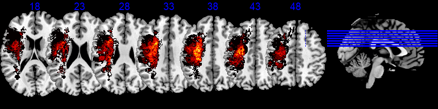
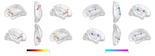
图2
两组右脑背侧通路fMRI功能连接差异"

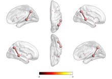
图3
两组右脑腹侧通路fMRI功能连接差异"

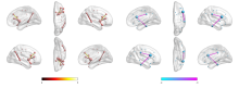
图4
两组右脑腹、背侧通路fMRI功能连接差异"

表2
两组右脑功能网络有差异的局部指标比较"
| 网络指标 | 脑区 | 对照组(n= 12) | 患者组(n= 10) | t值 | P值 |
|---|---|---|---|---|---|
| 介数中心性 | 后扣带回 | 1.63±1.46 | 4.85±4.31 | 2.442 | 0.024 |
| 眶部额上回 | 3.82±2.71 | 7.62±5.41 | 2.143 | 0.045 | |
| 度中心性 | 额中回 | 7.00±1.36 | 8.93±1.42 | 3.249 | 0.004 |
| 节点集聚系数 | 颞横回 | 0.23±0.08 | 0.14±0.08 | -2.689 | 0.014 |
| 节点效率 | 额中回 | 0.26±0.02 | 0.28±0.02 | 2.926 | 0.008 |
| 颞下回 | 0.29±0.02 | 0.26±0.02 | -2.315 | 0.031 | |
| 节点局部效率 | 颞横回 | 0.26±0.09 | 0.16±0.09 | -2.741 | 0.013 |
| 节点最短路径 | 额中回 | 0.65±0.07 | 0.59±0.04 | -2.417 | 0.025 |
表3
两组右脑功能网络全局指标比较"
| 网络指标 | 对照组(n= 12) | 患者组(n= 10) | t值 | P值 | |
|---|---|---|---|---|---|
| 同配性 | 同配性分数 | 0.04±0.05 | 0.04±0.06 | 0.068 | 0.947 |
| 同配性z分 | 1.94±0.73 | 2.21±1.13 | 0.682 | 0.503 | |
| 层级性 | 层级性分数 | 0.05±0.04 | 0.02±0.05 | -1.574 | 0.131 |
| 层级性z分 | -0.14±0.30 | -0.53±0.50 | -2.300 | 0.032 | |
| 网络效率 | 全局效率 | 0.23±0.01 | 0.23±0.01 | -1.047 | 0.308 |
| 局部效率 | 0.29±0.01 | 0.28±0.01 | -1.868 | 0.077 | |
| 小世界属性 | 聚集系数 | 0.23±0.01 | 0.23±0.02 | -0.047 | 0.963 |
| Gamma系数 | 0.59±0.07 | 0.52±0.06 | -2.558 | 0.019 | |
| Lambda系数 | 0.42±0.01 | 0.42±0.01 | -0.084 | 0.934 | |
| 最短路径 | 0.76±0.05 | 0.79±0.07 | 1.260 | 0.222 | |
| Sigma系数 | 0.56±0.06 | 0.49±0.06 | -2.493 | 0.022 | |
| 同步性 | 同步性分数 | 0.02±0.02 | 0.01±0.01 | -0.878 | 0.390 |
| 同步性z分 | -0.47±0.66 | -0.37±0.20 | 0.534 | 0.602 |
| [1] |
Doesborgh S J C, van de Sandt-Koenderman M W E, Dippel D W J, et al. Effects of semantic treatment on verbal communication and linguistic processing in aphasia after stroke: a randomized controlled trial[J]. Stroke, 2004, 35(1):141-146.
pmid: 14657447 |
| [2] |
Lomas J, Kertesz A. Patterns of spontaneous recovery in aphasic groups: a study of adult stroke patients[J]. Brain Lang, 1978, 5(3):388-401.
pmid: 656906 |
| [3] |
Teasell R, Bitensky J, Salter K, et al. The role of timing and intensity of rehabilitation therapies[J]. Top Stroke Rehabil, 2005, 12(3):46-57.
pmid: 16110427 |
| [4] |
Nouwens F, Visch-Brink E G, van de Sandt-Koenderman M M, et al. Optimal timing of speech and language therapy for aphasia after stroke: more evidence needed[J]. Expert Rev Neurother, 2015, 15(8):885-893.
doi: 10.1586/14737175.2015.1058161 pmid: 26088694 |
| [5] | Chen W, Ye Q, Ji X, et al. Mirror neuron system based therapy for aphasia rehabilitation[J]. Front Psychol, 2015, 6:1665. |
| [6] |
Norise C, Hamilton R H. Non-invasive brain stimulation in the treatment of post-stroke and neurodegenerative aphasia: parallels, differences, and lessons learned[J]. Front Hum Neurosci, 2016, 10:675.
doi: 10.3389/fpsyg.2019.00675 |
| [7] |
Thrane G, Friborg O, Anke A, et al. A meta-analysis of constraint-induced movement therapy after stroke[J]. J Rehabil Med, 2014, 46(9):833-842.
doi: 10.2340/16501977-1859 pmid: 25182341 |
| [8] |
Laska A C, Hellblom A, Murray V, et al. Aphasia in acute stroke and relation to outcome[J]. J Intern Med, 2001, 249(5):413-422.
pmid: 11350565 |
| [9] | Kerr A L, Cheng S Y, Jones T A. Experience-dependent neural plasticity in the adult damaged brain[J]. J Commun Disord, 2011, 44(5):538-548. |
| [10] |
Fridriksson J, Bonilha L, Baker J M, et al. Activity in preserved left hemisphere regions predicts anomia severity in aphasia[J]. Cereb. Cortex, 2010, 20(5):1013-1019.
doi: 10.1093/cercor/bhp160 |
| [11] |
Turkeltaub P E, Messing S, Norise C, et al. Are networks for residual language function and recovery consistent across aphasic patients?[J]. Neurology, 2011, 76(20):1726-1734.
doi: 10.1212/WNL.0b013e31821a44c1 pmid: 21576689 |
| [12] |
Fridriksson J, Richardson J D, Fillmore P, et al. Left hemisphere plasticity and aphasia recovery[J]. Neuroimage, 2012, 60(2):854-863.
doi: 10.1016/j.neuroimage.2011.12.057 pmid: 22227052 |
| [13] |
Meinzer M, Flaisch T, Breitenstein C, et al. Functional re-recruitment of dysfunctional brain areas predicts language recovery in chronic aphasia[J]. Neuroimage, 2008, 39(4):2038-2046.
pmid: 18096407 |
| [14] |
Miura K, Nakamura Y, Miura F, et al. Functional magnetic resonance imaging to word generation task in a patient with Broca's aphasia[J]. J Neurol, 1999, 246(10):939-942.
pmid: 10552242 |
| [15] |
Turkeltaub P E. Brain stimulation and the role of the right hemisphere in aphasia recovery[J]. Curr Neurol Neurosci Rep, 2015, 15(11):72.
doi: 10.1007/s11910-015-0593-6 |
| [16] |
Tourville J A, Guenther F H. The DIVA model: a neural theory of speech acquisition and production[J]. Lang Cogn Process, 2011, 26(7):952-981.
pmid: 23667281 |
| [17] | Brunswick N. The functional neuroanatomy of reading[J]. The Neural Basis of Reading, 2010, 1:79. |
| [18] |
Fridriksson J, Den Ouden D B, Hillis A E, et al. Anatomy of aphasia revisited[J]. Brain, 2018, 141(3):848-862.
doi: 10.1093/brain/awx363 pmid: 29360947 |
| [19] |
Liuzzi A G, Bruffaerts R, Peeters R, et al. Cross-modal representation of spoken and written word meaning in left pars triangularis[J]. Neuroimage, 2017, 150:292-307.
doi: 10.1016/j.neuroimage.2017.02.032 |
| [20] |
Northam G B, Adler S, Eschmann K C J, et al. Developmental conduction aphasia after neonatal stroke[J]. Ann Neurol, 2018, 83(4):664-675.
doi: 10.1002/ana.25218 pmid: 29572915 |
| [21] |
Hickok G, Poeppel D. The cortical organization of speech processing[J]. Nat Rev Neurosci, 2007, 8(5):393-402.
doi: 10.1038/nrn2113 |
| [22] |
Chang J, Zhang H, Tan Z, et al. Effect of electroacupuncture in patients with post-stroke motor aphasia: neurolinguistic and neuroimaging characteristics[J]. Wien Klin Wochenschr, 2017, 129(3-4):102-109.
doi: 10.1007/s00508-016-1070-1 |
| [23] |
Fernandez B, Cardebat D, Demonet J F, et al. Functional MRI follow-up study of language processes in healthy subjects and during recovery in a case of aphasia[J]. Stroke, 2004, 35(9):2171-2176.
pmid: 15297629 |
| [24] |
Murase N, Duque J, Mazzocchio R, et al. Influence of interhemispheric interactions on motor function in chronic stroke[J]. Ann Neurol, 2004, 55(3):400-409.
doi: 10.1002/(ISSN)1531-8249 |
| [25] |
Breier J I, Juranek J, Maher L M, et al. Behavioral and neurophysiologic response to therapy for chronic aphasia[J]. Arch Phys Med Rehabil, 2009, 90(12):2026-2033.
doi: 10.1016/j.apmr.2009.08.144 |
| [26] |
Zhuo C, Zhou C, Lin X, et al. Common and distinct global functional connectivity density alterations in drug-naïve patients with first-episode major depressive disorder with and without auditory verbal hallucination[J]. Prog Neuropsychopharmacol Biol Psychiatry, 2020, 96:109738.
doi: 10.1016/j.pnpbp.2019.109738 |
| [27] |
Baker C, Worrall L, Rose M, et al. A systematic review of rehabilitation interventions to prevent and treat depression in post-stroke aphasia[J]. Disabil Rehabil, 2018, 40(16):1870-1892.
doi: 10.1080/09638288.2017.1315181 |
| [28] |
Uchida-Ota M, Arimitsu T, Tsuzuki D, et al. Maternal speech shapes the cerebral frontotemporal network in neonates: a hemodynamic functional connectivity study[J]. Dev Cogn Neurosci, 2019, 39:100701.
doi: 10.1016/j.dcn.2019.100701 |
| [29] | Rosselli M, Ardila A, Bernal B. Angular gyrus connectivity model for language: a functional neuroimaging meta-analysis[J]. Rev Neurol, 2015, 60(11):495-503. |
| [30] |
Zhang Q, Wang H, Luo C, et al. The neural basis of semantic cognition in Mandarin Chinese: a combined fMRI and TMS study[J]. Hum Brain Mapp, 2019, 40(18):5412-5423.
doi: 10.1002/hbm.24781 pmid: 31507031 |
| [31] |
Manes J L, Parkinson A L, Larson C R, et al. Connectivity of the subthalamic nucleus and globus pallidus pars interna to regions within the speech network: a meta-analytic connectivity study[J]. Hum Brain Mapp, 2014, 35(7):3499-3516.
doi: 10.1002/hbm.22417 |
| [32] |
Treml E A, Halpin P N, Urban D L, et al. Modeling population connectivity by ocean currents, a graph-theoretic approach for marine conservation[J]. Landscape Ecology, 2008, 23(1):19-36.
doi: 10.1007/s10980-007-9138-y |
| [33] |
Zhou Q, Womer F Y, Kong L, et al. Trait-related cortical-subcortical dissociation in bipolar disorder: analysis of network degree centrality[J]. J Clin Psychiatry, 2017, 78(5):584-591.
doi: 10.4088/JCP.15m10091 |
| [34] |
Hager B, Yang A C, Brady R, et al. Neural complexity as a potential translational biomarker for psychosis[J]. J Affect Disord, 2017, 216:89-99.
doi: 10.1016/j.jad.2016.10.016 |
| [35] |
Goñi J, van den Heuvel M P, Avena-Koenigsberger A, et al. Resting-brain functional connectivity predicted by analytic measures of network communication[J]. Proc Natl Acad Sci USA, 2014, 111(2):833-838.
doi: 10.1073/pnas.1315529111 |
| [36] |
Zhu Y, Bai L, Liang P, et al. Disrupted brain connectivity networks in acute ischemic stroke patients[J]. Brain Imaging Behav, 2017, 11(2):444-453.
doi: 10.1007/s11682-016-9525-6 |
| [37] |
Zhang C, Peng G, Shao J, et al. Neural bases of congenital amusia in tonal language speakers[J]. Neuropsychologia, 2017, 97:18-28.
doi: 10.1016/j.neuropsychologia.2017.01.033 |
| [38] |
Lendrem W, Lincoln N B. Spontaneous recovery of language in patients with aphasia between 4 and 34 weeks after stroke[J]. J Neurol Neurosurg Psychiatry, 1985, 48(8):743-748.
doi: 10.1136/jnnp.48.8.743 |
| [1] | 林娜, 高菡璐, 卢惠苹, 陈燕清, 郑军凡, 陈述荣. 虚拟现实技术对脑卒中上肢功能影响的弥散张量成像研究[J]. 《中国康复理论与实践》, 2024, 30(1): 61-67. |
| [2] | 王昊懿, 史亚伟, 鲁俊, 许光旭. 主观垂直感知障碍对脑卒中患者功能影响的回顾性研究[J]. 《中国康复理论与实践》, 2024, 30(1): 68-73. |
| [3] | 陈珺雯, 陈谦, 陈程, 李淑月, 刘玲玲, 吴存书, 龚翔, 鲁俊, 许光旭. 改良八段锦身体活动对脑卒中患者心肺功能、运动功能和日常生活活动能力的效果[J]. 《中国康复理论与实践》, 2024, 30(1): 74-80. |
| [4] | 胡永林, 马颖, 窦超, 陆安民, 江小鸽, 宋新建, 肖玉华. 肩部控制训练联合神经松动术对脑卒中偏瘫患者肩痛及上肢功能的效果[J]. 《中国康复理论与实践》, 2024, 30(1): 81-86. |
| [5] | 王贺, 韩靓, 阚梦凡, 于少泓. 电刺激治疗脑卒中后肩手综合征有效性的系统评价与Meta分析[J]. 《中国康复理论与实践》, 2023, 29(9): 1048-1056. |
| [6] | 孙藤方, 任梦婷, 杨琳, 王耀霆, 王红雨, 闫兴洲. 高压氧治疗联合重复外周磁刺激干预脑卒中患者踝运动功能和平衡能力的效果[J]. 《中国康复理论与实践》, 2023, 29(8): 875-881. |
| [7] | 王亚楠, 刘西花. 脑卒中偏瘫患者主观和客观平衡功能测量的相关性及预测效能[J]. 《中国康复理论与实践》, 2023, 29(8): 890-895. |
| [8] | 王海云, 王寅, 周信杰, 何爱群. 基于“中枢-外周-中枢”理论的经颅直流电刺激结合针刺干预脑卒中患者中枢及上肢功能的效果[J]. 《中国康复理论与实践》, 2023, 29(8): 919-925. |
| [9] | 陈怡婷, 王倩, 崔慎红, 李映彩, 张思鈺, 魏衍旭, 任慧, 冷军, 陈斌. 双侧序贯重复经颅磁刺激干预脑卒中患者上肢运动功能的效果[J]. 《中国康复理论与实践》, 2023, 29(8): 926-932. |
| [10] | 李振亚, 孙洁, 郭鹏飞, 王光明. 脑卒中患者口期和咽期吞咽功能改变与误吸的相关性:基于电视透视吞咽检查[J]. 《中国康复理论与实践》, 2023, 29(8): 933-939. |
| [11] | 华玲, 张一楠, 郑玉, 孙俏仪, 房辉, 宋达. 手控节律音乐治疗对脑卒中后单侧空间忽略的效果[J]. 《中国康复理论与实践》, 2023, 29(7): 833-838. |
| [12] | 蒋孝翠, 刘臻, 苏清伦, 赵秦, 夏晓昧, 陆飞. 间歇性Theta节律经颅磁刺激对脑卒中后非流利性失语的影响[J]. 《中国康复理论与实践》, 2023, 29(7): 839-843. |
| [13] | 许苗苗, 李楠, 应颖, 杨凯翔, 杨婧瑞, 李杰, 邱彦群. 重复外周磁刺激对左右颈7神经交叉移位术后脑卒中患者上肢运动功能的效果[J]. 《中国康复理论与实践》, 2023, 29(6): 686-690. |
| [14] | 郑莉, 鲍治诚, 张琪, 任绪艳, 苏敏. 经皮耳迷走神经电刺激结合康复机器人训练对脑卒中患者上肢功能的效果[J]. 《中国康复理论与实践》, 2023, 29(6): 691-696. |
| [15] | 顾彬, 张津沁, 夏元浩, 胡靖然, 诸桥直纪, 黄富表. 反复促进疗法对脑卒中恢复期偏瘫患者手功能的影响[J]. 《中国康复理论与实践》, 2023, 29(6): 697-702. |
| 阅读次数 | ||||||
|
全文 |
|
|||||
|
摘要 |
|
|||||
|
||