


《中国康复理论与实践》 ›› 2023, Vol. 29 ›› Issue (10): 1147-1153.doi: 10.3969/j.issn.1006-9771.2023.10.005
黄志霖1, 徐发邵2, 施静2, 黄淦1, 刘梅芳2, 张霞辉2( )
)
收稿日期:2022-10-08
修回日期:2023-07-11
出版日期:2023-10-25
发布日期:2023-11-16
通讯作者:
张霞辉(1982-),男,硕士,主治医师,主要研究方向:卒中及相关并发症的针灸康复治疗。E-mail: 754258082@qq.com
作者简介:黄志霖(1997-),男,汉族,广东汕尾市人,硕士研究生,主要研究方向:针灸防治脑病的基础及临床应用研究。
基金资助:
HUANG Zhilin1, XU Fashao2, SHI Jing2, HUANG Gan1, LIU Meifang2, ZHANG Xiahui2()
Received:2022-10-08
Revised:2023-07-11
Published:2023-10-25
Online:2023-11-16
Contact:
ZHANG Xiahui, E-mail: 754258082@qq.com
Supported by:摘要:
目的 建立一种可行的卒中后吞咽障碍动物模型。
方法 清洁级Sprague-Dawley大鼠22只随机分为正常组(n = 11)和造模组(n = 11)。造模组采用线栓法制备卒中后吞咽障碍动物模型,正常组不予处理。分别于造模成功3 d、7 d后记录所有大鼠第1次吞咽的发作潜伏期和吞咽次数,采用TTC染色法检测脑组织梗死情况,采用TUNEL荧光染色法测定缺血脑组织神经细胞凋亡情况。
结果 与正常组比较,造模组造模3 d后吞咽潜伏期有所延长,吞咽次数减少,但均无显著性差异(P > 0.05);7 d后吞咽潜伏期延长(P < 0.05),吞咽次数有所减少,但无显著性差异(P > 0.05)。与正常组比较,造模组脑组织出现明显梗死灶和大量凋亡细胞,体质量减小(P < 0.05)。
结论 造模大鼠出现一定程度的吞咽障碍症状,有望成为脑卒中后吞咽障碍的转化模型。
中图分类号:
黄志霖, 徐发邵, 施静, 黄淦, 刘梅芳, 张霞辉. 线栓法建立卒中后吞咽障碍的大鼠模型[J]. 《中国康复理论与实践》, 2023, 29(10): 1147-1153.
HUANG Zhilin, XU Fashao, SHI Jing, HUANG Gan, LIU Meifang, ZHANG Xiahui. Establishment of rat model of dysphagia after stroke by thread embolism[J]. 《Chinese Journal of Rehabilitation Theory and Practice》, 2023, 29(10): 1147-1153.
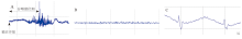
图1
典型肌电活动的EMG 注: A为大鼠咽喉区注射液体期间右下颌舌骨肌(产生吞咽动作)的肌电活动的典型例子;B为大鼠咽喉区未注射液体期间捕捉呼吸干扰的肌电活动;C为大鼠咽喉区未注射液体期间捕捉的心电干扰的肌电活动。"

表1
各组第1次吞咽发作潜伏期 单位:s"
| 组别 | n | 吞咽潜伏期 |
|---|---|---|
| 正常组 | 8 | 5.35(3.73, 5.58) |
| 造模组3 d后 | 7 | 17.00(5.00, 30.00) |
| 造模组7 d后 | 7 | 32.00(7.00, 34.00)a |
表2
各组吞咽次数比较 单位:次•(2 min)-1"
| 组别 | n | 吞咽次数 |
|---|---|---|
| 正常组 | 11 | 14.00(7.25, 40.00) |
| 造模组3 d后 | 10 | 4.50(1.75, 35.75) |
| 造模组7 d后 | 7 | 12.00(3.00, 12.00) |
表3
各组不同时间点体质量比较 单位:g"
| 组别 | n | 造模前 | 造模后第1天 | 造模后第2天 | 造模后第3天 | 造模后第4天 | 造模后第5天 | 造模后第6天 | 造模后第7天 |
|---|---|---|---|---|---|---|---|---|---|
| 正常组 | 11 | 196.15±5.85 | 204.26±9.93a | 220.53±14.18a | 227.33±13.11a | 226.86±11.50a | 246.90±12.76a | 251.31±15.57a | 264.81±13.26a |
| 造模组 | 7 | 198.83±10.92 | 181.79±13.48a,b | 172.81±20.99a,b | 169.22±28.39a,b | 168.00±33.96a,b | 176.08±35.77a,b | 179.22±35.87b | 191.09±39.77b |
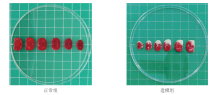
图2
各组脑组织TTC染色结果"

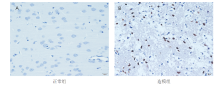
图3
各组凋亡细胞检测结果(TUNEL染色,×40)"

| [1] | 王陇德, 彭斌, 张鸿祺, 等. «中国脑卒中防治报告2020»概要[J]. 中国脑血管病杂志, 2022, 19(2): 136-144. |
| WANG L D, PENG B, ZHANG H Q, et al. Summary of China Stroke Prevention Report 2020[J]. Chin J Cerebrovasc Dis, 2022, 19(2): 136-144. | |
| [2] | 田婷, 关智媛, 石正洪, 等. 复发性缺血性脑卒中的危险因素、严重程度及短期预后分析[J]. 中国康复理论与实践, 2016, 22(2): 172-177. |
| TIAN T, GUAN Z Y, SHI Z H, et al. Analysis of risk factors, severity, and short-term prognosis of recurrent ischemic stroke[J]. Chin J Rehabil Theory Pract, 2016, 22(2): 172-177. | |
| [3] |
LEITE K, SASSI F C, MEDEIROS G C, et al. Clinical swallowing prognostic indicators in patients with acute ischemic stroke[J]. Arq Neuropsiquiatr, 2019, 77(7): 501-508.
doi: 10.1590/0004-282x20190080 |
| [4] |
SUGIYAMA N, NISHIYAMA E, NISHIKAWA Y, et al. A novel animal model of dysphagia following stroke[J]. Dysphagia, 2014, 29(1): 61-67.
doi: 10.1007/s00455-013-9481-x pmid: 23907747 |
| [5] |
YAMAMURA K, KUROSE M, OKAMOTO K. Guide to enhancing swallowing initiation: insights from findings in healthy subjects and dysphagic patients[J]. Curr Phys Med Rehabil Rep, 2018, 6(3): 178-185.
doi: 10.1007/s40141-018-0192-y |
| [6] | 方君辉, 宋丰军, 陈炳, 等. 颈项针联合电刺激对脑卒中吞咽障碍临床效果和脑血流的影响[J]. 中华中医药学刊, 2019, 37(2): 474-479. |
| FANG J H, SONG F J, CHEN B, et al. Effect of cervical acupuncture combined with electrical stimulation on clinical effects and cerebral blood flow in stroke[J]. Chin J Tradit Chin Med, 2019, 37(2): 474-479. | |
| [7] |
CHEN Y W, CHANG K H, CHEN H C, et al. The effects of surface neuromuscular electrical stimulation on post-stroke dysphagia: a systemic review and meta-analysis[J]. Clin Rehabil, 2016, 30(1): 24-35.
doi: 10.1177/0269215515571681 |
| [8] | 祁玉军, 潘秋银, 王文远, 等. 项丛刺疗法对脑卒中后吞咽障碍患者吞咽功能及呼吸功能的影响[J]. 中国针灸, 2021, 41(12): 1303-1307. |
| QI Y J, PAN Q Y, WANG W Y, et al. Effect of bush thorn therapy on swallowing function and respiratory function in patients with dysphagia after stroke[J]. Chin Acup, 2021, 41(12): 1303-1307. | |
| [9] | 冯声旺, 曹淑华, 杜淑佳, 等. 针刺配合吞咽训练治疗脑卒中后吞咽障碍:随机对照研究[J]. 中国针灸, 2016, 36(4): 347-350. |
| FENG S W, CAO S H, DU S J, et al. Acupuncture combined with swallowing training for post-stroke swallowing disorders: a randomized controlled study[J]. Chin Acup, 2016, 36 (4): 347-350. | |
| [10] | 李桂华, 张哲. 中医特色康复联合功能锻炼对缺血性卒中后吞咽障碍患者影响及BDNF研究[J]. 四川中医, 2020, 38(7): 205-208. |
| LI G H, ZHANG Z. Effect of combining functional exercise and rehabilitation of traditional Chinese medicine distinguishing feature for patients with dysphagia after ischemic stroke[J]. J Sichuan Tradit Chin Med, 2020, 38(7): 205-208. | |
| [11] |
JONES C A, COLLETTI C M, DING M C. Post-stroke dysphagia: recent insights and unanswered questions[J]. Curr Neurol Neurosci Rep, 2020, 20(12): 61.
doi: 10.1007/s11910-020-01081-z |
| [12] |
李斯锦, 李彦杰, 秦合伟, 等. 吞咽障碍动物模型研究进展[J]. 中国康复理论与实践, 2020, 26(11): 1311-1315.
doi: 10.3969/j.issn.1006-9771.2020.00.018 |
| LI S J, LI Y J, QIN H W, et al. Progress in animal models of swallowing disorder[J]. Chin Rehabil Theory Pract, 2020, 26 (11): 1311-1315. | |
| [13] |
ARCE-MCSHANE F I, ROSS C F, TAKAHASHI K, et al. Primary motor and sensory cortical areas communicate via spatiotemporally coordinated networks at multiple frequencies[J]. Proc Natl Acad Sci U S A, 2016, 113(18): 5083-5088.
doi: 10.1073/pnas.1600788113 |
| [14] |
BEST M D, NAKAMURA Y, KIJAK N A, et al. Semiautomatic marker tracking of tongue positions captured by videofluoroscopy during primate feeding[J]. Annu Int Conf IEEE Eng Med Biol Soc, 2015, 2015: 5347-5350.
doi: 10.1109/EMBC.2015.7319599 pmid: 26737499 |
| [15] |
INOKUCHI H, GONZÁLEZ-FERNÁNDEZ M, MATSUO K, et al. Electromyography of swallowing with fine wire intramuscular electrodes in healthy human: activation sequence of selected hyoid muscles[J]. Dysphagia, 2014, 29(6): 713-721.
doi: 10.1007/s00455-014-9566-1 pmid: 25142242 |
| [16] |
BLITZER A, CRUMLEY R L, DAILEY S H, et al. Recommendations of the Neurolaryngology Study Group on laryngeal electromyography[J]. Otolaryngol Head Neck Surg, 2009, 140(6): 782-793.
doi: 10.1016/j.otohns.2009.01.026 pmid: 19467391 |
| [17] |
GERMAN R Z, CROMPTON A W, GOULD F D, et al. Animal models for dysphagia studies: What have we learnt so far[J]. Dysphagia, 2017, 32(1): 73-77.
doi: 10.1007/s00455-016-9778-7 pmid: 28132098 |
| [18] | 李超, 叶秋平, 窦祖林, 等. 啮齿类动物吞咽障碍模型的研究及其进展[J]. 中华物理医学与康复杂志, 2021, 43(12): 1138-1141. |
| [19] |
HINKEL C J, SHARMA R, THAKKAR M M, et al. Neural mechanisms contributing to dysphagia in mouse models[J]. Otolaryngol Head Neck Surg, 2016, 155(2): 303-306.
doi: 10.1177/0194599816640261 pmid: 27048676 |
| [20] |
NAKAMURA Y, IRIARTE-DIAZ J, ARCE-MCSHANE F, et al. Sagittal plane kinematics of the jaw and hyolingual apparatus during swallowing in Macaca mulatta[J]. Dysphagia, 2017, 32(5): 663-677.
doi: 10.1007/s00455-017-9812-4 pmid: 28528492 |
| [21] |
DING P, FUNG G S, LIN M, et al. The effect of bilateral superior laryngeal nerve lesion on swallowing: a novel method to quantitate aspirated volume and pharyngeal threshold in videofluoroscopy[J]. Dysphagia, 2015, 30(1): 47-56.
doi: 10.1007/s00455-014-9572-3 pmid: 25270532 |
| [22] | 戴萌, 窦祖林, 卫小梅, 等. 吞咽造影的分析及应用进展[J]. 中国康复医学杂志, 2016, 31(11): 1269-1272. |
| [23] | LEVER T E, BRAUN S M, BROOKS R T, et al. Adapting human videofluoroscopic swallow study methods to detect and characterize dysphagia in murine disease models[J]. J Vis Exp, 2015(97): 52319. |
| [24] |
CULLINS M J, CONNOR N P. Reduced tongue force and functional swallowing changes in a rat model of post stroke dysphagia[J]. Brain Res, 2019, 1717: 160-166.
doi: S0006-8993(19)30224-0 pmid: 31022397 |
| [25] | DZIEWAS R, MICHOU E, TRAPL-GRUNDSCHOBER M, et al. European Stroke Organisation and European Society for Swallowing Disorders guideline for the diagnosis and treatment of post-stroke dysphagia[J]. Eur Stroke J, 2021, 6(3): LXXXIX-CXV. |
| [26] |
PATEL D A, KRISHNASWAMI S, STEGER E, et al. Economic and survival burden of dysphagia among inpatients in the United States[J]. Dis Esophagus, 2018, 31(1): 1-7.
doi: 10.1093/dote/dox131 pmid: 29155982 |
| [27] |
COHEN D L, ROFFE C, BEAVAN J, et al. Post-stroke dysphagia: a review and design considerations for future trials[J]. Int J Stroke, 2016, 11(4): 399-411.
doi: 10.1177/1747493016639057 pmid: 27006423 |
| [28] |
MCCARTY E B, CHAO T N. Dysphagia and swallowing disorders[J]. Med Clin North Am, 2021, 105(5): 939-954.
doi: 10.1016/j.mcna.2021.05.013 |
| [29] | 张耀文, 谢纯青, 万桂芳, 等. 神经肌肉电刺激对卒中后咽期吞咽启动延迟患者进食功能的即时效应观察[J]. 中华物理医学与康复杂志, 2020, 42(9): 797-800. |
| ZHANG Y W, XIE C Q, WAN G F, et al. The immediate effect of neuromuscular electrical stimulation on dysphagic stroke survivors' initiation of swallowing[J]. Chin J Phys Med Rehabil, 2020, 42(9): 797-800. | |
| [30] |
FARRELL Z, MURPHY E. A comment on "The natural history of dysphagia following a stroke" (Dysphagia 12:188-193, 1997)[J]. Dysphagia, 1998, 13(4): 230-231.
pmid: 9716756 |
| [31] |
SCHIMMEL M, VOEGELI G, DUVERNAY E, et al. Oral tactile sensitivity and masticatory performance are impaired in stroke patients[J]. J Oral Rehabil, 2017, 44(3): 163-171.
doi: 10.1111/joor.12482 pmid: 28075495 |
| [32] | KOKURA Y, KIMOTO K, OKADA Y, et al. The Controlling Nutritional Status score as a functional prognostic marker in patients with acute stroke: a multicenter retrospective cohort study[J]. Nutrition, 2020, 79-80: 110889. |
| [33] | 唐容, 段佳林, 李倩茜, 等. 脑卒中患者营养不良相关因素的系统评价[J]. 现代临床护理, 2022, 21(4): 66-76. |
| TANG R, DUAN J L, LI Q Q, et al. Influencing factors of malnutrition in stroke patients: a systematic evaluation and Meta-analysis[J]. Mod Clin Care, 2022, 21(4): 66-76. | |
| [34] |
SURA L, MADHAVAN A, CARNABY G, et al. Dysphagia in the elderly: management and nutritional considerations[J]. Clin Interv Aging, 2012, 7: 287-298.
doi: 10.2147/CIA.S23404 pmid: 22956864 |
| [35] |
DE COCK E, BATENS K, HEMELSOET D, et al. Dysphagia, dysarthria and aphasia following a first acute ischaemic stroke: incidence and associated factors[J]. Eur J Neurol, 2020, 27(10): 2014-2021.
doi: 10.1111/ene.v27.10 |
| [1] | 林娜, 高菡璐, 卢惠苹, 陈燕清, 郑军凡, 陈述荣. 虚拟现实技术对脑卒中上肢功能影响的弥散张量成像研究[J]. 《中国康复理论与实践》, 2024, 30(1): 61-67. |
| [2] | 王昊懿, 史亚伟, 鲁俊, 许光旭. 主观垂直感知障碍对脑卒中患者功能影响的回顾性研究[J]. 《中国康复理论与实践》, 2024, 30(1): 68-73. |
| [3] | 陈珺雯, 陈谦, 陈程, 李淑月, 刘玲玲, 吴存书, 龚翔, 鲁俊, 许光旭. 改良八段锦身体活动对脑卒中患者心肺功能、运动功能和日常生活活动能力的效果[J]. 《中国康复理论与实践》, 2024, 30(1): 74-80. |
| [4] | 胡永林, 马颖, 窦超, 陆安民, 江小鸽, 宋新建, 肖玉华. 肩部控制训练联合神经松动术对脑卒中偏瘫患者肩痛及上肢功能的效果[J]. 《中国康复理论与实践》, 2024, 30(1): 81-86. |
| [5] | 王贺, 韩靓, 阚梦凡, 于少泓. 电刺激治疗脑卒中后肩手综合征有效性的系统评价与Meta分析[J]. 《中国康复理论与实践》, 2023, 29(9): 1048-1056. |
| [6] | 孙藤方, 任梦婷, 杨琳, 王耀霆, 王红雨, 闫兴洲. 高压氧治疗联合重复外周磁刺激干预脑卒中患者踝运动功能和平衡能力的效果[J]. 《中国康复理论与实践》, 2023, 29(8): 875-881. |
| [7] | 王亚楠, 刘西花. 脑卒中偏瘫患者主观和客观平衡功能测量的相关性及预测效能[J]. 《中国康复理论与实践》, 2023, 29(8): 890-895. |
| [8] | 王海云, 王寅, 周信杰, 何爱群. 基于“中枢-外周-中枢”理论的经颅直流电刺激结合针刺干预脑卒中患者中枢及上肢功能的效果[J]. 《中国康复理论与实践》, 2023, 29(8): 919-925. |
| [9] | 陈怡婷, 王倩, 崔慎红, 李映彩, 张思鈺, 魏衍旭, 任慧, 冷军, 陈斌. 双侧序贯重复经颅磁刺激干预脑卒中患者上肢运动功能的效果[J]. 《中国康复理论与实践》, 2023, 29(8): 926-932. |
| [10] | 李振亚, 孙洁, 郭鹏飞, 王光明. 脑卒中患者口期和咽期吞咽功能改变与误吸的相关性:基于电视透视吞咽检查[J]. 《中国康复理论与实践》, 2023, 29(8): 933-939. |
| [11] | 李芳, 霍速, 杜巨豹, 刘秀贞, 李小爽, 宋为群. 经颅直流电刺激联合任务导向性康复训练对脊髓损伤大鼠前肢运动障碍的效果[J]. 《中国康复理论与实践》, 2023, 29(7): 777-781. |
| [12] | 华玲, 张一楠, 郑玉, 孙俏仪, 房辉, 宋达. 手控节律音乐治疗对脑卒中后单侧空间忽略的效果[J]. 《中国康复理论与实践》, 2023, 29(7): 833-838. |
| [13] | 蒋孝翠, 刘臻, 苏清伦, 赵秦, 夏晓昧, 陆飞. 间歇性Theta节律经颅磁刺激对脑卒中后非流利性失语的影响[J]. 《中国康复理论与实践》, 2023, 29(7): 839-843. |
| [14] | 许苗苗, 李楠, 应颖, 杨凯翔, 杨婧瑞, 李杰, 邱彦群. 重复外周磁刺激对左右颈7神经交叉移位术后脑卒中患者上肢运动功能的效果[J]. 《中国康复理论与实践》, 2023, 29(6): 686-690. |
| [15] | 郑莉, 鲍治诚, 张琪, 任绪艳, 苏敏. 经皮耳迷走神经电刺激结合康复机器人训练对脑卒中患者上肢功能的效果[J]. 《中国康复理论与实践》, 2023, 29(6): 691-696. |
| 阅读次数 | ||||||
|
全文 |
|
|||||
|
摘要 |
|
|||||
|
||