


《中国康复理论与实践》 ›› 2024, Vol. 30 ›› Issue (10): 1224-1231.doi: 10.3969/j.issn.1006-9771.2024.10.013
杨平1,2,3( ), 陈浩源2, 高睿馨2, 王欣平2
收稿日期:2024-07-24
修回日期:2024-08-12
出版日期:2024-10-25
发布日期:2024-11-08
通讯作者:
杨平(1983-),女,汉族,山东临沂市人,硕士,副研究员,主要研究方向:康复工程、生物力学。E-mail: yangping2005668@163.com
基金资助:
YANG Ping1,2,3(), CHEN Haoyuan2, GAO Ruixin2, WANG Xinping2, Philip ROWE3
Received:2024-07-24
Revised:2024-08-12
Published:2024-10-25
Online:2024-11-08
Contact:
YANG Ping, E-mail: yangping2005668@163.com
Supported by:摘要:
目的 设计一种基于低成本惯性测量单元和力敏电阻的便携式足部三维运动捕捉系统,探讨应用该系统评估裸足和穿鞋状态下足内小关节三维运动的可行性。
方法 该系统由数据采集、数据传输和数据处理部分组成。惯性测量单元用于足部小关节运动数据采集,力敏电阻用于检测足跟触地和足趾离地时间。所有数据通过无线局域网传输,数据处理由自主编写的宏程序完成。2024年1月至7月,2例健康成年女性穿戴该设备以自我感觉合适的速度在室内平地连续行走10 m。采用步态分析对受试者2进行行走一致性测试,并记录受试者1分别以裸足、穿两种护士鞋行走时,连续步态周期下第1跖骨相对于拇趾(Met-Ph)、中足相对于第1跖骨(Mid-Met)的背屈-跖屈、内收-外展和内旋-外旋运动曲线。
结果 该系统在行走时显示良好的测试一致性。Met-Ph和Mid-Met的背屈-跖屈、内收-外展和内旋-外旋活动范围在穿鞋时均较裸足时降低,使用带足弓支撑的鞋最低。
结论 本研究开发了一种低成本、便携式足部三维运动系统,可用于测量连续步态时足内小关节在裸足和穿鞋状态下的三维运动。
中图分类号:
杨平, 陈浩源, 高睿馨, 王欣平. 基于低成本惯性测量单元和力敏电阻的足部三维运动数据建模[J]. 《中国康复理论与实践》, 2024, 30(10): 1224-1231.
YANG Ping, CHEN Haoyuan, GAO Ruixin, WANG Xinping, Philip ROWE. Three-dimensional modeling of foot motion based on low-cost inertial measurement unit and force sensing resistor[J]. Chinese Journal of Rehabilitation Theory and Practice, 2024, 30(10): 1224-1231.
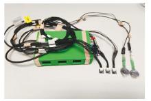
图1
控制盒"


图2
穿戴效果图"


图3
两种护士鞋"


图4
受试者2重复6次的裸足自由平地行走关节三维曲线"


图5
受试者1裸足、穿两种护士鞋自由行走的关节三维角度曲线"

| [1] |
MCNUTT E J, ZIPFEL B, DESILVA J M. The evolution of the human foot[J]. Evol Anthropol, 2018, 27(5): 197-217.
doi: 10.1002/evan.21713 pmid: 30242943 |
| [2] |
HORNESTAM J F, ARANTES P M M, SOUZA T R, et al. Foot pronation affects pelvic motion during the loading response phase of gait[J]. Braz J Phys Ther, 2021, 25(6): 727-734.
doi: 10.1016/j.bjpt.2021.04.005 pmid: 34020879 |
| [3] | 杨平, 蔡丽飞. 足过度旋前对人体力线的影响及治疗方法[J]. 中国康复理论与实践, 2016, 22(1): 72-74. |
| YANG P, CAI L F. Foot overpronation: influence on body alignment and managements[J]. Chin J Rehabil Theory Pract, 2016, 22(1): 72-74. | |
| [4] | LEARDINI A, STEBBINS J, HILLSTROM H, et al. ISB recommendations for skin-marker-based multi-segment foot kinematics[J]. J Biomech, 2021, 125: 110581. |
| [5] |
LEARDINI A, CARAVAGGI P, THEOLOGIS T, et al. Multi-segment foot models and their use in clinical populations[J]. Gait Posture, 2019, 69: 50-59.
doi: S0966-6362(18)31705-3 pmid: 30665039 |
| [6] | BAUER L, HAMBERGER M A, BÖCKER W, et al. Development of an IMU based 2-segment foot model for an applicable medical gait analysis[J]. BMC Musculoskelet Disord, 2024, 25(1): 606. |
| [7] | CAMPBELL K J, WILSON K J, LAPRADE R F, et al. Normative rearfoot motion during barefoot and shod walking using biplane fluoroscopy[J]. Knee Surg Sports Traumatol Arthrosc, 2016, 24(4): 1402-1408. |
| [8] | LENZ A L, STROBEL M A, ANDERSON A M, et al. Assignment of local coordinate systems and methods to calculate tibiotalar and subtalar kinematics: a systematic review[J]. J Biomech, 2021, 120: 110344. |
| [9] | CHAN P H, STEBBINS J, ZAVATSKY A B. Efficacy of quantifying marker-cluster rigidity in a multi-segment foot model: a Monte-Carlo based global sensitivity analysis and regression model[J]. Comput Methods Biomech Biomed Eng, 2022, 25(3): 308-319. |
| [10] | STONE A, STENDER C J, WHITTAKER E C, et al. Ability of a multi-segment foot model to measure kinematic differences in cavus, neutrally aligned, asymptomatic planus, and symptomatic planus foot types[J]. Gait Posture, 2024, 113: 452-461. |
| [11] |
SIMON J, DOEDERLEIN L, MCINTOSH A S, et al. The Heidelberg foot measurement method: development, description and assessment[J]. Gait Posture, 2006, 23(4): 411-424.
doi: 10.1016/j.gaitpost.2005.07.003 pmid: 16157483 |
| [12] |
RANKINE L, LONG J, CANSECO K, et al. Multisegmental foot modeling: a review[J]. Crit Rev Biomed Eng, 2008, 36(2-3): 127-181.
doi: 10.1615/critrevbiomedeng.v36.i2-3.30 pmid: 19740070 |
| [13] | SCHALLIG W, VAN DEN NOORT J C, PIENING M, et al. The Amsterdam Foot Model: a clinically informed multi-segment foot model developed to minimize measurement errors in foot kinematics[J]. J Foot Ankle Res, 2022, 15(1): 46. |
| [14] | ZHU S, JENKYN T. Development of a clinically useful multi-segment kinetic foot model[J]. J Foot Ankle Res, 2023, 16(1): 86. |
| [15] |
SCHALLIG W, VAN DEN NOORT J C, MCCAHILL J, et al. Comparing the kinematic output of the Oxford and Rizzoli Foot Models during normal gait and voluntary pathological gait in healthy adults[J]. Gait Posture, 2020, 82: 126-132.
doi: S0966-6362(20)30524-5 pmid: 32920448 |
| [16] | TEIXEIRA B G, ARAÚJO V L, SANTOS T R T, et al. Comparison between the Rizzoli and Oxford foot models with independent and clustered tracking markers[J]. Gait Posture, 2022, 91: 48-51. |
| [17] |
BALSDON M E R, DOMBROSKI C E. Reliability of a multi-segment foot model in a neutral cushioning shoe during treadmill walking[J]. J Foot Ankle Res, 2018, 11: 60.
doi: 10.1186/s13047-018-0301-2 pmid: 30473733 |
| [18] |
SHULTZ R, JENKYN T. Determining the maximum diameter for holes in the shoe without compromising shoe integrity when using a multi-segment foot model[J]. Med Eng Phys, 2012, 34(1): 118-122.
doi: 10.1016/j.medengphy.2011.06.017 pmid: 21890394 |
| [19] |
SHULTZ R, KEDGLEY A E, JENKYN T R. Quantifying skin motion artifact error of the hindfoot and forefoot marker clusters with the optical tracking of a multi-segment foot model using single-plane fluoroscopy[J]. Gait Posture, 2011, 34(1): 44-48.
doi: 10.1016/j.gaitpost.2011.03.008 pmid: 21498078 |
| [20] | SCHALLIG W, STREEKSTRA G J, HULSHOF C M, et al. The influence of soft tissue artifacts on multi-segment foot kinematics[J]. J Biomech, 2021, 120: 110359. |
| [21] | 张发宁, 叶东强, 孙晓乐, 等. 着鞋与裸足对跑步时第1跖趾关节的在体运动学影响[J]. 中国运动医学杂志, 2022, 41(8): 617-624. |
| ZHANG F N, YE D Q, SUN X L, et al. Effects of shoe-wearing and barefoot on the in vivo kinematics of the first metatarsophalangeal joint during running[J]. Chin J Sports Med, 2022, 41(8): 617-624. | |
| [22] | MCHENRY B D, EXTEN E, LONG J T, et al. Sagittal fluoroscopy for the assessment of hindfoot kinematics[J]. J Biomech Eng, 2016, 138(3): 4032445. |
| [23] |
MCHENRY B D, EXTEN E L, LONG J, et al. Sagittal subtalar and talocrural joint assessment with weight-bearing fluoroscopy during barefoot ambulation[J]. Foot Ankle Int, 2015, 36(4): 430-435.
doi: 10.1177/1071100714559540 pmid: 25380773 |
| [24] |
MCHENRY B D, EXTEN E L, CROSS J A, et al. Sagittal subtalar and talocrural joint assessment during ambulation with controlled ankle movement (CAM) boots[J]. Foot Ankle Int, 2017, 38(11): 1260-1266.
doi: 10.1177/1071100717723129 pmid: 28800714 |
| [25] |
MCHENRY B D, KRUGER K M, EXTEN E L, et al. Sagittal subtalar and talocrural joint assessment between barefoot and shod walking: a fluoroscopic study[J]. Gait Posture, 2019, 72: 57-61.
doi: S0966-6362(18)31123-8 pmid: 31151088 |
| [26] | 张发宁, 孙晓乐, 张燊, 等. 不同跑姿对第1跖趾关节在体6自由度的影响[J]. 医用生物力学, 2021, 36(S1): 341. |
| [27] | ZHANG F, YE D, ZHANG X, et al. Influence of shod and barefoot running on the in vivo kinematics of the first metatarsophalangeal joint[J]. Front Bioeng Biotechnol, 2022, 10: 892760. |
| [28] |
OKKALIDIS N, MARINAKIS G, GATT A, et al. A multi-segment modelling approach for foot trajectory estimation using inertial sensors[J]. Gait Posture, 2020, 75: 22-27.
doi: S0966-6362(19)30087-6 pmid: 31590066 |
| [29] | 张怡颖. 基于IMU的人体全身运动捕捉技术与装置研究[D]. 杭州: 浙江大学, 2018. |
| ZHANG Y Y. Research for human body motion caputure technology and device based on IMU[D]. Hangzhou: Zhejing University, 2018. | |
| [30] | ROUHANI H, FAVRE J, CREVOISIER X, et al. Measurement of multi-segment foot joint angles during gait using a wearable system[J]. J Biomech Eng, 2012, 134(6): 061006. |
| [31] | SWANSON E C, WEATHERSBY E J, CAGLE J C, et al. Evaluation of force sensing resistors for the measurement of interface pressures in lower limb prosthetics[J]. J Biomech Eng, 2019, 141(10): 1010091-10100913. |
| [32] | YANG P, ROWE P. A new, simple, inexpensive system for measuring foot movement with widespread applications in the rehabilitation clinic[C]. Pathum Thani, Thailand:Proceedings of the 16th International Convention on Rehabilitation Engineering and Assistive Technology, 2024: 33-36. |
| [33] | GROOD E S, SUNTAY W J. A joint coordinate system for the clinical description of three-dimensional motions: application to the knee[J]. J Biomech Eng, 1983, 105(2): 136-144. |
| [34] | ALLAN J J, MCCLELLAND J A, MUNTEANU S E, et al. First metatarsophalangeal joint range of motion is associated with lower limb kinematics in individuals with first metatarsophalangeal joint osteoarthritis[J]. J Foot Ankle Res, 2020, 13(1): 33. |
| [35] | WEGENER C, GREENE A, BURNS J, et al. In-shoe multi-segment foot kinematics of children during the propulsive phase of walking and running[J]. Hum Mov Sci, 2015, 39: 200-211. |
| [36] | ROOT M W J, ORIEN W. Normal and abnormal function of the foot[M]. Los Angeles: Clinical Biomechanics Corporation, 1977. |
| [37] | HOLLANDER K, HEIDT C, BC V D Z, et al. Long-term effects of habitual barefoot running and walking: a aystematic review[J]. Med Sci Sports Exerc, 2017, 49(4): 752-762. |
| [38] | REINSTEIN M, WEISMAN A, MASHARAWI Y. Barefoot walking is beneficial for individuals with persistent plantar heel pain: a single-blind randomized controlled trial[J]. Ann Phys Rehabil Med, 2024, 67(2): 101786. |
| [39] | STOLT M, SUHONEN R, KIELO E, et al. Foot health of nurses: a cross-sectional study[J]. Int J Nurs Pract, 2017, 23(4): e12560. |
| [40] | BERNARDES R A, PARREIRA P, SOUSA L B, et al. Foot disorders in nursing standing environments: a scoping review protocol[J]. Nurs Rep, 2021, 11(3): 584-589. |
| [41] | MBUE N D, WANG W. Nurses' experience with chronic foot pain and their job: the national science foundation foot health survey[J]. Heliyon, 2023, 9(3): e14485. |
| [42] | 陈佳丽, 谢静颖, 李佩芳, 等. 四川省三级医院护士足部健康现状及影响因素[J]. 护理研究, 2020, 34(23): 4275-4280. |
| CHEN J L, XIE J Y, LI P F, et al. Status quo and influencing factors of foot health of nurses in tertiary hospitals in Sichuan province[J]. Chin Nurs Res, 2020, 34(23): 4275-4280. | |
| [43] | JACQUIER-BRET J, GORCE P. Prevalence of body area work-related musculoskeletal disorders among healthcare professionals: a systematic review[J]. Int J Environ Res Public Health, 2023, 20(1): 841. |
| [44] | YAWAR A, LIEBERMAN D E. Effects of shoe heel height on ankle dynamics in running[J]. Sci Rep, 2024, 14(1): 17959. |
| [45] | JAKOBSEN L, LYSDAL F G, BAGEHORN T, et al. The effect of footwear outsole material on slip resistance on dry and contaminated surfaces with geometrically controlled outsoles[J]. Ergonomics, 2023, 66(3): 322-329. |
| [46] |
TIRTASHI F H, ESLAMI M, TAGHIPOUR M. Effect of shoe insole on the dynamics of lower extremities in individuals with leg length discrepancy during walking[J]. J Bodyw Mov Ther, 2022, 31: 51-56.
doi: 10.1016/j.jbmt.2022.03.006 pmid: 35710221 |
| [47] | LIU Z, NIE J, YANG F, et al. Influence of shoe upper structure on shoe microclimate and human physiological characteristics during running[J]. Technol Health Care, 2024, 32(S1): 487-499. |
| [48] | SPENCER S. Biomechanical effects of shoe gear on the lower extremity[J]. Clin Podiatr Med Surg, 2020, 37(1): 91-99. |
| [1] | 李艳丽, 刘兰群, 徐基民, 王海芳. 脑卒中后足下垂相关研究的文献计量分析[J]. 《中国康复理论与实践》, 2024, 30(6): 686-692. |
| [2] | 马玉宝, 黄志彬, 李依格, 樊志娇, 张丽华, 孙凤龙. 佩戴软式支具对慢性踝关节不稳患者足底动力学的效果[J]. 《中国康复理论与实践》, 2024, 30(5): 613-620. |
| [3] | 马圣楠, 柯竟悦, 董洪铭, 李建萍, 张洪浩, 刘超, 沈双, 李古强. 核心稳定性训练干预前交叉韧带重建术后动态平衡及表面肌电的效果[J]. 《中国康复理论与实践》, 2023, 29(8): 882-889. |
| [4] | 张意彬, 吕杰, 喻洪流. 基于模糊逻辑算法的智能膝关节假肢步态相位识别[J]. 《中国康复理论与实践》, 2023, 29(8): 896-902. |
| [5] | 余中起, 王超, 贺刚, 刁连福, 刘梦婷, 于瑶, 张梁, 王瑞艳. 三种足内在肌训练对扁平足患者拇外展肌横截面积和足形态的效果[J]. 《中国康复理论与实践》, 2023, 29(8): 961-966. |
| [6] | 韩亚兵, 刘少青, 李新通, 梁馨文, 罗敬, 李婷, 潘玮敏. 髋部神经肌肉训练对女子足球运动员前交叉韧带损伤风险的影响[J]. 《中国康复理论与实践》, 2023, 29(7): 770-776. |
| [7] | 余中起, 王超, 贺刚, 张梁, 王瑞艳. 短足训练对成年扁平足患者干预效果的系统综述[J]. 《中国康复理论与实践》, 2023, 29(5): 551-557. |
| [8] | 王芳, 杨涛, 何耀广, 曹子君, 刘国庆, 胡军, 张建国, 樊瑜波. 基于糖尿病患者步态周期足底压力的变刚度鞋垫设计[J]. 《中国康复理论与实践》, 2023, 29(4): 408-415. |
| [9] | 黄承兰, 侯俞彤, 杨云霄, 曾红, 张子砚, 赵文宽, 王赞博, 单春雷, 戴尅戎, 蔡斌, 王金武. 3D打印矫形鞋垫在扁平足中应用的系统综述[J]. 《中国康复理论与实践》, 2023, 29(4): 416-422. |
| [10] | 马玉宝,王晨曦,高维广,樊志娇,马全胜,孙凤龙. 体表感觉训练对前交叉韧带重建术后患者足偏角和足底冲量的影响[J]. 《中国康复理论与实践》, 2022, 28(9): 1096-1103. |
| [11] | 杨佳曼,王毅,毛志涛,刘通,范德辉. 足姿指数评价青少年特发性脊柱侧弯患者足部位置的信度[J]. 《中国康复理论与实践》, 2022, 28(8): 909-913. |
| [12] | 高维广,刘淑惠,马玉宝,娄亚兵. 软式支具对慢性踝关节不稳患者的即时疗效[J]. 《中国康复理论与实践》, 2022, 28(7): 783-788. |
| [13] | 王琪,毛敏,孙威,宋祺鹏. 老年人本体感觉、足底触觉和肌肉力量与姿势稳定性的关系[J]. 《中国康复理论与实践》, 2022, 28(4): 373-378. |
| [14] | 黄兆欣,张艺,崔晨曦,祝晓静,肖晓飞. 基于ICF的青年女性“内八字”步态生物力学分析[J]. 《中国康复理论与实践》, 2022, 28(12): 1459-1465. |
| [15] | 王建国,唐佳,董继革,陈亚平. 功能性踝关节不稳足底压力分析[J]. 《中国康复理论与实践》, 2022, 28(10): 1217-1223. |
| 阅读次数 | ||||||
|
全文 |
|
|||||
|
摘要 |
|
|||||
|
||