


《中国康复理论与实践》 ›› 2025, Vol. 31 ›› Issue (11): 1256-1264.doi: 10.3969/j.issn.1006-9771.2025.11.002
王辉煌1, 李雪静2( ), 刘福1, 陈思航1, 李疏影1, 蒲昱帆1
收稿日期:2025-07-14
修回日期:2025-09-16
出版日期:2025-11-25
发布日期:2025-11-26
通讯作者:
李雪静
E-mail:lixuejing914@163.com
作者简介:王辉煌(2001-),女,汉族,江苏如皋市人,硕士研究生,主要研究方向:神经康复。
基金资助:
WANG Huihuang1, LI Xuejing2(), LIU Fu1, CHEN Sihang1, LI Shuying1, PU Yufan1
Received:2025-07-14
Revised:2025-09-16
Published:2025-11-25
Online:2025-11-26
Contact:
LI Xuejing
E-mail:lixuejing914@163.com
Supported by:摘要:
目的 采用静息态功能磁共振成像(rs-fMRI)探究缺血性脑卒中手功能障碍患者大脑自发功能活动的特征。
方法 2024年8月至2025年6月,选取徐州医科大学附属淮安医院缺血性脑卒中手功能障碍患者23例(患者组),并招募年龄相匹配的健康成年人10例作为健康对照组。收集患者组Fugl-Meyer运动功能量表上肢部分(FMA-UE)、Wolf运动功能测试量表(WMFT)和改良Lindmark量表评分。所有受试者均进行rs-fMRI检查,比较两组低频振幅(ALFF)、比率低频振幅(fALFF)和局部一致性(ReHo)的z转换值。基于独立成分分析构建静息态功能网络,进行功能网络连接分析。对患者组rs-fMRI的所有指标与临床量表评分进行相关性分析。
结果 与健康对照组相比,患者组左(患)侧中央前回zALFF值降低(P < 0.05,FWE校正),右(健)侧Cerebelum-Crus2-R的zfALFF值降低(P < 0.05,FWE校正),右(健)侧豆状壳核ReHo值降低(P < 0.05,Alpha Sim校正);患者组在执行网络的右(健)侧岛盖部额下回(IFGoperc.R)功能连接(FC)增强(P < 0.05,Alpha Sim校正),在背侧注意网络(DAN)的左(患)侧枕中回(MOG.L)的FC减弱(P < 0.05,Alpha Sim校正)。患者组DAN中MOG.L的FC与FMA-UE评分(r = 0.439, P = 0.036)、WMFT评分(r = 0.516, P = 0.012)和改良Lindmark量表评分(r = 0.425, P = 0.043)相关。
结论 缺血性脑卒中手功能障碍患者在静息状态下,大脑、小脑和基底节部分脑区自发活动减弱,部分脑网络连接异常,且MOG.L的FC可能成为评价患者手功能障碍的客观影像学依据。
中图分类号:
王辉煌, 李雪静, 刘福, 陈思航, 李疏影, 蒲昱帆. 缺血性脑卒中手功能障碍患者静息态脑功能的活动特征[J]. 《中国康复理论与实践》, 2025, 31(11): 1256-1264.
WANG Huihuang, LI Xuejing, LIU Fu, CHEN Sihang, LI Shuying, PU Yufan. Characteritics of resting state brain functional activities in patients with hand dysfunction after ischemic stroke[J]. Chinese Journal of Rehabilitation Theory and Practice, 2025, 31(11): 1256-1264.

图1
两组ALFF差异脑区"
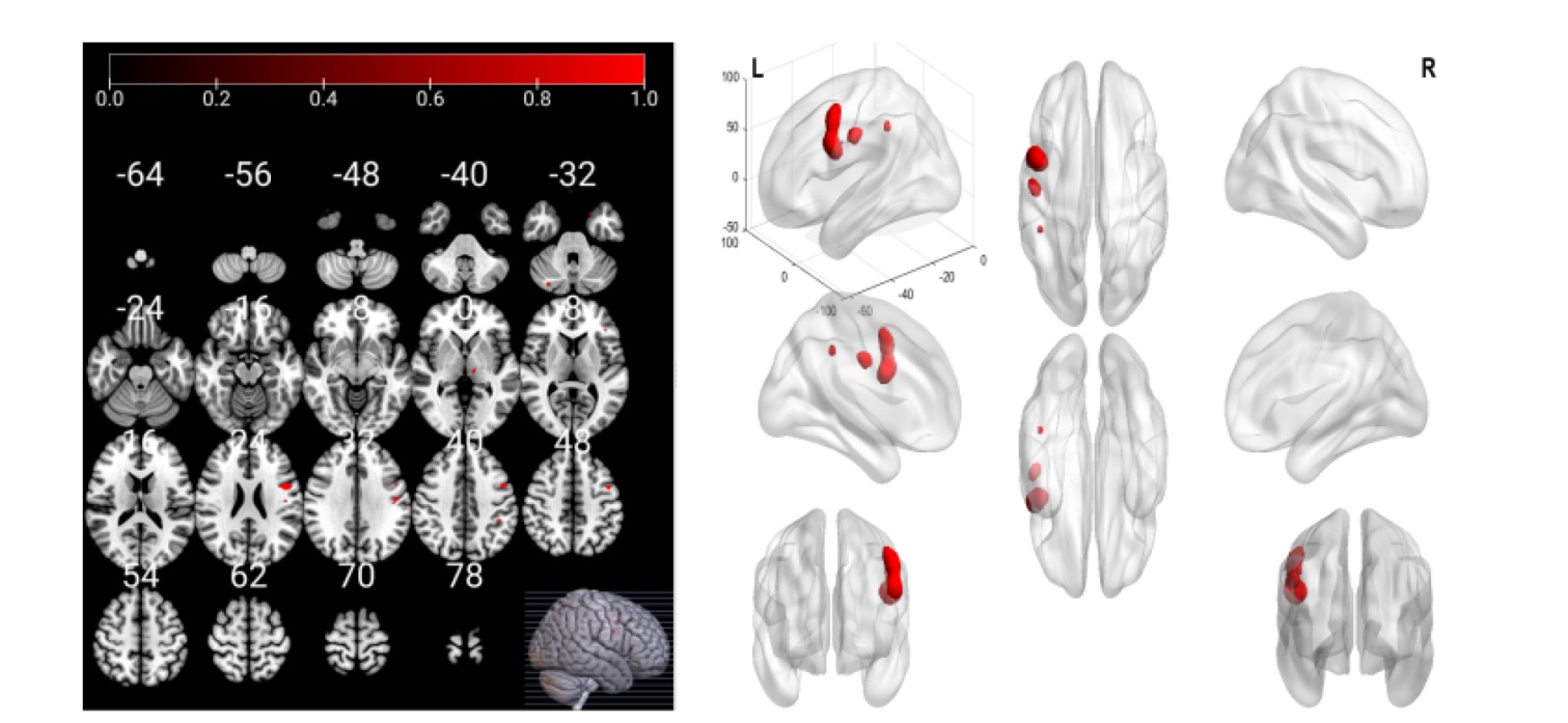
表1
两组差异脑区"
| 指标 | 脑区 | 半球 | AAL | MNI峰值坐标 | t值 | 体素数 | ||
|---|---|---|---|---|---|---|---|---|
| x | y | z | ||||||
| ALFF | 中央前回 | 左 | 1 | -48 | 3 | 45 | 5.354 | 76 |
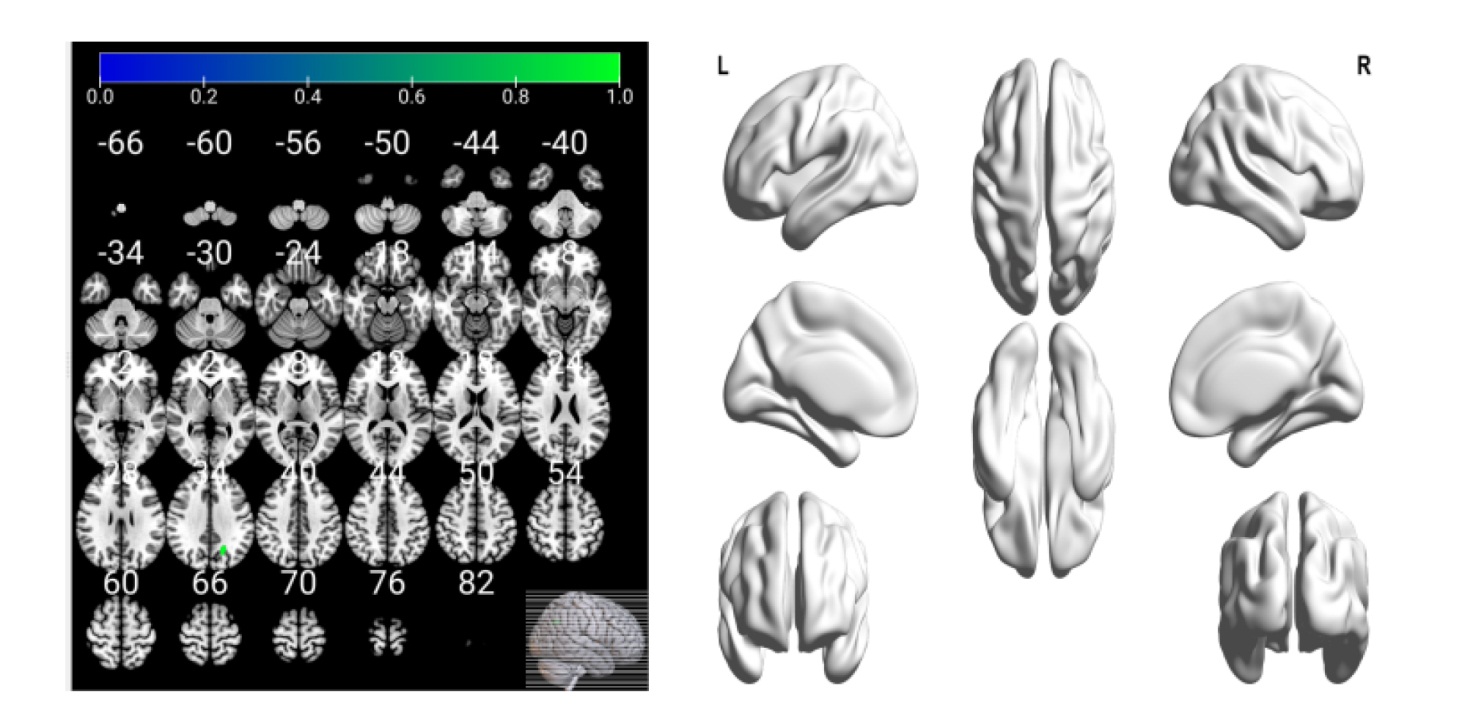
| fALFF | Cerebelum-Crus2-R | 右 | 94 | 48 | -78 | -36 | 4.578 | 161 |
| ReHo | 豆状壳核 | 右 | 74 | 24 | 0 | 6 | 3.881 | 186 |
| RECN | IFGoperc.R | 右 | 12 | 33 | 3 | 27 | 3.762 | 6 |
| DAN | MOG.L | 左 | 51 | -21 | -63 | 36 | 3.857 | 27 |
图2
两组fALFF差异脑区"
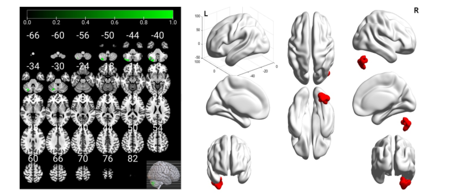
图3
两组ReHo差异脑区"
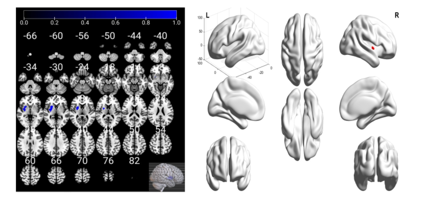
图4
两组RECN差异脑区"

图5
两组DAN差异脑区"
| [1] |
WANG F, WANG J, HAN Y, et al. Triglyceride-glucose index and stroke recurrence in elderly patients with ischemic stroke[J]. Front Endocrinol (Lausanne), 2022, 13: 1005614.
doi: 10.3389/fendo.2022.1005614 |
| [2] | 张志颖, 李春光, 苏敏. 脑机接口对缺血性脑卒中患者上肢功能影响的近红外脑功能成像研究[J]. 中华物理医学与康复杂志, 2025, 47(4): 300-306. |
| ZHANG Z Y, LI C G, SU M. A brain-computer interface can improve upper limb function after an ischemic stroke[J]. Chin J Phys Med Rehabil, 2025, 47(4): 300-306. | |
| [3] | 朱冬燕, 王梁, 吉桧媛, 等. 多模态软体手功能康复机器人对脑卒中患者手功能的影响[J]. 中华物理医学与康复杂志, 2025, 47(2): 108-111. |
| [4] | 谢晓明, 韩会建, 刘宏亮, 等. 经颅直流电刺激治疗脑卒中后认知功能障碍的静息态功能性磁共振研究[J]. 中华物理医学与康复杂志, 2020, 42(5): 392-396. |
| XIE X M, HAN H J, LIU H L, et al. Transcranial direct current stimulation promotes recovery of cognitive function after a stroke[J]. Chin J Phys Med Rehabil, 2020, 42(5): 392-396. | |
| [5] |
WANG J, MAO L, ZHOU H, et al. Altered functional connectivity in acute ischemic post-stroke non-fluent aphasia based on fMRI-EEG multimodal fusion[J]. IEEE Trans Neural Syst Rehabil Eng, 2025, 33: 2428-2438.
doi: 10.1109/TNSRE.2025.3580069 |
| [6] |
ZHANG K, DING L, WANG X, et al. Evidence of mirror therapy for recruitment of ipsilateral motor pathways in stroke recovery: a resting fMRI study[J]. Neurotherapeutics, 2024, 21(2): e00320.
doi: 10.1016/j.neurot.2024.e00320 |
| [7] | 何万林, 李谨利, 冯莉, 等. 高原藏族2型糖尿病患者静息态磁共振成像研究:基于低频振幅和比率低频振幅[J]. 磁共振成像, 2023, 14(5): 72-78, 122. |
| HE W L, LI J L, FENG L, et al. Study on resting-state functional magnetic resonance imaging in plateau Tibetan with type 2 diabetes mellitus: amplitude of low-frequency fluctuations and fractional amplitude of low-frequency fluctuations[J]. Chin J Magn Reson Imaging, 2023, 14(5): 72-78, 122. | |
| [8] | 谢芳芳, 顾元嘉, 管翀, 等. 比率低频振幅技术在延年九转法治疗慢性疲劳综合征伴焦虑抑郁中的应用价值[J]. 磁共振成像, 2024, 15(7): 58-63. |
| XIE F F, GU Y J, GUAN C, et al. The application value of fractional amplitude of low frequency fluctuation in the treatment of chronic fatigue syndrome with anxiety depression by Prolong Life with Nine Turn method[J]. Chin J Magn Reson Imaging, 2024, 15(7): 58-63. | |
| [9] |
MA Z Z, WU J J, CAO Z, et al. Motor imagery-based brain-computer interface rehabilitation programs enhance upper extremity performance and cortical activation in stroke patients[J]. J Neuroeng Rehabil, 2024, 21(1): 91.
doi: 10.1186/s12984-024-01387-w |
| [10] |
WANG Y, WANG C, WEI Y, et al. Abnormal functional connectivities patterns of multidomain cognitive impairments in pontine stroke patients[J]. Hum Brain Mapp, 2022, 43(15): 4676-4688.
doi: 10.1002/hbm.v43.15 |
| [11] |
CAI H, ZHANG N, JIANG Y, et al. State-dependent functional network connectivity alterations in post-stroke dementia with subcortical lesions[J]. J Alzheimers Dis, 2025, 103(4): 1245-1256.
doi: 10.1177/13872877241313056 |
| [12] |
KIMBERLEY T J, CRAMER S C, WOLF S L, et al. Long-term outcomes of vagus nerve stimulation paired with upper extremity rehabilitation after stroke[J]. Stroke, 2025, 56(8): 2255-2265.
doi: 10.1161/STROKEAHA.124.050479 |
| [13] |
WANG A, TIAN X, JIANG D, et al. Rehabilitation with brain-computer interface and upper limb motor function in ischemic stroke: a randomized controlled trial[J]. Med, 2024, 5(6): 559-569.e4.
doi: 10.1016/j.medj.2024.02.014 pmid: 38642555 |
| [14] |
DAWSON J, LIU C Y, FRANCISCO G E, et al. Vagus nerve stimulation paired with rehabilitation for upper limb motor function after ischemic stroke (VNS-REHAB): a randomised, blinded, pivotal, device trial[J]. Lancet, 2021, 397(10284): 1545-1553.
doi: 10.1016/S0140-6736(21)00475-X |
| [15] | 中华医学会神经病学分会, 中华医学会神经病学分会脑血管病学组. 中国各类主要脑血管病诊断要点2019[J]. 中华神经科杂志, 2019, 52(9): 710-715. |
| Chinese Society of Neurology, Chinese Society of Stroke. Diagnostic criteria of cerebrovascular diseases in China (version 2019)[J]. Chin J Neurol, 2019, 52(9): 710-715. | |
| [16] | 王睿月, 齐丽娜, 陈琳渝, 等. 左右制衡机制下不同电刺激对脑卒中后偏瘫患者大脑皮质兴奋性和上肢运动功能的影响[J]. 中华物理医学与康复杂志, 2025, 47(1): 19-24. |
| WANG R Y, QI L N, CHEN L Y, et al. The effects of electrical stimulation on upper limb motor function, left-right coordination and balance after a stroke[J]. Chin J Phys Med Rehabil, 2025, 47(1): 19-24. | |
| [17] | 蔡倩, 徐亮, 孙武东, 等. 重复外周磁刺激联合重复经颅磁刺激对脑卒中后上肢功能障碍的影响[J]. 中华物理医学与康复杂志, 2024, 46(5): 412-416. |
| CAI Q, XU L, SUN W D, et al. The effects of combining repetitive peripheral magnetic stimulation with repetitive transcranial magnetic stimulation in treating upper limb dysfunction after a stroke[J]. Chin J Phys Med Rehabil, 2024, 46(5): 412-416. | |
| [18] |
GONG X, JIN S, ZHOU Y, et al. Curative effect of medicine cake sticking ultrasound drug penetration combined with body training on hemiplegia after stroke: an in vitro ultrasound targeted drug controlled release technology[J]. Prev Med, 2023, 173: 107600.
doi: 10.1016/j.ypmed.2023.107600 |
| [19] | 刘若一. 基于脑连接探究针刺治疗中风偏瘫效应机制的多模态磁共振研究[D]. 北京: 北京中医药大学, 2023. |
| [20] |
WANG Y, CHEN H, WANG C, et al. Static and dynamic interactions within the triple-network model in stroke patients with multidomain cognitive impairments[J]. Neuroimage Clin, 2024, 43: 103655.
doi: 10.1016/j.nicl.2024.103655 |
| [21] |
ZUO X N, DI M A, KELLY C, et al. The oscillating brain: complex and reliable[J]. Neuroimage, 2010, 49(2): 1432-1445.
doi: 10.1016/j.neuroimage.2009.09.037 |
| [22] |
DING L, ZHANG K, WANG X, et al. Functional reorganization of white matter supporting the transhemispheric mechanism of mirror therapy after stroke: a multimodal MRI study[J]. IEEE Trans Neural Syst Rehabil Eng, 2025, 33: 1126-1134.
doi: 10.1109/TNSRE.2025.3549380 |
| [23] | 彭源, 张熙斌, 梅伟文, 等. 不同治疗时程的重复经颅磁刺激对脑卒中患者上肢运动功能及脑功能连接的影响[J]. 中国康复医学杂志, 2024, 39(10): 1436-1442. |
| PENG Y, ZHANG X B, MEI W W, et al. Effects of low frequency repetitive transcranial magnetic stimulation at different stimulating sessions on upper limb motor functionand brain functional connectivity in stroke patients[J]. Chin J Rehabil Med, 2024, 39(10): 1436-1442. | |
| [24] | 李冉, 刘素娟, 侯亚静, 等. 脑卒中6个月内上肢运动康复轨迹的纵向生存数据研究[J]. 中国康复医学杂志, 2025, 40(7): 1025-1032. |
| LI R, LIU S J, HOU Y J, et al. Longitudinal survival data study on upper extremity motor rehabilitation trajectory within 6months of stroke[J]. Chin J Rehabil Med, 2025, 40(7): 1025-1032. | |
| [25] | 修雨薇, 郭川, 范磊, 等. 躯干控制对卒中后上肢功能障碍的康复研究进展[J]. 临床神经病学杂志, 2025, 38(3): 222-225. |
| XIU Y W, GUO C, FAN L, et al. Research progress on trunk control for post-stroke upper limb dysfunction rehabilitation[J]. J Clin Neurol, 2025, 38(3): 222-225. | |
| [26] | 沈芳, 刘虎, 顾旭东, 等. 动作观察疗法对缺血性脑卒中患者上肢运动功能恢复的影响[J]. 中华物理医学与康复杂志, 2017, 39(3): 184-188. |
| SHEN F, LIU H, GU X D, et al. Action observation therapy can improve upper extremity motor function after stroke[J]. Chin J Phys Med Rehabil, 2017, 39(3): 184-188. | |
| [27] | 易小琦, 黄俊浩, 陈暇女, 等. 针刺促进缺血性脑卒中功能恢复的静息态功能连接研究[J]. 中国康复医学杂志, 2021, 36(4): 383-387. |
| YI X Q, HUANG J H, CHEN X N, et al. Effects of acupuncture in functional recovery of ischemic stroke: a resting-state fMRI study[J]. Chin J Rehabil Med, 2021, 36(4): 383-387. | |
| [28] |
PELLICCIARI M, BONNI S, PONZO V, et al. Dynamic reorganization of TMS-evoked activity in subcortical stroke patients[J]. Neuroimage, 2018, 175: 365-378.
doi: S1053-8119(18)30299-4 pmid: 29635028 |
| [29] | 钟燕彪. 单相脉冲电针干预脑卒中患者预测上肢运动功能恢复能力及机制研究[D]. 上海: 上海中医药大学, 2020. |
| [30] | 李鹏岳. 应用DTI技术对卒中患者运动康复中脑白质结构的可塑性研究[D]. 重庆: 第三军医大学, 2016. |
| LI P Y. Study on the plasticity of the white matter during motor recovery of stroke patients by diffusion tensor imaging[D]. Chongqing: Third Military Medical University, 2016. | |
| [31] | HANNANU F F, ZEFFIRO T A, LAMALLE L, et al. Parietal operculum and motor cortex activities predict motor recovery in moderate to severe stroke[J]. Neuroimage Clin, 2017, 4: 518-529. |
| [32] | 钟佳利, 景小珊, 梁莹. VMHC与ReHo在评价tDCS改善脑卒中后认知障碍中的应用价值[J]. 磁共振成像, 2024, 15(2): 129-134. |
| ZHONG J L, JING X S, LIANG Y. Application value of VMHC and ReHo in evaluating tDCS in improving cognitive impairment after stroke[J]. Chin J Magn Reson Imaging, 2024, 15(2): 129-134. | |
| [33] |
YEO B, KRIENEN F, SEPULCRE J, et al. The organization of the human cerebral cortex estimated by intrinsic functional connectivity[J]. J Neurophysiol, 2011, 106(3): 1125-1165.
doi: 10.1152/jn.00338.2011 pmid: 21653723 |
| [34] | 刘越, 王梦星, 杜小霞, 等. 基于独立成分分析的遗尿症儿童脑功能网络研究[J]. 中国医学物理学杂志, 2021, 38(3): 382-386. |
| LIU Y, WANG M X, DU X X, et al. Brain functional network of pediatric patients with enuresis: a research based on independent component analysis[J]. Chin J Med Phys, 2021, 38(3): 382-386. | |
| [35] | 傅凯丽, 赵洪力, 赵曼丽, 等. 基于静息态功能磁共振技术探讨针刺对脑卒中后认知障碍患者脑功能影响[J]. 光明中医, 2025, 40(4): 650-654. |
| [36] |
CORBETTA M, SHULMAN G L. Control of goal-directed and stimulus-driven attention in the brain[J]. Nat Rev Neurosci, 2002, 3(3): 201-215.
doi: 10.1038/nrn755 pmid: 11994752 |
| [37] |
SEELEY W W, MENON V, SCHATZBERG A F, et al. Dissociable intrinsic connectivity networks for salience processing and executive control[J]. J Neurosci, 2007, 27(9): 2349-2356.
doi: 10.1523/JNEUROSCI.5587-06.2007 pmid: 17329432 |
| [38] | 马震震. 脑机接口联合电针调控促进卒中后上肢运动功能恢复的临床机制研究[D]. 上海: 上海中医药大学, 2021. |
| MA Z Z. Research on influence and brain mechanism of Braincomputer interface and scalp acupuncture therapy on rehabilitation of motor recovery after stroke[D]. Shanghai: Shanghai University of Traditional Chinese Medicine, 2021. | |
| [39] | 苏彬. 重复经颅磁刺激在脑卒中患者下肢运动功能障碍的应用及神经重塑机制[D]. 上海: 上海体育大学, 2023. |
| SU B. The effect of repetitive transcranial magnetic stimulation on lower limb motor dysfunction and neural remodeling mechanism in stroke patients[D]. Shanghai: Shanghai University of Sport, 2023. | |
| [40] | 陆梦馨. 基于多模态磁共振和机器学习探究中风偏瘫后针刺脑效应机制的研究[D]. 北京: 北京中医药大学, 2023. |
| [41] | 杨浩, 余秋蓉, 魏彧, 等. 脑卒中运动功能障碍的局部一致性fMRI研究[J]. 中国康复医学杂志, 2020, 35(1): 10-16. |
| YANG H, YU Q R, WEI Y, et al. Brain regional homogeneity alterations at resting state in the patients with motor deficits after stroke[J]. Chin J Rehabil Med, 2020, 35(1): 10-16. | |
| [42] |
KUMAR V, MANG S, GRODD W. Direct diffusion-based parcellation of the human thalamus[J]. Brain Struct Funct, 2015, 220(3): 1619-1635.
pmid: 24659254 |
| [1] | 邹聪聪, 王潇珺, 马锦蓉, 鲁商波, 丁勇, 王哈妮, 宋建飞. 耳迷走神经电刺激联合双任务训练对缺血性脑卒中患者上肢功能的效果[J]. 《中国康复理论与实践》, 2025, 31(5): 513-519. |
| [2] | 刘鹏程, 屈萌艰, 龙黎萍, 王亚琳, 阳明珠, 刘培勇, 周君, 刘静. 多重感觉刺激模态的气电手训练系统联合低频重复经颅磁刺激对脑卒中患者手部运动和触压觉的效果[J]. 《中国康复理论与实践》, 2025, 31(4): 458-465. |
| [3] | 王潇珺, 王哈妮, 俞红, 李元梅, 周煜达. 高精度经颅直流电刺激联合上肢机器人对缺血性脑卒中上肢功能的效果[J]. 《中国康复理论与实践》, 2025, 31(2): 218-224. |
| [4] | 田富宝, 李泓钰, 田洋, 许宁, 李珂, 白川萍, 杨彩军. 基于双峰平衡恢复模型的经颅直流电刺激对缺血性脑卒中患者上肢功能障碍的效果[J]. 《中国康复理论与实践》, 2025, 31(11): 1271-1278. |
| [5] | 于婷婷, 蔡福良, 缪桂华, 顾晨, 彭媛. 基于个性优势的结构化治疗与教育对缺血性脑卒中康复效果的随机对照试验[J]. 《中国康复理论与实践》, 2024, 30(8): 965-971. |
| [6] | 陈晨, 孟兆祥, 杨康, 张敏杰, 左亚南, 王奎, 张熙斌, 全逸峰, 金星. 智能镜像手套任务导向性训练联合低频重复经颅磁刺激对脑卒中患者手功能效果的随机对照试验[J]. 《中国康复理论与实践》, 2024, 30(7): 831-838. |
| [7] | 高修明, 孟文文, 杨娜, 雷兆峰. 作业疗法基础上结合镜像疗法对脑卒中后异己手综合征的效果[J]. 《中国康复理论与实践》, 2024, 30(11): 1359-1427. |
| [8] | 田沛, 范荣富, 王洪岩, 彭明丽. 缺血性脑卒中患者并发尿路感染的病原菌分布、临床特点及危险因素[J]. 《中国康复理论与实践》, 2024, 30(10): 1179-1186. |
| [9] | 顾彬, 张津沁, 夏元浩, 胡靖然, 诸桥直纪, 黄富表. 反复促进疗法对脑卒中恢复期偏瘫患者手功能的影响[J]. 《中国康复理论与实践》, 2023, 29(6): 697-702. |
| [10] | 邓婷, 陈敬绵, 刘小蒙, 姚晓华, 刘芦姗, 何威, 张通, 芦海涛. 轻中度急性缺血性脑卒中并发卒中相关性肺炎的危险因素分析[J]. 《中国康复理论与实践》, 2023, 29(6): 708-713. |
| [11] | 李芳, 张通, 李冰洁, 赵军, 张豪杰. 运动表象训练对脑卒中患者手功能和运动表象能力的影响[J]. 《中国康复理论与实践》, 2023, 29(4): 479-484. |
| [12] | 张春龙, 刘福亮, 商娜, 李芳, 刘慧珍. 血清脂联素、超敏C-反应蛋白与急性缺血性脑卒中短期预后的相关性[J]. 《中国康复理论与实践》, 2023, 29(10): 1221-1226. |
| [13] | 刘明月, 李哲, 曹永生, 郝道剑, 宋薛艺. 基于运动表象的脑机接口训练对亚急性期脑卒中患者手功能康复的效果[J]. 《中国康复理论与实践》, 2023, 29(1): 71-76. |
| [14] | 孙凤宝, 章晓峰, 刘勇, 徐智韬, 金振华. 多模态镜像疗法对脑卒中患者上肢及手功能的效果[J]. 《中国康复理论与实践》, 2023, 29(1): 77-81. |
| [15] | 赖海芳,顾琳,纵亚,牛传欣,谢青. 采用多层感知器神经网络构建亚急性期缺血性脑卒中患者短期预后的预测模型[J]. 《中国康复理论与实践》, 2022, 28(3): 335-339. |
| 阅读次数 | ||||||
|
全文 |
|
|||||
|
摘要 |
|
|||||
|
||