| [1] |
Alzheimer's Disease International (ADI). World Alzheimer Report 2023[EB/OL]. (2023-09-21)[2025-02-02]. https://www.alzint.org/resource/world-alzheimer-report-2023.
|
| [2] |
CHANG J, LI Y, SHAN X, et al. Neural stem cells promote neuroplasticity: a promising therapeutic strategy for the treatment of Alzheimer's disease[J]. Neural Regen Res, 2024, 19(3): 619-628.
doi: 10.4103/1673-5374.380874
pmid: 37721293
|
| [3] |
KIM S, NAM Y, JEONG Y O, et al. Topographical visualization of the reciprocal projection between the medial septum and the hippocampus in the 5xfad mouse model of Alzheimer's disease[J]. Int J Mol Sci, 2019, 20(16): 3992.
doi: 10.3390/ijms20163992
|
| [4] |
GARRIDO S, DUNNE L, CHANG E, et al. The use of music playlists for people with dementia: a critical synthesis[J]. J Alzheimers Dis, 2017, 60(3): 1129-1142.
doi: 10.3233/JAD-170612
pmid: 28984606
|
| [5] |
SÄRKÄMÖ T, ALTENMÜLLER E, RODRÍGUEZ-FORNELLS A, et al. Editorial: music, brain, and rehabilitation: emerging therapeutic applications and potential neural mechanisms[J]. Front Hum Neurosci, 2016, 10: 103.
doi: 10.3389/fnhum.2016.00103
pmid: 27014034
|
| [6] |
HOFBAUER L M, ROSS S D, RODRIGUEZ F S. Music-based interventions for community-dwelling people with dementia: a systematic review[J]. Health Soc Care Commun, 2022, 30(6): 2186-2201.
doi: 10.1111/hsc.v30.6
|
| [7] |
TICHKO P, KIM J C, ZAPPI V, et al. Integrating music‐based interventions with gamma‐frequency stimulation for subjective cognitive decline and mild cognitive impairment[J]. Alzheimers Dement, 2022, 18(S11): e062532.
doi: 10.1002/alz.v18.S11
|
| [8] |
JACOBSEN J H, STELZER J, FRITZ T H, et al. Why musical memory can be preserved in advanced Alzheimer's disease[J]. Brain, 2015, 138(8): 2438-2450.
doi: 10.1093/brain/awv135
|
| [9] |
张勇, 王森莉, 黄荣华, 等. 音乐治疗对阿尔茨海默病患者干预效果的Meta分析[J]. 中国全科医学, 2024, 27(12): 1511-1518.
doi: 10.12114/j.issn.1007-9572.2023.0452
|
|
ZHANG Y, WANG S L, HUANG R H, et al. Intervention effect of music therapy on patients with Alzheimers disease: a meta-analysis[J]. Chin Gener Pract, 2024, 27(12): 1511-1518.
|
| [10] |
胡月青, 吕继辉, 王嫱, 等. 音乐疗法联合强光治疗对阿尔茨海默病患者睡眠障碍的疗效观察[J]. 首都医科大学学报, 2021, 42(3): 367-372.
doi: 10.3969/j.issn.1006-7795.2021.03.005
|
|
HU Y Q, LÜ J H, WANG Q, et al. Efficacy of music therapy combined with bright light therapy on sleep disorders in patients with Alzheimer's disease[J]. J Cap Med Univ, 2021, 42(3): 367-372.
|
| [11] |
FANG R, YE S, HUANGFU J, et al. Music therapy is a potential intervention for cognition of Alzheimer's disease: a mini-review[J]. Transl Neurodegener, 2017, 6: 2.
doi: 10.1186/s40035-017-0073-9
pmid: 28149509
|
| [12] |
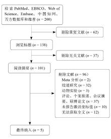
邓沁丽, 陈俊. 基于坐标的脑成像元分析方法:激活似然性评估[J]. 心理科学, 2015, 38(1): 216-220.
|
|
DENG Q L, CHEN J. A coordinate-based meta-analysis in brain imaging study: activation likelihood estimation[J]. J Psychol Sci, 2015, 38(1): 216-220.
|
| [13] |
TURKELTAUB P E, EICKHOFF S B, LAIRD A R, et al. Minimizing within‐experiment and within‐group effects in activation likelihood estimation meta-analyses[J]. Hum Brain Mapp, 2012, 33(1): 1-13.
doi: 10.1002/hbm.v33.1
|
| [14] |
FOX P T, LAIRD A R, EICKHOFF S B, et al. User manual for GingerALE 2.3[EB/OL]. (2013-06-13)[2024-05-23]. https://brainmap.org/ale/.
|
| [15] |
FISCHER C E, CHURCHILL N, LEGGIERI M, et al. Long-known music exposure effects on brain imaging and cognition in early-stage cognitive decline: a pilot study[J]. J Alzheimers Dis, 2021, 84(2): 819-833.
doi: 10.3233/JAD-210610
pmid: 34602475
|
| [16] |
GOLDEN H L, AGUSTUS J L, NICHOLAS J M, et al. Functional neuroanatomy of spatial sound processing in Alzheimer's disease[J]. Neurobiol Aging, 2016, 39: 154-164.
doi: 10.1016/j.neurobiolaging.2015.12.006
pmid: 26923412
|
| [17] |
KING J B, JONES K G, GOLDBERG E, et al. Increased functional connectivity after listening to favored music in adults with Alzheimer dementia[J]. J Prev Alzheimers Dis, 2019, 6(1): 56-62.
doi: 10.14283/jpad.2018.19
pmid: 30569087
|
| [18] |
SLATTERY C F, AGUSTUS J L, PATERSON R W, et al. The functional neuroanatomy of musical memory in Alzheimer's disease[J]. Cortex, 2019, 115: 357-370.
doi: S0010-9452(19)30054-1
pmid: 30846199
|
| [19] |
SATOH M, YUBA T, TABEI K I, et al. Music therapy using singing training improves psychomotor speed in patients with Alzheimer's disease: a neuropsychological and fMRI study[J]. Dement Geriatr Cogn Dis Extra, 2015, 5(3): 296-308.
doi: 10.1159/000436960
pmid: 26483829
|
| [20] |
SACHDEV P S. The default mode network, depression and Alzheimer's disease[J]. Int Psychogeriatr, 2022, 34(8): 675-678.
doi: 10.1017/S1041610222000539
pmid: 35918182
|
| [21] |
BREWER J A, GARRISON K A, WHITFIELD-GABRIELI S. What about the "self" is processed in the posterior cingulate cortex?[J]. Front Hum Neurosci, 2013, 7: 647.
doi: 10.3389/fnhum.2013.00647
pmid: 24106472
|
| [22] |
DADARIO N B, SUGHRUE M E. The functional role of the precuneus[J]. Brain, 2023, 146(9): 3598-3607.
doi: 10.1093/brain/awad181
|
| [23] |
CHEUNG V K M, MEYER L, FRIEDERICI A D, et al. The right inferior frontal gyrus processes nested non-local dependencies in music[J]. Sci Rep, 2018, 8(1): 3822.
doi: 10.1038/s41598-018-22144-9
pmid: 29491454
|
| [24] |
PUGLISI G, HOWELLS H, SCIOTINO T, et al. Frontal pathways in cognitive control: direct evidence from intraoperative stimulation and diffusion tractography[J]. Brain, 2019, 142(8): 2451-2465.
doi: 10.1093/brain/awz178
pmid: 31347684
|
| [25] |
NISSIM N R, O'SHEA A M, BRYANT V, et al. Frontal structural neural correlates of working memory performance in older adults[J]. Front Aging Neurosci, 2017, 8: 328.
|
| [26] |
ROTH R H, DING J B. Cortico-basal ganglia plasticity in motor learning[J]. Neuron, 2024, 112(15): 2486-2502.
doi: 10.1016/j.neuron.2024.06.014
|
| [27] |
KITTLESON A R, WOODWARD N D, HECKERS S, et al. The insula: leveraging cellular and systems-level research to better understand its roles in health and schizophrenia[J]. Neurosci Biobehav Rev, 2024, 160: 105643.
doi: 10.1016/j.neubiorev.2024.105643
|
| [28] |
ZHANG R, GAN X, XU T, et al. A neurofunctional signature of affective arousal generalizes across valence domains and distinguishes subjective experience from autonomic reactivity[J]. Nat Commun, 2025, 16(1): 6492.
doi: 10.1038/s41467-025-61706-0
|
| [29] |
ZHOU Y N, JIANG L, ZHANG Y, et al. Anti-LINGO-1 antibody protects neurons and synapses in the medial prefrontal cortex of APP/PS1 transgenic mice[J]. Neurosci Res, 2023, 193: 28-40.
doi: 10.1016/j.neures.2023.02.005
|
| [30] |
FABORODE O S, DALLE E, MABANDLA M V. Inescapable footshocks induce molecular changes in the prefrontal cortex of rats in an amyloid-beta-42 model of Alzheimer's disease[J]. Behav Brain Res, 2022, 419: 113679.
doi: 10.1016/j.bbr.2021.113679
|
| [31] |
JONES S A, MORALES A M, HOLLEY A L, et al. Default mode network connectivity is related to pain frequency and intensity in adolescents[J]. Neuroimage Clin, 2020, 27: 102326.
doi: 10.1016/j.nicl.2020.102326
|
| [32] |
FLEISCHER V, MUTHURAMAN M, ANWAR A R, et al. Continuous reorganization of cortical information flow in multiple sclerosis: a longitudinal fMRI effective connectivity study[J]. Sci Rep, 2020, 10: 806.
doi: 10.1038/s41598-020-57895-x
pmid: 31964982
|
 ), 刘清栾2, 万嘉敏1