


《中国康复理论与实践》 ›› 2020, Vol. 26 ›› Issue (4): 382-387.doi: 10.3969/j.issn.1006-9771.2020.04.003
李雅静1,2,3,4,5,李建军1,2,3,4,5( ),高峰1,2,3,4,5,郭韵6,刘俊1,2,3,4,5,徐珮珮1,2,3,4,5
收稿日期:2019-11-24
修回日期:2020-03-06
出版日期:2020-04-25
发布日期:2020-04-27
通讯作者:
李建军
E-mail:crrc100@163.com
作者简介:李雅静(1996-),女,汉族,安徽蚌埠市人,硕士研究生,主要研究方向:脊柱脊髓损伤的康复与治疗。
基金资助:
LI Ya-jing1,2,3,4,5,LI Jian-jun1,2,3,4,5(),GAO Feng1,2,3,4,5,GUO Yun6,LIU Jun1,2,3,4,5,XU Pei-pei1,2,3,4,5
Received:2019-11-24
Revised:2020-03-06
Published:2020-04-25
Online:2020-04-27
Contact:
LI Jian-jun
E-mail:crrc100@163.com
Supported by:摘要:
目的 观察脊髓损伤患者大脑中央前回初级运动皮质(M1)代谢变化。方法 2018年12月至2019年10月,选择住院脊髓损伤患者20例(患者组)和健康人15例(对照组),使用磁共振波谱(MRS)观察大脑左侧M1感兴趣区氮-乙酰天门冬氨酸(NAA)、胆碱(Cho)、肌酸(Cr)和肌醇(MI)浓度。结果 患者组MI浓度较对照组明显增高(t = 3.745, P < 0.01),NAA、Cho、Cr、NAA/Cr、Cho/Cr、Cho/NAA与对照组相比无显著性差异( t < 1.431, P > 0.05)。 结论 脊髓损伤患者M1区可能有胶质细胞增生,提示发生代偿性修复。
中图分类号:
李雅静,李建军,高峰,郭韵,刘俊,徐珮珮. 脊髓损伤患者大脑中央前回的代谢改变[J]. 《中国康复理论与实践》, 2020, 26(4): 382-387.
LI Ya-jing,LI Jian-jun,GAO Feng,GUO Yun,LIU Jun,XU Pei-pei. Metabolism Levels in Precentral Gyrus of Patients after Spinal Cord Injury[J]. 《Chinese Journal of Rehabilitation Theory and Practice》, 2020, 26(4): 382-387.

图1
大脑M1区矢状面、横断面、冠状面定位"
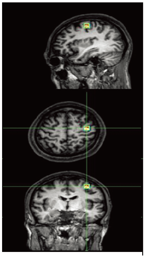
表1
两组左侧M1区代谢物比较"
| 代谢物 | 对照组 (n = 15) | 患者组 (n = 20) | t值 | P值 |
|---|---|---|---|---|
| NAA(mmol/kg) | 0.10±0.01 | 0.09±0.03 | -0.645 | 0.524 |
| Cho(mmol/kg) | 0.04±0.01 | 0.04±0.01 | 0.776 | 0.443 |
| MI(mmol/kg) | 0.01±0.01 | 0.02±0.01 | 3.745 | 0.001 |
| Cr(mmol/kg) | 0.05±0.01 | 0.05±0.02 | 1.267 | 0.214 |
| NAA/Cr | 2.04±0.55 | 2.03±0.68 | -0.033 | 0.974 |
| CHO/Cr | 0.66±0.24 | 0.92±0.83 | 1.140 | 0.262 |
| CHO/NAA | 0.34±0.14 | 0.42±0.16 | 1.431 | 0.162 |
| [1] |
Rouanet C, Reges D, Rocha E, et al. Traumatic spinal cord injury: current concepts and treatment update[J]. Arq Neuropsiquiatr, 2017, 75(6):387-393.
doi: 10.1590/0004-282x20170048 |
| [2] |
Crowley S T, Fukushima Y, Uchida S, et al. Enhancement of motor function recovery after spinal cord injury in mice by delivery of brain-derived neurotrophic factor mRNA[J]. Mol Ther Nucleic Acids, 2019, 17:465-476.
doi: 10.1016/j.omtn.2019.06.016 |
| [3] |
Freund P, Weiskopf N, Ward N S, et al. Disability, atrophy and cortical reorganization following spinal cord injury[J]. Brain, 2011, 134(6):1610-1622.
doi: 10.1093/brain/awr093 |
| [4] |
Turner J A, Lee J S, Martinez O, et al. Somatotopy of the motor cortex after long-term spinal cord injury or amputation[J]. IEEE Trans Neural Sys Rehabil Engineering, 2001, 9(2):154-160.
doi: 10.1109/TNSRE.7333 |
| [5] | Zheng W, Chen Q, Chen X, et al. Brain white matter impairment in patients with spinal cord injury[J]. Neural Plast, 2017, 2017:1-8. |
| [6] |
Jurkiewicz M T, Mikulis D J, Mcilroy W E, et al. Sensorimotor cortical plasticity during recovery following spinal cord injury: a longitudinal fMRI study[J]. Neurorehabil Neural Repair, 2007, 21(6):527-538.
doi: 10.1177/1545968307301872 |
| [7] |
Wrigley P J, Press S R, Gustin S M, et al. Neuropathic pain and primary somatosensory cortex reorganization following spinal cord injury[J]. Pain, 2009, 141(1):52-59.
doi: 10.1016/j.pain.2008.10.007 |
| [8] |
Freund P, Schneider T, Nagy Z, et al. Degeneration of the injured cervical cord is associated with remote changes in corticospinal tract integrity and upper limb impairment[J]. PLoS One, 2012, 7(12):e51729.
doi: 10.1371/journal.pone.0051729 |
| [9] |
Freund P, Weiskopf N, Ashburner J, et al. MRI investigation of the sensorimotor cortex and the corticospinal tract after acute spinal cord injury: a prospective longitudinal study[J]. Lancet Neurol, 2013, 12(9):873-881.
doi: 10.1016/S1474-4422(13)70146-7 |
| [10] |
Freund P, Wheeler-Kingshott C A, Nagy Z, et al. Axonal integrity predicts cortical reorganisation following cervical injury[J]. J Neurol Neurosurg Psychiatry, 2012, 83(6):629-637.
doi: 10.1136/jnnp-2011-301875 |
| [11] |
Hou J M, Yan R B, Xiang Z M, et al. Brain sensorimotor system atrophy during the early stage of spinal cord injury in humans[J]. Neuroscience, 2014, 266:208-215.
doi: 10.1016/j.neuroscience.2014.02.013 pmid: 24561217 |
| [12] |
Jurkiewicz M T, Crawley A P, Verrier M C, et al. Somatosensory cortical atrophy after spinal cord injury: a voxel-based morphometry study[J]. Neurology, 2006, 66(5):762-764.
pmid: 16534122 |
| [13] |
Hains B C, Black J A, Waxman S G. Primary cortical motor neurons undergo apoptosis after axotomizing spinal cord injury[J]. J Comp Neurol, 2003, 462(3):328-341.
doi: 10.1002/(ISSN)1096-9861 |
| [14] |
Lee B H, Lee K H, Kim U J, et al. Injury in the spinal cord may produce cell death in the brain[J]. Brain Res, 2004, 1020(1-2):37-44.
doi: 10.1016/j.brainres.2004.05.113 |
| [15] |
Choe A S, Belegu V, Yoshida S, et al. Extensive neurological recovery from a complete spinal cord injury: a case report and hypojournal on the role of cortical plasticity[J]. Front Hum Neurosci, 2013, 7:290.
doi: 10.3389/fnhum.2013.00290 pmid: 23805087 |
| [16] |
Crawley A P, Jurkiewicz M T, Yim A, et al. Absence of localized grey matter volume changes in the motor cortex following spinal cord injury[J]. Brain Res, 2004, 1028(1):19-25.
pmid: 15518637 |
| [17] |
Henderson L A, Gustin S M, Macey P M, et al. Functional reorganization of the brain in humans following spinal cord injury: evidence for underlying changes in cortical anatomy[J]. J Neurosci, 2011, 31(7):2630-2637.
doi: 10.1523/JNEUROSCI.2717-10.2011 |
| [18] |
Kim B G, Dai H, Mcatee M, et al. Remodeling of synaptic structures in the motor cortex following spinal cord injury[J]. Exp Neurol, 2006, 198(2):401-415.
doi: 10.1016/j.expneurol.2005.12.010 |
| [19] |
Moxon K A, Oliviero A, Aguilar J, et al. Cortical reorganization after spinal cord injury: always for good?[J]. Neuroscience, 2014, 283:78-94.
doi: 10.1016/j.neuroscience.2014.06.056 pmid: 24997269 |
| [20] |
Nardone R, Höller Y, Brigo F, et al. Functional brain reorganization after spinal cord injury: systematic review of animal and human studies[J]. Brain Res, 2013, 1504:58-73.
doi: 10.1016/j.brainres.2012.12.034 |
| [21] | Peng X, Tan Y, He L, et al. Alterations of functional connectivity between thalamus and cortex before and after decompression in cervical spondylotic myelopathy patients[J]. NeuroReport, 2019: 1-7. |
| [22] |
Matsubayashi K, Nagoshi N, Komaki Y, et al. Assessing cortical plasticity after spinal cord injury by using resting-state functional magnetic resonance imaging in awake adult mice[J]. Sci Rep, 2018, 8(1):14406.
doi: 10.1038/s41598-018-32766-8 pmid: 30258091 |
| [23] |
Xu A K, Gong Z, He Y Z, et al. Comprehensive therapeutics targeting the corticospinal tract following spinal cord injury[J]. J Zhejiang Univ Sci B, 2019, 20(3):205-218.
doi: 10.1631/jzus.B1800280 |
| [24] |
Oz G, Alger J R, Barker P B, et al. Clinical proton MR spectroscopy in central nervous system disorders[J]. Radiology, 2014, 270(3):658-679.
doi: 10.1148/radiol.13130531 |
| [25] |
Lin A, Tran T, Bluml S, et al. Guidelines for Acquiring and Reporting Clinical Neurospectroscopy[J]. Semin Neurol, 2013, 32(4):432-453.
doi: 10.1055/s-00000071 |
| [26] |
Soares D P, Law M. Magnetic resonance spectroscopy of the brain: review of metabolites and clinical applications[J]. Clin Radiol, 2009, 64(1):12-21.
doi: 10.1016/j.crad.2008.07.002 pmid: 19070693 |
| [27] |
Wyss P O, Hock A, Kollias S. The application of human spinal cord magnetic resonance spectroscopy to clinical studies: a review[J]. Semin Ultrasound CT MRI, 2017, 38(2):153-162.
doi: 10.1053/j.sult.2016.07.005 |
| [28] |
Bellenberg B, Busch M, Trampe N, et al. 1H-magnetic resonance spectroscopy in diffuse and focal cervical cord lesions in multiple sclerosis[J]. Eur Radiol, 2013, 23(12):3379-3392.
doi: 10.1007/s00330-013-2942-7 pmid: 23884299 |
| [29] | Ellingson B M, Salamon N, Hardy A J, et al. Prediction of neurological impairment in cervical spondylotic myelopathy using a combination of diffusion MRI and proton MR spectroscopy[J]. PLoS One, 2015, 10(10):e139451. |
| [30] | Hock A. Spinal cord MR spectroscopy in neoplastic lesions[C]. Proceedings of the International Society for Magnetic Resonance in Medicine, 2012: 838. |
| [31] |
Iltis I, Hutter D, Bushara K O, et al. 1H MR spectroscopy in Friedreich's ataxia and ataxia with oculomotor apraxia type 2[J]. Brain Res, 2010, 1358:200-210.
doi: 10.1016/j.brainres.2010.08.030 |
| [32] |
van den Berg M E L, Castellote J M, de Pedro-Cuesta J, et al. Survival after spinal cord injury: a systematic review[J]. J Neurotrauma, 2010, 27(8):1517-1528.
doi: 10.1089/neu.2009.1138 |
| [33] |
Jazayeri S B, Jazayeri S B, Beygi S, et al. Incidence of traumatic spinal cord injury worldwide: a systematic review[J]. Eur Spine J, 2015, 24(5):905-918.
doi: 10.1007/s00586-014-3424-6 pmid: 24952008 |
| [34] | Akinwunmi Oni-Orisan M K W L, Douglas Ward A V B K. Alterations in cortical sensorimotor connectivity following complete cervical spinal cord injury: a prospective resting-state fMRI study[J]. PLoS One, 2016, 3(11):e0150351. |
| [35] |
Kaushal M, Oni-Orisan A, Chen G, et al. Evaluation of whole-brain resting-state functional connectivity in spinal cord injury: a large-scale network analysis using network-based statistic[J]. J Neurotrauma, 2017, 34(6):1278-1282.
doi: 10.1089/neu.2016.4649 |
| [36] | Athanasiou A, Arfaras G, Xygonakis I, et al. Commercial BCI control and functional brain networks in spinal cord injury: a proof-of-concept[C]. Thessaloniki, Greece: IEEE 30th International Symposium on Computer-Based Medical Systems, 2017: 262-267. |
| [37] |
Jutzeler C R, Huber E, Callaghan M F, et al. Association of pain and CNS structural changes after spinal cord injury[J]. Sci Rep, 2016, 6:18534.
doi: 10.1038/srep18534 |
| [38] | Guo Y, Gao F, Liu Y, et al. White matter microstructure alterations in patients with spinal cord injury assessed by diffusion tensor imaging[J]. Front Human Neurosci, 2019, 13:11. |
| [39] |
Wrigley P J, Gustin S M, Macey P M, et al. Anatomical changes in human motor cortex and motor pathways following complete thoracic spinal cord injury[J]. Cerebral Cortex, 2008, 19(1):224-232.
doi: 10.1093/cercor/bhn072 |
| [40] |
Alizadeh A, Karimi-Abdolrezaee S. Microenvironmental regulation of oligodendrocyte replacement and remyelination in spinal cord injury[J]. J Physiol, 2016, 594(13):3539-3552.
doi: 10.1113/JP270895 |
| [41] |
Haggerty A E, Marlow M M, Oudega M. Extracellular matrix components as therapeutics for spinal cord injury[J]. Neurosci Lett, 2017, 652:50-55.
doi: S0304-3940(16)30736-4 pmid: 27702629 |
| [42] |
Okada S, Hara M, Kobayakawa K, et al. Astrocyte reactivity and astrogliosis after spinal cord injury[J]. Neurosci Res, 2018, 126:39-43.
doi: 10.1016/j.neures.2017.10.004 |
| [43] |
Orr M B, Gensel J C. Spinal cord injury scarring and inflammation: therapies targeting glial and inflammatory responses[J]. Neurotherapeutics, 2018, 15(3):541-553.
doi: 10.1007/s13311-018-0631-6 |
| [44] |
Wang Y, Wang Y, Gao Z, et al. Attenuated reactive gliosis and enhanced functional recovery following spinal cord injury in null mutant mice of platelet-activating factor receptor[J]. Mol Neurobiol, 2016, 53(5):3448-3461.
doi: 10.1007/s12035-015-9263-6 |
| [45] |
Gujar S K, Maheshwari S, Bjorkman-Burtscher I, et al. Magnetic resonance spectroscopy[J]. J Neuroophthalmol, 2005, 25(3):217-226.
doi: 10.1097/01.wno.0000177307.21081.81 |
| [46] |
de Stefano N, Filippi M. MR spectroscopy in multiple sclerosis[J]. J Neuroimaging, 2007, 17(2):31S-35S.
doi: 10.1111/jon.2007.17.issue-s1 |
| [47] |
Pfrieger F W, Barres B A. Synaptic efficacy enhanced by glial cells in vitro[J]. Science, 1997, 277(5332):1684-1687.
doi: 10.1126/science.277.5332.1684 |
| [48] |
Barros L F, Brown A, Swanson R A. Glia in brain energy metabolism: a perspective[J]. Glia, 2018, 66(6):1134-1137.
doi: 10.1002/glia.v66.6 |
| [49] |
Ohtake Y, Smith G M, Li S. Reactive astrocyte scar and axon regeneration:suppressor or facilitator?[J]. Neural Regen Res, 2016, 11(7):1050-1051.
doi: 10.4103/1673-5374.187022 |
| [50] |
Jones L L, Sajed D, Tuszynski M H. Axonal regeneration through regions of chondroitin sulfate proteoglycan deposition after spinal cord injury: a balance of permissiveness and inhibition[J]. J Neurosci, 2003, 23(28):9276-9288.
doi: 10.1523/JNEUROSCI.23-28-09276.2003 |
| [51] | Morgenstern D A, Asher R A, Fawcett J W. Chapter 22 Chondroitin sulphate proteoglycans in the CNS injury response[M]// Mckerracher L, Doucet G, Rossignol S. Progress in Brain Research. Amsterdam: Elsevier, 2002: 137,313-332. |
| [52] |
Windle W F, Chambers W W. Regeneration in the spinal cord of the cat and dog[J]. J Comp Neurol, 1950, 93(2):241-257.
doi: 10.1002/(ISSN)1096-9861 |
| [53] |
Yuan Y, He C. The glial scar in spinal cord injury and repair[J]. Neurosci Bull, 2013, 29(4):421-435.
doi: 10.1007/s12264-013-1358-3 |
| [54] |
Georgieva M, Wei Y, Dumitrascuta M, et al. Fatty acid suppression of glial activation prevents central neuropathic pain after spinal cord injury[J]. Pain, 2019, 160(12):2724-2742.
doi: 10.1097/j.pain.0000000000001670 pmid: 31365471 |
| [55] |
Anderson M A, Burda J E, Ren Y, et al. Astrocyte scar formation aids central nervous system axon regeneration[J]. Nature, 2016, 532(7598):195-200.
doi: 10.1038/nature17623 pmid: 27027288 |
| [56] |
Liddelow S A, Guttenplan K A, Clarke L E, et al. Neurotoxic reactive astrocytes are induced by activated microglia[J]. Nature, 2017, 541(7638):481-487.
doi: 10.1038/nature21029 pmid: 28099414 |
| [57] |
Goncalves S, Stevens T K, Doyle-Pettypiece P, et al. N-acetylaspartate in the motor and sensory cortices following functional recovery after surgery for cervical spondylotic myelopathy[J]. J Neurosurg Spine, 2016, 25(4):436-443.
doi: 10.3171/2016.2.SPINE15944 |
| [1] | 周治宁, 周容, 肖燕文, 王博文, 吕娇娇, 刘宇. 多靶区经颅直流电刺激对健康成年人工作记忆-姿势控制双任务表现的影响[J]. 《中国康复理论与实践》, 2024, 30(1): 21-28. |
| [2] | 刘冬, 徐子涵, 李江, 鞠萍. M1区联合背外侧前额叶高频重复经颅磁刺激对脊髓损伤后神经病理性疼痛患者脑电图θ振幅的效果[J]. 《中国康复理论与实践》, 2024, 30(1): 87-94. |
| [3] | 李芳, 霍速, 杜巨豹, 刘秀贞, 李小爽, 宋为群. 经颅直流电刺激联合任务导向性康复训练对脊髓损伤大鼠前肢运动障碍的效果[J]. 《中国康复理论与实践》, 2023, 29(7): 777-781. |
| [4] | 刘宁, 刘雨泉, 祝斌, 于凌佳, 谭海宁, 杨雍, 李想. 脊髓损伤神经学分类国际标准国内应用情况的文献计量学研究[J]. 《中国康复理论与实践》, 2023, 29(7): 808-815. |
| [5] | 王一吉, 周红俊, 何泽佳, 刘根林, 郑樱, 郝春霞, 卫波, 康海琼, 张缨, 逯晓蕾, 袁媛, 蒙倩茹. 不完全性脊髓损伤患者运动功能对称性与步态对称性的关系[J]. 《中国康复理论与实践》, 2023, 29(6): 639-645. |
| [6] | 袁媛, 周红俊, 丛欣莹, 刘根林, 卫波, 郑樱, 郝春霞, 张缨, 王一吉, 康海琼, 逯晓蕾, 蒙倩茹. 创伤性颈脊髓损伤程度与磁共振成像的关系[J]. 《中国康复理论与实践》, 2023, 29(6): 725-730. |
| [7] | 蒋乐, 杜良杰, 黄富表. 完全性脊髓损伤患者的情绪及认知行为分析[J]. 《中国康复理论与实践》, 2023, 29(5): 576-581. |
| [8] | 郭霜, 谢咏祺, 张良, 张春佳, 彭润, 杨德刚, 杨明亮. 舞蹈致儿童无骨折脱位型脊髓损伤神经学预后的影响因素及预测模型[J]. 《中国康复理论与实践》, 2023, 29(5): 582-589. |
| [9] | 张园, 杨剑. 基于ICD-11和ICF脊髓损伤患者运动康复干预方案及其健康效益:系统综述的系统综述[J]. 《中国康复理论与实践》, 2023, 29(12): 1377-1385. |
| [10] | 石孝宇, 杨剑. 脊髓损伤患者适应性身体活动及其健康效益:基于ICF的Scoping综述[J]. 《中国康复理论与实践》, 2023, 29(12): 1395-1404. |
| [11] | 刘根林,周红俊,李建军,卫波,郑樱,郝春霞,张缨,王一吉,康海琼,逯晓蕾,袁媛,蒙倩茹. 伴并发症脊髓损伤的神经学分类研究进展[J]. 《中国康复理论与实践》, 2022, 28(8): 934-938. |
| [12] | 康海琼,周红俊,刘根林,卫波,郑樱,张缨,郝春霞,王一吉,逯晓蕾,袁媛,蒙倩茹. 脊髓损伤患者股骨远端和胫骨近端骨密度的变化[J]. 《中国康复理论与实践》, 2022, 28(7): 855-858. |
| [13] | 张妙媛,何英,李晓霞,彭敏,张蕾,刘姝颖,孔瑛. 脊髓损伤清洁间歇导尿患者自我管理现状及影响因素[J]. 《中国康复理论与实践》, 2022, 28(6): 716-724. |
| [14] | 周小珏,冯婧,庞日朝,刘捷,张安仁. 隔日限食减轻脊髓损伤大鼠炎症反应的芳香烃受体/细胞因子信号传导抑制因子2/核转录因子-κB信号通路机制[J]. 《中国康复理论与实践》, 2022, 28(5): 544-551. |
| [15] | 魏娟芳,王琳杰,崔艳如,岑秋宇,张安仁. 骨髓间充质干细胞衍生外泌体对脊髓损伤动物治疗作用的系统综述[J]. 《中国康复理论与实践》, 2022, 28(5): 585-592. |
| 阅读次数 | ||||||
|
全文 |
|
|||||
|
摘要 |
|
|||||
|
||