


《Chinese Journal of Rehabilitation Theory and Practice》 ›› 2020, Vol. 26 ›› Issue (4): 472-478.doi: 10.3969/j.issn.1006-9771.2020.04.016
Previous Articles Next Articles
YAO Tao-tao1,2,CHEN Zhuo-ming3( ),ZHANG Shu-chen4
Received:2019-06-20
Revised:2019-07-19
Published:2020-04-25
Online:2020-04-27
Contact:
CHEN Zhuo-ming
E-mail:tchzm@21cn.com
Supported by:CLC Number:
YAO Tao-tao,CHEN Zhuo-ming,ZHANG Shu-chen. Imaging Features of Abnormal Neural Connectivity in Autistic Children[J]. 《Chinese Journal of Rehabilitation Theory and Practice》, 2020, 26(4): 472-478.
"
| 组别 | n | 性别(男/女,n) | 年龄(岁) | 头围(cm) |
|---|---|---|---|---|
| 对照组 | 8 | 6/2 | 3.7±2.2 | 50.4±1.4 |
| 孤独症组 | 14 | 12/2 | 4.4±1.9 | 51.1±1.6 |
| χ2/t值 | 0.393 | 0.809 | 1.033 | |
| P值 | 0.602 | 0.428 | 0.314 |
"
| 部位 | 代谢物 | 对照组 | 孤独症组 | F值a | P值a | F值b | P值b | t值a | P值a | t值b | P值b | ||
|---|---|---|---|---|---|---|---|---|---|---|---|---|---|
| 左 | 右 | 左 | 右 | ||||||||||
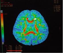
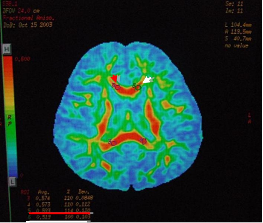
| 额叶 | FA | 0.298±0.038 | 0.347±0.022 | 0.272±0.108 | 0.284±0.129 | 0.464 | 0.510 | 0.883 | 0.368 | -1.641 | 0.199 | -0.857 | 0.411 |
| ADC | 8.715±0.319 | 8.552±0.167 | 8.912±0.581 | 8.985±0.472 | 0.553 | 0.473 | 2.423 | 0.148 | 0.689 | 0.540 | -0.372 | 0.718 | |
| 海马 | FA | 0.503±0.032 | 0.489±0.042 | 0.396±0.074 | 0.408±0.076 | 5.922 | 0.033 | 4.022 | 0.070 | 0.998 | 0.392 | -0.511 | 0.621 |
| ADC | 9.340±0.423 | 8.962±0.141 | 9.034±0.729 | 9.000±0.539 | 0.480 | 0.503 | 0.090 | 0.770 | 1.433 | 0.247 | 0.135 | 0.895 | |
| 小脑 | FA | 0.395±0.120 | 0.406±0.107 | 0.392±0.158 | 0.439±0.127 | 0.002 | 0.964 | 0.181 | 0.679 | -0.820 | 0.472 | -1.621 | 0.136 |
| ADC | 8.797±0.764 | 8.645±0.774 | 7.988±0.761 | 8.071±0.751 | 3.227 | 0.100 | 0.747 | 0.406 | 2.845 | 0.065 | -0.768 | 0.460 | |
| 胼胝体膝部 | FA | 0.642±0.143 | 0.619±0.138 | 0.478±0.209 | 0.547±0.223 | 1.423 | 0.258 | 0.213 | 0.654 | 1.975 | 0.143 | -2.335 | 0.042 |
| ADC | 8.675±0.364 | 8.490±0.193 | 8.554±2.874 | 8.321±2.808 | 0.009 | 0.925 | 0.013 | 0.913 | 0.871 | 0.448 | 1.383 | 0.197 | |
| 胼胝体压部 | FA | 0.679±0.171 | 0.702±0.138 | 0.656±0.103 | 0.646±0.105 | 0.024 | 0.881 | 0.732 | 0.411 | -0.542 | 0.625 | 0.354 | 0.730 |
| ADC | 9.472±0.217 | 9.133±0.365 | 9.607±0.256 | 9.235±0.473 | 0.881 | 0.368 | 0.258 | 0.621 | 2.205 | 0.115 | 3.520 | 0.006 | |
"
| 部位 | 代谢物 | 对照组 | 孤独症组 | F值a | P值a | F值b | P值b | t值c | P值c | t值d | P值d | ||
|---|---|---|---|---|---|---|---|---|---|---|---|---|---|
| 左 | 右 | 左 | 右 | ||||||||||
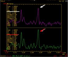
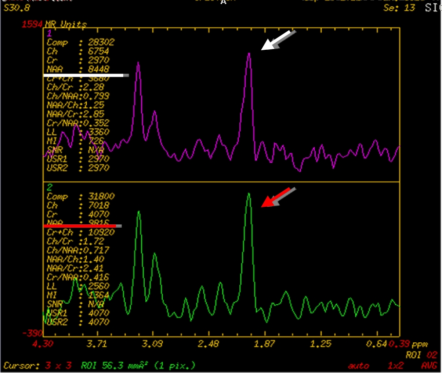
| 额叶 | NAA(mmol/kg) | 10915.50±2304.02 | 11260.50±2374.15 | 7716.00±5290.04 | 8425.71±5854.92 | 1.100 | 0.308 | 0.866 | 0.364 | -2.021 | 0.083 | -2.807 | 0.015 |
| Cho(mmol/kg) | 8662.50±1931.34 | 8784.88±2263.00 | 5729.43±4493.54 | 6494.71±4534.03 | 0.882 | 0.187 | 0.847 | 0.370 | -0.533 | 0.610 | -2.804 | 0.015 | |
| Cr(mmol/kg) | 5984.00±1163.30 | 5929.00±1506.78 | 4224.00±2595.13 | 4735.57 ±2818.30 | 2.123 | 0.162 | 0.613 | 0.444 | 0.206 | 0.843 | -2.648 | 0.020 | |
| NAA/Cr | 1.758±0.155 | 1.927±0.249 | 1.780±0.247 | 1.780±0.401 | 0.415 | 0.523 | 0.416 | 0.527 | -1.981 | 0.088 | 0.005 | 0.996 | |
| Cho/Cr | 1.472±0.368 | 1.507±0.354 | 1.305±0.274 | 1.349 ±0.200 | 0.619 | 0.442 | 0.839 | 0.372 | -0.526 | 0.615 | -0.609 | 0.553 | |
| 海马 | NAA(mmol/kg) | 6705.00±1459.01 | 7662.00±2789.01 | 5160.5±2070.00 | 5555.14±2480.99 | 2.978 | 0.102 | 4.029 | 0.060 | -1.034 | 0.335 | -1.123 | 0.282 |
| Cho(mmol/kg) | 6674.25±1021.25 | 6954.75±1387.34 | 5107.36±2236.16 | 5254.07±2304.16 | 3.033 | 0.099 | 3.272 | 0.087 | -0.675 | 0.521 | -0.695 | 0.499 | |
| Cr(mmol/kg) | 4955.50±1403.07 | 4972.00±1525.66 | 3424.07±1394.31 | 3474.43±1641.15 | 6.502 | 0.020 | 4.715 | 0.044 | -0.041 | 0.968 | -0.184 | 0.857 | |
| NAA/Cr | 1.397±0.273 | 1.547±0.317 | 1.610±0.517 | 1.640±0.392 | 1.621 | 0.219 | 0.210 | 0.652 | -0.795 | 0.453 | -0.236 | 0.817 | |
| Cho/Cr | 1.423±0.368 | 1.466±0.359 | 1.566±0.500 | 1.597±0.467 | 0.785 | 0.387 | 0.632 | 0.437 | -0.456 | 0.662 | -0.268 | 0.793 | |
| 小脑 | NAA(mmol/kg) | 8600.00±4215.58 | 9866.00±4509.24 | 5305.71±6420.88 | 5803.21±6493.00 | 0.905 | 0.356 | 1.592 | 0.225 | -2.347 | 0.066 | -1.417 | 0.180 |
| Cho(mmol/kg) | 8138.17±3011.96 | 8046.50±3551.14 | 4482.71±5795.56 | 4785.79±5463.38 | 1.325 | 0.267 | 1.333 | 0.265 | 0.153 | 0.885 | -1.120 | 0.283 | |
| Cr(mmol/kg) | 7206.83±2751.87 | 7434.17±2739.97 | 4051.93±4554.73 | 3367.57±3830.18 | 1.815 | 0.197 | 4.251 | 0.056 | -0.361 | 0.733 | 2.032 | 0.063 | |
| NAA/Cr | 0.887±0.600 | 0.967±0.610 | 1.054±0.636 | 1.565±1.365 | 0.380 | 0.545 | 1.153 | 0.297 | -1.202 | 0.268 | -1.639 | 0.125 | |
| Cho/Cr | 0.869±0.573 | 0.793±0.499 | 0.864±0.531 | 1.189±0.783 | 0.020 | 0.890 | 1.455 | 0.243 | 0.877 | 0.410 | -2.225 | 0.044 | |
| 胼胝体膝部 | NAA(mmol/kg) | 3900.00±4348.98 | 4232.00±4872.09 | 3465.43±5067.72 | 3769.71±5460.74 | 0.135 | 0.718 | 0.150 | 0.704 | -1.023 | 0.353 | -1.411 | 0.182 |
| Cho(mmol/kg) | 3818.83±4298.59 | 3976.50±4589.77 | 3559.29±5370.07 | 3667.71±5341.55 | 0.063 | 0.805 | 0.079 | 0.783 | -0.776 | 0.473 | -0.764 | 0.458 | |
| Cr(mmol/kg) | 2035.00±2386.77 | 2319.17±2699.86 | 2203.93±3184.85 | 2218.86±3076.47 | <0.001 | 0.994 | 0.051 | 0.825 | -1.032 | 0.349 | -0.182 | 0.858 | |
| NAA/Cr | 0.766±1.106 | 0.708±1.009 | 0.667±0.807 | 0.694±0.868 | 0.348 | 0.563 | 0.114 | 0.739 | 1.353 | 0.218 | -0.581 | 0.571 | |
| Cho/Cr | 0.757±1.110 | 0.667±0.953 | 0.655±0.808 | 0.676±0.836 | 0.266 | 0.612 | 0.045 | 0.834 | 1.360 | 0.216 | -0.734 | 0.476 | |
| 胼胝体压部 | NAA(mmol/kg) | 10051.33±5322.88 | 10752.00±5691.77 | 7377.57±7158.37 | 8255.14±7925.93 | 1.389 | 0.257 | 1.791 | 0.201 | -1.934 | 0.111 | -3.076 | 0.009 |
| Cho(mmol/kg) | 6538.33±3509.77 | 6992.33±3793.72 | 5224.21±4929.72 | 5637.50±5309.14 | 1.109 | 0.309 | 1.224 | 0.286 | -1.332 | 0.240 | -1.539 | 0.148 | |
| Cr(mmol/kg) | 4563.67±2259.11 | 4686.00±2354.55 | 3311.00±3246.49 | 3501.14±3533.40 | 1.757 | 0.205 | 1.868 | 0.192 | -1.021 | 0.354 | -0.934 | 0.367 | |
| NAA/Cr | 1.401±1.172 | 1.428±1.198 | 1.294±1.192 | 1.393±1.274 | 0.004 | 0.952 | 0.005 | 0.943 | -0.374 | 0.719 | -1.079 | 0.300 | |
| Cho/Cr | 1.013±0.910 | 0.936±0.806 | 0.936±0.873 | 0.978±0.928 | 0.032 | 0.860 | 0.076 | 0.786 | 0.896 | 0.400 | -0.478 | 0.641 | |
"
"
| [1] |
Shimony J S, McKinstry R C, Akbudak E, et al. Quantitative diffusion-tensor anisot ropy brain MR imaging: normative human data and anatomic analysis[J]. Radiology, 1999, 212(3):770-784.
pmid: 10478246 |
| [2] | 张玉, 刘芸, 黄浩宇. 孤独症病因学的研究进展[J]. 中国全科医学, 2017, 20(11):1392-1397. |
| [3] |
Fernell E, Gillberg C. Autism spectrum disorder diagnoses in Stockholm preschoolers[J]. Res Dev Disabil, 2010, 31(3):680-685.
doi: 10.1016/j.ridd.2010.01.007 |
| [4] |
Tyszka J M, Kennedy D P, Paul L K, et al. Largely typical patterns of resting-state functional connectivity in high-functioning adults with autism[J]. Cereb Cortex, 2014, 24(7):1894-1905.
doi: 10.1093/cercor/bht040 |
| [5] | Cociu B A, Das S, Billeci L, et al. Multimodal functional and structural brain connectivity analysis in autism: a preliminary integrated approach with EEG, fMRI and DTI[J]. IEEE Trans Cogn Dev Sys, 2017, 99:1. |
| [6] |
Anagnostou E, Taylor M J. Review of neuroimaging in autism spectrum disorders: what have we learned and where we go from here[J]. Mol Autism, 2011, 2(1):1-9.
doi: 10.1186/2040-2392-2-1 |
| [7] | 卢佳, 王佳, 崔洪雨, 等. 孤独症的脑影像学研究[J]. 脑与神经疾病杂志, 2017, 25(6):394-396. |
| [8] |
Jeong J W, Tiwari V N, Behen M E, et al. In vivo detection of reduced Purkinje cell fibers with diffusion MRI tractography in children with autistic spectrum disorders[J]. Front Hum Neurosci, 2014, 8:110.
doi: 10.3389/fnhum.2014.00110 pmid: 24592234 |
| [9] |
Olivito G, Clausi S, Laghi F, et al. Resting-state functional connectivity changes between dentate nucleus and cortical social brain regions in autism spectrum disorders[J]. Cerebellum, 2017, 16(2):283-292.
doi: 10.1007/s12311-016-0795-8 pmid: 27250977 |
| [10] | Crippa A, Del Vecchio G, Busti Ceccarelli S, et al. Cortico-cerebellar connectivity in autism spectrum disorder: what do we know so far?[J]. Front Psychiatry, 2016, 7:20. |
| [11] |
Fletcher P T, Whitaker R T, Tao R, et al. Microstructural connectivity of the arcuate fasciculus in adolescents with high-functioning autism[J]. Neuroimage, 2010, 51(3):1117-1125.
doi: 10.1016/j.neuroimage.2010.01.083 pmid: 20132894 |
| [12] |
Knaus T A, Silver A M, Kennedy M, et al. Language laterality in autism spectrum disorder and typical controls: a functional, volumetric, and diffusion tensor MRI study[J]. Brain Lang, 2010, 112(2):113-120.
doi: 10.1016/j.bandl.2009.11.005 |
| [13] |
Perkins T J, Stokes M A, McGillivray J A, et al. Increased left hemisphere impairment in high-functioning autism: a tract based spatial statistics study[J]. Psychiatry Res, 2014, 224(2):119-123.
doi: 10.1016/j.pscychresns.2014.08.003 |
| [14] | Li Y, Fang H, Zheng W, et al. A fiber tractography study of social-emotional related fiber tracts in children and adolescents with autism spectrum disorder[J]. Neurosci Bull, 2017, 7(10):155-159. |
| [15] |
Blanken L M E, Muetzel R L, Jaddoe V W V, et al. White matter microstructure in children with autistic traits[J]. Psychiatry Res, 2017, 263(4):127-134.
doi: 10.1016/j.pscychresns.2017.03.015 |
| [16] | Igelström K M, Webb T W, Graziano M S. Functional connectivity between the temporoparietal cortex and cerebellum in autism spectrum disorder[J]. Cerebral Cortex, 2016, 27(4):2617-2627. |
| [17] |
Hanaie R, Mohri I, Kagitani-Shimono K, et al. Abnormal corpus callosum connectivity socio-communicative deficits and motor deficits in children with autism spectrum disorder: a diffusion tensor imaging study[J]. Autism Dev Disord, 2014, 44(9):2209-2220.
doi: 10.1007/s10803-014-2096-8 |
| [18] | Chang C, Qiu N N, Xiao T. Structural change of the corpus callosum fibers in toddlers with autism spectrum disorder:two-year follow-up[J]. Zhonghua Er Ke Za Zhi, 2017, 55(12):920-930. |
| [19] |
Hong S, Ke X, Tang T, et al. Detecting abnormalities of corpus callosum connectivity in autism using magnetic resonance imaging and diffusion tensor tractography[J]. Psychiatry Res, 2011, 194(3):333-339.
doi: 10.1016/j.pscychresns.2011.03.009 |
| [20] |
Zhang F, Savadjiev P, Cai W, et al. Whole brain white matter connectivity analysis using machine learning:An application to autism[J]. Neuroimage, 2018, 172:826-837.
doi: S1053-8119(17)30856-X pmid: 29079524 |
| [21] | 李想, 杜明君, 何益群. 孤独症患者脑功能影像学研究进展[J]. 中国临床医学影像杂志, 2015, 26(3):206-208. |
| [22] |
Jou R J, Mateljevic N, Kaiser M D, et al. Structural neural phenotype of autism: preliminary evidence from a diffusion tensor imaging study using tract-based spatial statistics[J]. Am J Neuroradiol, 2011, 32(9):1607-1613.
doi: 10.3174/ajnr.A2558 pmid: 21799040 |
| [23] |
Cardon G J, Hepburn S, Rojas D C. Structural covariance of sensory networks, the cerebellum, and amygdala in autism spectrum disorder[J]. Front Neurol, 2017, 8:615.
doi: 10.3389/fneur.2017.00615 |
| [1] | SHAO Weiting, LEI Jianghua. Effect of response interruption and redirection as a behavioral intervention on vocal stereotypy in children with autism spectrum disorder: a scoping review [J]. 《Chinese Journal of Rehabilitation Theory and Practice》, 2024, 30(1): 10-20. |
| [2] | LIN Na, GAO Hanlu, LU Huiping, CHEN Yanqing, ZHENG Junfan, CHEN Shurong. Effect of virtual reality on upper limb function after stroke: a study of diffusion tensor imaging [J]. 《Chinese Journal of Rehabilitation Theory and Practice》, 2024, 30(1): 61-67. |
| [3] | WEI Xiaowei, YANG Jian, WEI Chunyan. Psychological and behavioral benefits of adapted yoga exercise for children with autism spectrum disorder in special education schools: a systematic review [J]. 《Chinese Journal of Rehabilitation Theory and Practice》, 2023, 29(9): 1021-1028. |
| [4] | GENG Limeng, LIU Congcong, LI Ling, LÜ Panpan, WANG Xin, LIU Fang. Psychological and behavioral characteristics of children with autism spectrum disorder using Psycho-educational Profile (Third Edition) [J]. 《Chinese Journal of Rehabilitation Theory and Practice》, 2023, 29(9): 1035-1039. |
| [5] | ZHU Kaixuan, WANG Yuxiang, WANG Xianna, ZHANG Yan, WANG Yunlei, ZHANG Haojie, BAI Chen, LI Xingzhu, ZHANG Tong. Sleep disturbance and association with social behavior in preschool children with autism spectrum disorder [J]. 《Chinese Journal of Rehabilitation Theory and Practice》, 2023, 29(5): 608-614. |
| [6] | REN Dengfeng, GUO Qiang, ZHAO Hang, LIU Qiaoyun. Comprehension of Chinese spoken garden path sentences for children with high-functioning autism [J]. 《Chinese Journal of Rehabilitation Theory and Practice》, 2023, 29(2): 243-248. |
| [7] | HU Jiaquan, ZHU Liling, JIANG Zhimei. Early screening tools for autism spectrum disorder in the past two decades: a visualized analysis [J]. 《Chinese Journal of Rehabilitation Theory and Practice》, 2023, 29(11): 1304-1315. |
| [8] | WANG Xinting, YANG Jian. Structured physical activity programs for children with autism spectrum disorders and their health benefits: a systematic review [J]. 《Chinese Journal of Rehabilitation Theory and Practice》, 2023, 29(10): 1117-1124. |
| [9] | YUE Jiaojiao, LIU Qiaoyun, LIU Min, LU Haidan, ZHAO Hang, LI Ping, ZHANG Yan. Spatial language understanding and expression in children with autism: a study of “(go/come) to” sentence in Chinese [J]. 《Chinese Journal of Rehabilitation Theory and Practice》, 2023, 29(1): 1-5. |
| [10] | LÜ Panpan, XU Chongfeng, GAO Wenting, LIU Congcong, GENG Limeng, LIU Fang. Typical behavior analysis of children with autism in symbolic play [J]. 《Chinese Journal of Rehabilitation Theory and Practice》, 2023, 29(1): 6-11. |
| [11] | YUAN Kexin,FEI Zhixing,CHEN Siqi,ZHANG Xueru,LI Ping,LIU Qiaoyun. Turn-taking behavior in operational games for autistic children with low language function [J]. 《Chinese Journal of Rehabilitation Theory and Practice》, 2022, 28(12): 1452-1458. |
| [12] | SONG Beibei,WANG Yunting,WANG Dongming,BAI Kaixiang. Functional effects of physical activity on children and adolescents with autism based on ICF-CY: a systematic review [J]. 《Chinese Journal of Rehabilitation Theory and Practice》, 2022, 28(11): 1309-1317. |
| [13] | LAN Wanting,ZHONG Jiugen,SHEN Yingying,GONG Jiaheng,ZOU Zhi,HOU Xiaohui. Mechanism of exercise reducing neuroinflammation in autism spectrum disorder: a visualized analysis [J]. 《Chinese Journal of Rehabilitation Theory and Practice》, 2022, 28(10): 1190-1197. |
| [14] | WANG Jin-fang,SHI Qing-li,CHEN Hong-yan,WANG Shi-nan,YAO Jing-fan,FENG Li,ZHANG Yu-mei. Relationship between Small-world Network and Cognitive Impairment for Patients with White Matter Lesions Based On Diffusion Tensor Imaging [J]. 《Chinese Journal of Rehabilitation Theory and Practice》, 2021, 27(7): 780-784. |
| [15] | Chen-chen XU,Ming-yan YAO,Fu-bing QIU,Chuan-ping HAO,An-qiao LI,Wen YU,Yue-shuai JIANG,Ting ZHU. Adapted Rhythmic Gymnastics Based on ICF-CY for Children with Low Function Autism Spectrum Disorder [J]. 《Chinese Journal of Rehabilitation Theory and Practice》, 2021, 27(4): 412-419. |
| Viewed | ||||||
|
Full text |
|
|||||
|
Abstract |
|
|||||
|
||