


《Chinese Journal of Rehabilitation Theory and Practice》 ›› 2020, Vol. 26 ›› Issue (5): 592-596.doi: 10.3969/j.issn.1006-9771.2020.05.020
Previous Articles Next Articles
LIU Yu1,CAO Zhi-hong2a,LUO Yi-feng2a( ),SHAN Hai-rong2a,XIE Wen-chao2a,ZHANG Xiao-jie2b,WANG Wen-yun2b
Received:2019-07-21
Revised:2019-10-17
Published:2020-05-25
Online:2020-05-29
Contact:
LUO Yi-feng
E-mail:luoyifeng1207@163.com
Supported by:CLC Number:
LIU Yu,CAO Zhi-hong,LUO Yi-feng,SHAN Hai-rong,XIE Wen-chao,ZHANG Xiao-jie,WANG Wen-yun. Hippocampal Subfield Volumes before and after Treatment for Mild Alzheimer's Disease: Study with Magnetic Resonance Imaging[J]. 《Chinese Journal of Rehabilitation Theory and Practice》, 2020, 26(5): 592-596.
"
| 组别 | n | 性别 (男/女, n) | 年龄(岁) | 教育程度(年) |
|---|---|---|---|---|
| 对照组 | 25 | 13/12 | 62.3±10.4 | 5.6±1.3 |
| 轻度AD组 | 25 | 14/11 | 63.8±10.6 | 6.3±2.0 |
| χ2/t值 | 0.081 | 0.505 | 1.467 | |
| P值 | 0.777 | 0.616 | 0.149 |
"

"
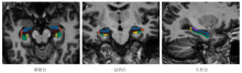
| 组别 | n | 左CA1 | 左CA2-3 | 左CA4-DG | 左下托 | 左旁下托 | 左海马伞 |
|---|---|---|---|---|---|---|---|
| 对照组 | 25 | 296.38±36.46 | 980.32±94.72 | 554.21±53.44 | 620.32±52.78 | 443.31±51.13 | 67.32±15.98 |
| 轻度AD组 | 25 | 269.36±35.88 | 915.45±93.18 | 517.37±55.03 | 612.75±50.22 | 429.17±53.08 | 61.62±15.90 |
| t值 | 2.641 | 2.441 | 2.401 | 0.520 | 0.959 | 1.264 | |
| P值 | 0.011 | 0.018 | 0.020 | 0.606 | 0.342 | 0.212 | |
| 组别 | n | 右CA1 | 右CA2-3 | 右CA4-DG | 右下托 | 右旁下托 | 右海马伞 |
| 对照组 | 25 | 304.50±36.92 | 983.59±102.39 | 565.04±57.79 | 625.99±54.32 | 466.83±49.79 | 65.16±16.58 |
| 轻度AD组 | 25 | 280.16±38.11 | 916.21±99.82 | 550.32±56.90 | 630.21±55.12 | 455.76±47.10 | 63.77±15.68 |
| t值 | 2.294 | 2.356 | 0.908 | 0.273 | 0.808 | 0.305 | |
| P值 | 0.026 | 0.023 | 0.369 | 0.786 | 0.423 | 0.762 |
"
| 组别 | n | 左CA1 | 左CA2-3 | 左CA4-DG | 左下托 | 左旁下托 | 左海马伞 |
|---|---|---|---|---|---|---|---|
| 安慰剂组 | 25 | 271.14±34.97 | 916.32±94.21 | 516.42±55.12 | 616.45±51.06 | 427.19±55.12 | 61.45±16.07 |
| 治疗组 | 25 | 290.10±33.91 | 966.74±93.26 | 550.38±54.21 | 615.48±53.18 | 432.18±52.07 | 64.31±16.71 |
| t值 | 1.946 | 1.902 | 2.196 | 0.066 | 0.329 | 0.617 | |
| P值 | 0.058 | 0.063 | 0.033 | 0.948 | 0.744 | 0.540 | |
| 组别 | n | 右CA1 | 右CA2-3 | 右CA4-DG | 右下托 | 右旁下托 | 右海马伞 |
| 安慰剂组 | 25 | 281.13±37.48 | 922.23±98.07 | 549.42±55.82 | 632.37± 55.33 | 457.88±48.90 | 63.99±16.34 |
| 治疗组 | 25 | 302.13±36.89 | 973.73±94.81 | 555.32±54.81 | 631.22±55.47 | 461.30±49.11 | 64.82±16.73 |
| t值 | 1.997 | 1.888 | 0.377 | 0.073 | 0.247 | 0.178 | |
| P值 | 0.052 | 0.065 | 0.708 | 0.942 | 0.806 | 0.860 |
"
| 组别 | n | 治疗前 | 治疗后 |
|---|---|---|---|
| 安慰剂组 | 25 | 20.87±3.92 | 21.51±4.12 |
| 治疗组 | 25 | 21.38±4.03 | 24.83±4.37 |
| t值 | 0.454 | 2.764 | |
| P值 | 0.652 | 0.008 |
| [1] |
Lane C A, Hardy J, Schott J M. Alzheimer's disease[J]. Eur J Neurol, 2018, 25(1):59-70.
doi: 10.1111/ene.13439 pmid: 28872215 |
| [2] |
Grabher B J. Effects of Alzheimer disease on patients and their family[J]. J Nucl Med Technol, 2018, 46(4):335-340.
doi: 10.2967/jnmt.118.218057 |
| [3] |
Selkoe D J. Resolving controversies on the path to Alzheimer's therapeutics[J]. Nature Med, 2011, 17(9):1060-1065.
doi: 10.1038/nm.2460 |
| [4] |
Strohle A, Schmidt D K, Schultz F, et al. Drug and exercise treatment of Alzheimer disease and mild cognitive impairment: a systematic review and meta-analysis of effects on cognition in randomized controlled trials[J]. Am J Geriatr Psychiatry, 2015, 23(12):1234-1249.
doi: 10.1016/j.jagp.2015.07.007 |
| [5] |
Aschenbrenner A J, Gordon B A, Benzinger T, et al. Influence of tau PET, amyloid PET, and hippocampal volume on cognition in Alzheimer disease[J]. Neurology, 2018, 91(9):e859-e866.
doi: 10.1212/WNL.0000000000006075 |
| [6] | Frisoni G B, Fox N C, Jack C J, et al. The clinical use of structural MRI in Alzheimer disease[J]. Nat Rev Neurol, 2010, 6(2):67-77. |
| [7] |
Hata K, Nakamoto K, Nunomura A, et al. Automated volumetry of medial temporal lobe subregions in mild cognitive impairment and Alzheimer disease[J]. Alzheimer Dis Assoc Disord, 2019, 33(3):206-211.
doi: 10.1097/WAD.0000000000000318 |
| [8] | 赵亮, 王冬青, 朱颖, 等. 阿尔茨海默病患者海马形态、体积变化的影像学特征研究[J]. 中华行为医学与脑科学杂志, 2010, 19(3):200-202. |
| [9] | 刘瑜, 金洁, 罗一烽, 等. 轻度认知损害及轻中度阿尔茨海默病海马亚区MRI体积的研究[J]. 临床放射学杂志, 2014, 33(7):972-975. |
| [10] |
McKhann G M, Knopman D S, Chertkow H, et al. The diagnosis of dementia due to Alzheimer's disease: recommendations from the National Institute on Aging-Alzheimer's Association workgroups on diagnostic guidelines for Alzheimer's disease[J]. Alzheimer Dement, 2011, 7(3):263-269.
doi: 10.1016/j.jalz.2011.03.005 |
| [11] | Creavin S T, Wisniewski S, Noel-Storr A H, et al. Mini-Mental State Examination (MMSE) for the detection of dementia in clinically unevaluated people aged 65 and over in community and primary care populations[J]. Cochrane Database Syst Rev, 2016(1):D11145. |
| [12] |
Reisberg B, Ferris S H, de Leon M J, et al. The Global Deterioration Scale for assessment of primary degenerative dementia[J]. Am J Psychiatry, 1982, 139(9):1136-1139.
doi: 10.1176/ajp.139.9.1136 |
| [13] |
Lima A, Castilhos R, Chaves M. The use of the Clinical Dementia Rating Scale sum of boxes scores in detecting and staging cognitive impairment/dementia in Brazilian patients with low educational attainment[J]. Alzheimer Dis Assoc Disord, 2017, 31(4):322-327.
doi: 10.1097/WAD.0000000000000205 |
| [14] |
van Leemput K, Bakkour A, Benner T, et al. Automated segmentation of hippocampal subfields from ultra-high resolution in vivo MRI[J]. Hippocampus, 2009, 19(6):549-557.
doi: 10.1002/hipo.20615 pmid: 19405131 |
| [15] |
Shi F, Liu B, Zhou Y, et al. Hippocampal volume and asymmetry in mild cognitive impairment and Alzheimer's disease: meta-analyses of MRI studies[J]. Hippocampus, 2009, 19(11):1055-1064.
doi: 10.1002/hipo.v19:11 |
| [16] |
Theysohn J M, Kraff O, Maderwald S, et al. The human hippocampus at 7 T: in vivo MRI[J]. Hippocampus, 2009, 19(1):1-7.
doi: 10.1002/hipo.20487 pmid: 18727048 |
| [17] |
Wisse L E, Biessels G J, Heringa S M, et al. Hippocampal subfield volumes at 7 T in early Alzheimer's disease and normal aging[J]. Neurobiol Aging, 2014, 35(9):2039-2045.
doi: 10.1016/j.neurobiolaging.2014.02.021 |
| [18] |
Kerchner G A, Berdnik D, Shen J C, et al. APOE epsilon4 worsens hippocampal CA1 apical neuropil atrophy and episodic memory[J]. Neurology, 2014, 82(8):691-697.
doi: 10.1212/WNL.0000000000000154 |
| [19] |
Yassa M A, Stark S M, Bakker A, et al. High-resolution structural and functional MRI of hippocampal CA3 and dentate gyrus in patients with amnestic mild cognitive impairment[J]. Neuroimage, 2010, 51(3):1242-1252.
doi: 10.1016/j.neuroimage.2010.03.040 |
| [20] |
Chen X Q, Mobley W C. Alzheimer disease pathogenesis: insights from molecular and cellular biology studies of oligomeric Abeta and tau species[J]. Front Neurosci, 2019, 13(1):659.
doi: 10.3389/fnins.2019.00659 |
| [1] | ZHANG Ruiming, WANG Kai, YIN Rui, WANG Hailiang, MO Yan. Evaluation method of assistance effect of extravehicular activity glove exoskeleton under weightlessness [J]. 《Chinese Journal of Rehabilitation Theory and Practice》, 2023, 29(7): 856-861. |
| [2] | YUAN Yuan, ZHOU Hongjun, CONG Xinying, LIU Genlin, WEI Bo, ZHENG Ying, HAO Chunxia, ZHANG Ying, WANG Yiji, KANG Haiqiong, LU Xiaolei, MENG Qianru. Relationship between impairment and magnetic resonance imaging finding in patients with traumatic cervical spinal cord injury after surgery [J]. 《Chinese Journal of Rehabilitation Theory and Practice》, 2023, 29(6): 725-730. |
| [3] | SONG Beibei, LIU Hui, BAI Kaixiang. Content and psychological measurement index of ICF-CY-based motor function evaluation scale for children and adolescents with disabilities: a scoping review [J]. 《Chinese Journal of Rehabilitation Theory and Practice》, 2023, 29(2): 182-192. |
| [4] | CHENG Siman, XIN Rong, ZHAO Yan, LIU Qingyu, XIE Jiale, LIU Peng, WANG Pu. Functional magnetic resonance imaging study about repetitive transcranial magnetic stimulation for dysfunction after stroke: a scoping review [J]. 《Chinese Journal of Rehabilitation Theory and Practice》, 2023, 29(2): 193-204. |
| [5] | LIU Xiaoxie, CHU Hongling, LIU Mei, GUO Aixin, WANG Siyuan, ZENG Fanshuo, JIANG Shan, XIE Yuxiao, ZHOU Mouwang. Scientific, transparent and applicable rankings of Chinese guidelines and consensus of rehabilitation medicine published in medical journals in 2022 [J]. 《Chinese Journal of Rehabilitation Theory and Practice》, 2023, 29(12): 1365-1376. |
| [6] | WANG Xiaohe, XIAO Shuqin, ZHANG Lu. Communication and cooperation within a stroke rehabilitation team: a qualitative research [J]. 《Chinese Journal of Rehabilitation Theory and Practice》, 2023, 29(12): 1473-1480. |
| [7] | ZHANG Xiaoyu,YANG Fan,WEN Jianzhong,YU Weiyong. Application of resting-state functional magnetic resonance imaging in acute mild traumatic brain injury [J]. 《Chinese Journal of Rehabilitation Theory and Practice》, 2022, 28(9): 1084-1088. |
| [8] | YIN Minmin,LING Xing,YANG Yaru,DAI Hengyu,GE Shengnan,HUANG Zhaoming. Development of diagnosis, assessment and intervention solution for speech fluency disorder using WHO-FICs [J]. 《Chinese Journal of Rehabilitation Theory and Practice》, 2022, 28(6): 630-636. |
| [9] | GE Shengnan,WANG Yongli,YIN Minmin,WAN Qin,YANG Yaru,Lancy Lantin HUANG,HUANG Zhaoming. Diagnosis, assessment and rehabilitation of speech disorders after cerebral palsy using WHO-FICs [J]. 《Chinese Journal of Rehabilitation Theory and Practice》, 2022, 28(6): 637-645. |
| [10] | CAI Guiyan,CHEN Ruilin,XU Shurui,TAO Jing,LIU Jiao. Characteristics of amplitude of low frequency fluctuation in patients with knee osteoarthritis and low back pain [J]. 《Chinese Journal of Rehabilitation Theory and Practice》, 2022, 28(5): 602-608. |
| [11] | QIU Zhuoying, KWOK Joseph Kin Fun, SUN Hongwei, WU Shicai, LÜ Jun, YAO Meilin, WANG Guoxiang, QIU Fubing, LU Yan, XU Guangxu, JI Linhong, JING Qi, CHEN Di, HAO Chuanping, LI Anqiao, WANG Shaopu, XIAO Xiaofei. Systematic implementation of World Health Organization rehabilitation competency framework in rehabilitation: conceptual framework, approaches and application [J]. 《Chinese Journal of Rehabilitation Theory and Practice》, 2022, 28(3): 265-274. |
| [12] | DUAN Mingxue, JING Qi, QIU Zhuoying, SUN Hongwei, LI Anqiao, WU Fang, CAI Weiqin, GAO Qianqian, ZHENG Wengui. Performance evaluation of rehabilitation workers using World Health Organization rehabilitation competency framework [J]. 《Chinese Journal of Rehabilitation Theory and Practice》, 2022, 28(3): 285-294. |
| [13] | WANG Longping,YANG Xuening,ZENG Bin,XU Guangqing. Application of six-minute walk test in preoperative evaluation of patients undergoing pulmonary lobectomy [J]. 《Chinese Journal of Rehabilitation Theory and Practice》, 2022, 28(2): 242-248. |
| [14] | YE Lü,WANG Bin. Monitoring and evaluation of health-promoting schools and physical activity from the perspective of health services [J]. 《Chinese Journal of Rehabilitation Theory and Practice》, 2022, 28(12): 1400-1407. |
| [15] | CHEN Siqi,ZHAO Hang,WANG Jie,XIA Bin,LIU Qiaoyun. Development and standardrization of communication function scale for two to six years old children using ICF-CY [J]. 《Chinese Journal of Rehabilitation Theory and Practice》, 2022, 28(12): 1444-1451. |
| Viewed | ||||||
|
Full text |
|
|||||
|
Abstract |
|
|||||
|
||