


《Chinese Journal of Rehabilitation Theory and Practice》 ›› 2020, Vol. 26 ›› Issue (6): 662-667.doi: 10.3969/j.issn.1006-9771.2020.06.007
Previous Articles Next Articles
GONG Shi-li1,2,YANG Cui-cui2,HU Chao-ying2,WANG Ming-yang2,ZHANG Lan1,2( )
)
Received:2019-12-24
Revised:2020-01-17
Published:2020-06-25
Online:2020-06-29
Contact:
ZHANG Lan
E-mail:lanizhg@126.com
Supported by:CLC Number:
GONG Shi-li,YANG Cui-cui,HU Chao-ying,WANG Ming-yang,ZHANG Lan. Promotion of Analgecine on Polarization of M1 Microglia to M2 and Inhibition on Secretion of Inflammatory Factors in Vitro[J]. 《Chinese Journal of Rehabilitation Theory and Practice》, 2020, 26(6): 662-667.
"
| 组别 | n | TNF-α | IL-6 |
|---|---|---|---|
| 对照1组 | 3 | 48.70±1.637 | 5.093±0.5215 |
| AGC1组 | 3 | 25.73±5.660 | 1.863±0.635 |
| OGD/R组 | 3 | 342.3±22.070a | 31.13±3.003b |
| L1组 | 3 | 212.3±14.560c | 7.148±1.924c |
| M1组 | 3 | 138.4±14.280c | 6.127±1.966c |
| H1组 | 3 | 74.85±9.630d | 3.793±0.463d |
| F值 | 83.600 | 40.840 | |
| P值 | <0.0001 | <0.0001 |
"

"
"
"
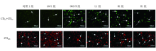
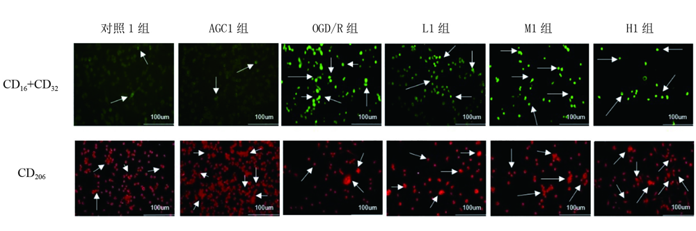
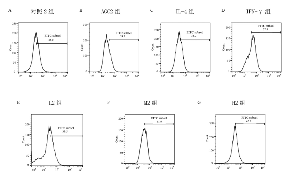
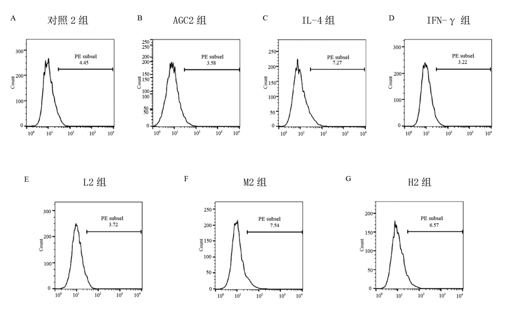
| 组别 | n | CD16+CD32 | CD206 |
|---|---|---|---|
| 对照2组 | 4 | 46.60±5.796 | 5.093±0.5215 |
| AGC2组 | 4 | 26.60±8.821 | 1.863±0.6350 |
| IL-4组 | 4 | 39.00±8.256 | 7.163±2.115 |
| IFN-γ组 | 4 | 55.00±9.906 | 2.567±0.9602 |
| L2组 | 4 | 39.65±10.98 | 4.437±1.865 |
| M2组 | 4 | 39.40±11.120 | 7.353±2.969 |
| H2组 | 4 | 40.97±11.180 | 6.037±0.3755a |
| F值 | 0.8271 | 0.917 | |
| P值 | 0.5582 | 0.508 |
| [1] |
Thion M S, Ginhoux F, Garel S. Microglia and early brain development: an intimate journey[J]. Science, 2018, 362(6411):185-189.
doi: 10.1126/science.aat0474 |
| [2] | Chhor V, Le Charpentier T, Lebon S, et al. Characterization of phenotype markers and neuronotoxic potential of polarized primary microglia in vitro[J]. Brain Behave Immun, 2013, 32:70-85. |
| [3] |
Hu X M, Li P Y, Guo Y L, et al. Microglia/macrophage polarization dynamics reveal novel mechanism of injury expansion after focal cerebral ischemia[J]. Stroke, 2012, 43(11):3063-3070.
doi: 10.1161/STROKEAHA.112.659656 |
| [4] |
Cherry J D, Olschowka J A, O'Banion M K. Neuroinflammation and M2 microglia: the good, the bad, and the inflamed[J]. J Neuroinflammation, 2014, 11:98.
doi: 10.1186/1742-2094-11-98 |
| [5] |
Tang Y, Le W D. Differential Roles of M1 and M2 Microglia in neurodegenerative diseases[J]. Mol Neurobiol, 2016, 53(2):1181-1194.
doi: 10.1007/s12035-014-9070-5 pmid: 25598354 |
| [6] |
Carlo P, Stefano F, Maria-Grazia D S. Temporal pattern of expression and colocalization of microglia/macrophage phenotype markers following brain ischemic injury in mice[J]. J Neuroinflammation, 2011, 8:174.
doi: 10.1186/1742-2094-8-174 |
| [7] | Yang X M, He R G. Effect of extracts from rabbit skin inflamed by vaccinia virus in the management of postherpetic neuralgia and on serum interleukin-6 level in aged patients[J]. Nan Fang Yi Ke Da Xue Xue Bao, 2007, 27(12):1941-1943. |
| [8] | 费秋惠. 恩再适治疗带状疱疹后遗神经痛的可行性和安全性[J]. 医学理论与实践, 2019, 32(4):557-559. |
| [9] | 陈和星. 恩再适在治疗带状疱疹后遗神经痛中的可行性及对脑源性神经营养因子的表达意义[J]. 江西医药, 2014, 49(11):1293-1295. |
| [10] |
Dong J, Tu H P, Ding W Y, et al. Analgecine, the extracts of Vaccinia-inoculated rabbit skin, effectively alleviates the chronic low back pain with little side effect: a randomized multi-center double-blind placebo-controlled phase 3 clinical trial[J]. Contemp Clin Trials Commun, 2016, 2:16-24.
doi: 10.1016/j.conctc.2015.11.002 |
| [11] |
Poomarin S, Passakorn S, Nattayaporn A, et al. Role of microglia under cardiac and cerebral ischemia/reperfusion (I/R) injury[J]. Metab Brain Dis, 2018, 33(4):1019-1030.
doi: 10.1007/s11011-018-0232-4 |
| [12] |
Hu L, He D, Bai Y. Microglia-mediated inflammation and neurodegenerative disease[J]. Mol Neurobiol, 2016, 53(10):6709-6715.
doi: 10.1007/s12035-015-9593-4 |
| [13] |
Hu J J, Qin L J, Liu Z Y, et al. MiR-15a regulates oxygen glucose deprivation/ reperfusion (OGD/R)-induced neuronal injury by targeting BDNF[J]. Kaohsiung J Med Sci, 2020, 36(1):27-34.
doi: 10.1002/kjm2.v36.1 |
| [14] |
Li H, Ma J W, Fang Q, et al. Botch protects neurons from ischemic insult by antagonizing Notch-mediated neuroinflammation[J]. Exp Neurol, 2019, 321:113028.
doi: 10.1016/j.expneurol.2019.113028 |
| [15] |
Dugan L L, Kim-Han J S. Astrocyte mitochondria in in vitro models of ischemia[J]. J Bioenerg Biomembr, 2004, 36(4):317-321.
doi: 10.1023/B:JOBB.0000041761.61554.44 |
| [16] | Ryou M G, Mallet R T. An in vitro oxygen-glucose deprivation model for studying ischemia-reperfusion injury of neuronal cells[J]. Methods Mol Biol, 2018, 1717:229-235. |
| [17] |
Kroner A, Greenhalgh A D, Zarruk J G, et al. TNF and increased intracellular iron alter macrophage polarization to a detrimental M1 phenotype in the injured spinal cord[J]. Neuron, 2014, 83(5):1098-1116.
doi: 10.1016/j.neuron.2014.07.027 pmid: 25132469 |
| [18] | Bernstein J E, Savla P, Dong F, et al. Inflammatory markers and severity of intracerebral hemorrhage[J]. Cureus, 2018, 10(10):e3529. |
| [19] |
Kettenmann H, Hanisch U K, Noda M, et al. Physiology of microglia[J]. Physiol Rev, 2011, 91(2):461-553.
doi: 10.1152/physrev.00011.2010 pmid: 21527731 |
| [20] |
Ghosh S, Castillo E, Frias E S, et al. Bioenergetic regulation of microglia[J]. Glia, 2018, 66(6):1200-1212.
doi: 10.1002/glia.v66.6 |
| [21] | Varnum M M, Ikezu T. The classification of microglial activation phenotypes on neurodegeneration and regeneration in Alzheimer's disease brain[J]. Arch Immunol Ther Exp (Warsz), 2012, 60(4):252-266. |
| [22] | Li C W, Bian Y Q, Feng Y, et al. Neuroprotective effects of BHDPC, a novel neuroprotectant, on experimental stroke by modulating microglia polarization[J]. ACS Chem Neurisci, 2019, 10(5):2434-2449. |
| [23] |
Ma Y Y, Wang J X, Wang Y T, et al. The biphasic function of microglia in ischemic stroke[J]. Prog Neurobiol, 2017, 157:247-272.
doi: 10.1016/j.pneurobio.2016.01.005 |
| [24] |
Colonna M, Butovsky O. Microglia function in the central nervous system during health and neurodegeneration[J]. Annu Rev Immunol, 2017, 35:441-468.
doi: 10.1146/annurev-immunol-051116-052358 pmid: 28226226 |
| [25] |
Orihuela R, McPherson C A, Harry G J. Microglia M1/M2 polarization and metabolic states[J]. Br J Pharmacol, 2016, 173(4):649-665.
doi: 10.1111/bph.v173.4 |
| [26] |
Kigerl K A, Gensel J C, Ankeny D P, et al. Identification of two distinct macrophage subsets with divergent effectscausing either neurotoxicity or regeneration in the injured mouse spinal cord[J]. J Neurosci, 2009, 29(43):13435-13444.
doi: 10.1523/JNEUROSCI.3257-09.2009 |
| [1] | DENG Ting, CHEN Jingmian, LIU Xiaomeng, YAO Xiaohua, LIU Lushan, HE Wei, ZHANG Tong, LU Haitao. Risk factors of stroke-associated pneumonia for patients with mild to moderate acute ischemic stroke [J]. 《Chinese Journal of Rehabilitation Theory and Practice》, 2023, 29(6): 708-713. |
| [2] | ZHANG Chunlong, LIU Fuliang, SHANG Na, LI Fang, LIU Huizhen. Association of serum adiponectin and high sensitivity C-reactive protein levels to short-term outcome in patients with acute ischemic stroke [J]. 《Chinese Journal of Rehabilitation Theory and Practice》, 2023, 29(10): 1221-1226. |
| [3] | HAN Kaiyue,LIU Guangliang,SU Wenlong,TANG Zhiqing,ZHANG Hao. Effects of intelligent aerobic bicycle training on ischemic stroke patients at different disease courses [J]. 《Chinese Journal of Rehabilitation Theory and Practice》, 2022, 28(7): 822-827. |
| [4] | ZHOU Xiaojue,FENG Jing,PANG Rizhao,LIU Jie,ZHANG Anren. Every-other-day fasting attenuated inflammation in rats after spinal cord injury via the aryl hydrocarbon receptor/suppressor of cytokine signaling 2/nuclear transcription factor-κB signaling pathway [J]. 《Chinese Journal of Rehabilitation Theory and Practice》, 2022, 28(5): 544-551. |
| [5] | LAI Haifang,GU Lin,ZONG Ya,NIU Chuanxin,XIE Qing. Prediction of short-term outcome after subacute ischemic stroke using multiple layer perceptron neural network [J]. 《Chinese Journal of Rehabilitation Theory and Practice》, 2022, 28(3): 335-339. |
| [6] | ZHU Hui,XIA Youbing,GONG Zunke,WANG Shiyan,MA Ke,YAN Jinqiu. Effects of high-frequency repetitive transcranial magnetic stimulation on central facial paralysis after ischemic stroke [J]. 《Chinese Journal of Rehabilitation Theory and Practice》, 2022, 28(2): 199-203. |
| [7] | LAN Wanting,ZHONG Jiugen,SHEN Yingying,GONG Jiaheng,ZOU Zhi,HOU Xiaohui. Mechanism of exercise reducing neuroinflammation in autism spectrum disorder: a visualized analysis [J]. 《Chinese Journal of Rehabilitation Theory and Practice》, 2022, 28(10): 1190-1197. |
| [8] | Na WANG,Pei-lan LI,Lu-shan LIU,Feng-rong WANG. Prediction of Short- and Long-term Death in Patients with Acute Ischemic Stroke Using Various Scoring Systems [J]. 《Chinese Journal of Rehabilitation Theory and Practice》, 2021, 27(3): 256-260. |
| [9] | YANG Zhao-yu,LI Pei-jun,LI Jian,LIU Xiao-dan,WU Wei-bing. Effect of Exercise on Chronic Obstructive Pulmonary Disease Systemic Inflammation and Skeletal Muscle Dysfunction: A Systematic Review [J]. 《Chinese Journal of Rehabilitation Theory and Practice》, 2021, 27(12): 1443-1449. |
| [10] | WANG Na-na, DU Xiao-zheng, HE Wen-jie, ZHANG Xin-yu, ZHENG Xin, LI Meng-xin. Roles of Notch Signaling Pathway in Acupuncture for Ischemic Stroke (review) [J]. 《Chinese Journal of Rehabilitation Theory and Practice》, 2021, 27(1): 67-70. |
| [11] | CHEN Xiao-ping,WANG Ming-yang,MA Deng-lei,GONG Shi-li,ZHANG Li,LI Ya-li,LI Lin,HU Chao-ying,ZHANG Lan. Effects of Analgecine on Apoptosis and Neuroinflammation in Cerebral Ischemia-reperfusion Injury Rats [J]. 《Chinese Journal of Rehabilitation Theory and Practice》, 2020, 26(9): 1038-1044. |
| [12] | LIU Jian-hua,WEI Qing-chuan,HU Xiu-ru,YE Sai-qing,YAN Zhi-yu,GAO Qiang. Relationship between Fractional Anisotropy of Diffusion Tensor Imaging and Motor Function after Ischemic Stroke [J]. 《Chinese Journal of Rehabilitation Theory and Practice》, 2020, 26(7): 749-752. |
| [13] | WEI Hai-ping,GUO Jia,WANG Huan,GE Zhao-ming. Effects of Vagus Nerve Stimulation on Adenosine Monophosphate Activated Protein Kinase-Silent Mating Type Information Regulation 2 Homolog 1 Pathway in Ischemia-reperfusion Rats [J]. 《Chinese Journal of Rehabilitation Theory and Practice》, 2020, 26(7): 775-779. |
| [14] | LIU Hui-zhen,GUO Shu-bin,SHANG Na,LI Fang,LIU Lu-shan,LI Pei-lan,CHEN Jing-mian,WANG Feng-rong,LI Jun-yu. Prediction of Serum 25-Hydroxyvitamin D for Outcome of Acute Ischemic Stroke in Emergency [J]. 《Chinese Journal of Rehabilitation Theory and Practice》, 2020, 26(7): 830-835. |
| [15] | WANG Chuan-jie,WU Yi,TAO Feng,YANG Lei. Effects of Enriched Environment on Cognition and Expression of Bcl-2/Bax Protein in Hippocampus after Ischemic Stroke in Mice [J]. 《Chinese Journal of Rehabilitation Theory and Practice》, 2020, 26(5): 539-543. |
| Viewed | ||||||
|
Full text |
|
|||||
|
Abstract |
|
|||||
|
||