


《Chinese Journal of Rehabilitation Theory and Practice》 ›› 2021, Vol. 27 ›› Issue (6): 653-660.doi: 10.3969/j.issn.1006-9771.2021.06.005
Previous Articles Next Articles
Jing-yi WANG1,Jie YIN1,Jian-cheng LIU2,Ri-zhao PANG2,Wen-chun WANG2( )
)
Received:2021-01-04
Revised:2021-03-10
Published:2021-06-25
Online:2021-06-21
Contact:
Wen-chun WANG
E-mail:852900340@qq.com
Supported by:CLC Number:
Jing-yi WANG,Jie YIN,Jian-cheng LIU,Ri-zhao PANG,Wen-chun WANG. Effect of Iridoid-rich Fraction from Valeriana Jatamansi Jones on Neuron Pyroptosis in Rats with Acute Spinal Cord Injury[J]. 《Chinese Journal of Rehabilitation Theory and Practice》, 2021, 27(6): 653-660.
"
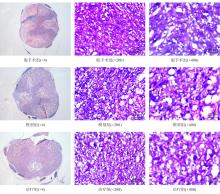
| 组别 | n | 组织残余面积 |
|---|---|---|
| 假手术组 | 4 | 96.392±2.987 |
| 模型组 | 4 | 76.773±8.641a |
| 治疗组 | 4 | 88.130±6.083b |
| F值 | 9.655 | |
| P值 | 0.006 |
"

"
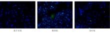
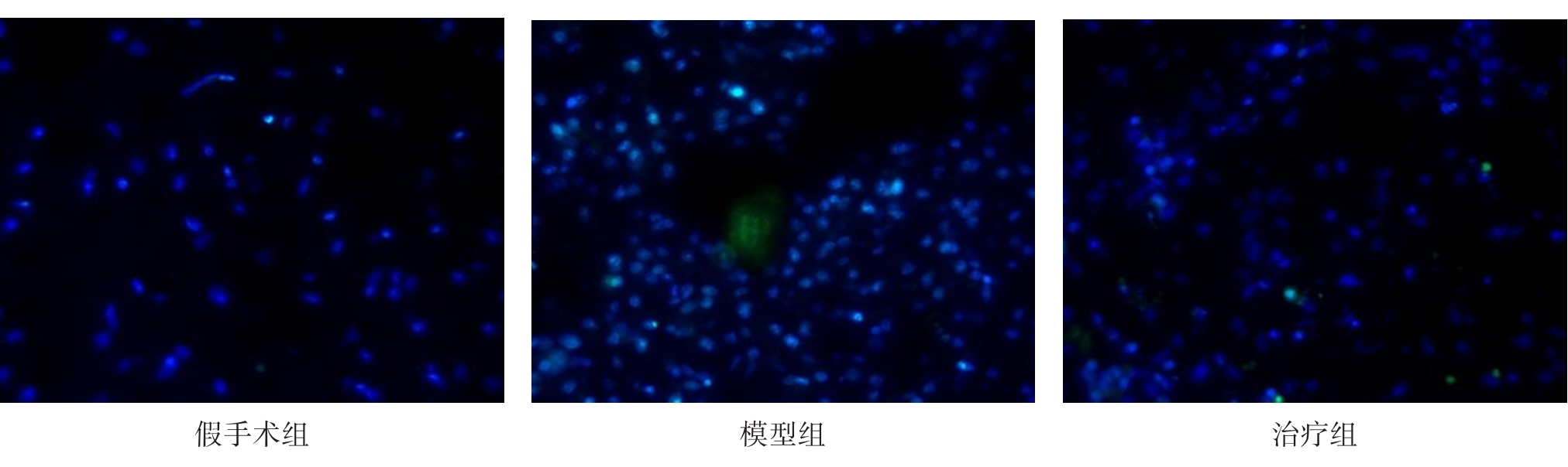
| 组别 | n | 阳性细胞率 |
|---|---|---|
| 假手术组 | 4 | 0.382 ± 0.322 |
| 模型组 | 4 | 10.921 ± 3.583a |
| 治疗组 | 4 | 6.873 ± 0.95a,b |
| F值 | 24.501 | |
| P值 | < 0.001 |
"
"
| 组别 | n | IL-1β | IL-18 |
|---|---|---|---|
| 假手术组 | 4 | 2.714±0.245 | 10.769±1.011 |
| 模型组 | 4 | 4.544±0.119a | 14.255±1.976a |
| 治疗组 | 4 | 3.046±0.142a,b | 11.512±1.753b |
| F值 | 121.169 | 5.058 | |
| P值 | < 0.001 | 0.034 |

"
"
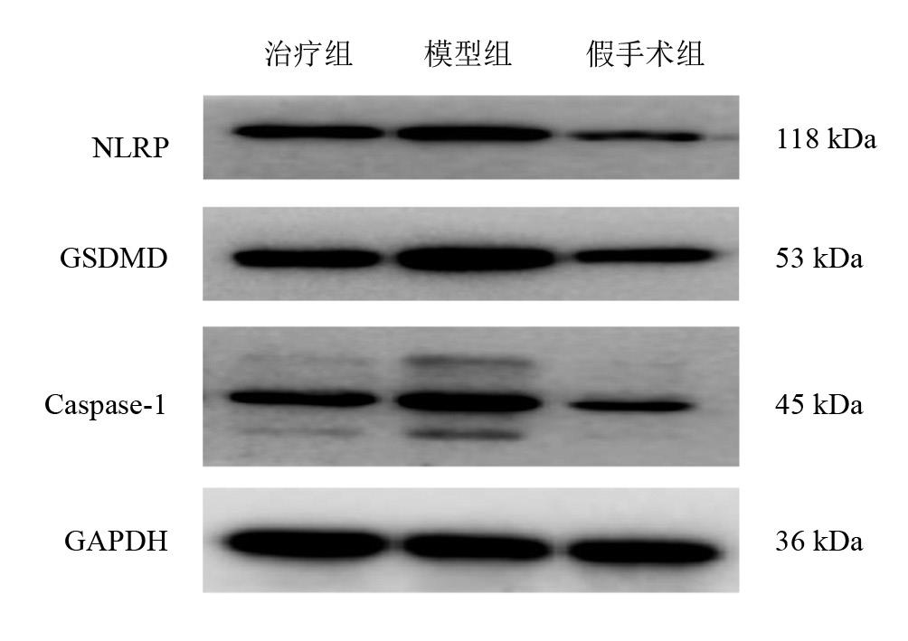
| 组别 | n | NLRP3 | GSDMD | Caspase-1 |
|---|---|---|---|---|
| 假手术组 | 4 | 0.412±0.100 | 0.792±0.161 | 0.388±0.078 |
| 模型组 | 4 | 0.901±0.223a | 1.414±0.283a | 0.970±0.241a |
| 治疗组 | 4 | 0.634±0.111b | 1.026±0.162b | 0.691±0.109a,b |
| F值 | 10.002 | 8.940 | 13.339 | |
| P值 | 0.005 | 0.007 | 0.002 |
| 1 | ROPPER A E, ROPPER A H. Acute spinal cord compression [J]. N Engl J Med, 2017, 376(14): 1358-1369. |
| 2 | IHALAINEN T, RINTA-KIIKKA I, LUOTO T M, et al. Traumatic cervical spinal cord injury: a prospective clinical study of laryngeal penetration and aspiration [J]. Spinal Cord, 2017, 55(11): 979-984. |
| 3 | REBECCA T, JASON L, CRAVEN B C. Diagnostic accuracy and feasibility of depression screening in spinal cord injury: a systematic review [J]. J Spinal Cord Med, 2019, 42(): 99-107. |
| 4 | BÖKEL A, EGEN C, GUTENBRUNNER C, et al. [Spinal cord injury in Germany: a survey on the living and care situation of people with spinal cord injury] [J]. [in German]. Rehabilitation (Stuttg), 2020, 59(4): 205-221. |
| 5 | JAZAYERI S B, BEYGI S, SHOKRANEH F, et al. Incidence of traumatic spinal cord injury worldwide: a systematic review [J]. Eur Spine J, 2015, 24(5): 905-918. |
| 6 | RAMER L M, RAMER M S, BRADBURY E J. Restoring function after spinal cord injury: towards clinical translation of experimental strategies [J]. Lancet Neurol, 2014, 13(12): 1241-1256. |
| 7 | 李庆杰,王琦,都帅,等. 蜘蛛香的化学成分及其抗焦虑作用研究进展[J]. 吉林中医药, 2020, 40(9): 1254-1256. |
| LI Q J, WANG Q, DU S, et al. Advances in study on the chemical constituents and antianxiety activity of Valeriana jatamansi Jones [J]. Jilin J Chin Med, 2020, 40(9): 1254-1256. | |
| 8 | MAURMANN N, REOLON G K, RECH S B, et al. A valepotriate fraction of Valeriana glechomifolia shows sedative and anxiolytic properties and impairs recognition but not aversive memory in mice [J]. Evid Based Complement Alternat Med, 2011, 2011: 720853. |
| 9 | 孙勇. 基于脑肠轴探讨蜘蛛香环烯醚萜类有效部位抗抑郁作用及其机制[D]. 成都:西南交通大学, 2019. |
| SUN Y. Studies on antidepressant effect and mechanism of the total Irioids of roots and rhizomes of Valeriana jatamansi Jones based on Gut-Brain Axis [D]. Chengdu: Southwest Jiaotong University, 2019. | |
| 10 | LIN Y, XU K K, CHEN C Y, et al. A study of the substance dependence effect of the ethanolic extract and iridoid-rich fraction from Valeriana jatamansi Jones in mice [J]. Pharmacogn Mag, 2015, 11(44): 745-749. |
| 11 | SRIDHARAN S, MOHANKUMAR K, JEEPIPALLI S P, et al. Neuroprotective effect of Valeriana wallichii rhizome extract against the neurotoxin MPTP in C57BL/6 mice [J]. Neurotoxicology, 2015, 51: 172-183. |
| 12 | 熊德启. 蜘蛛香环烯醚萜类有效部位对急性脊髓损伤大鼠运动功能的影响及相关机制探讨[D]. 成都:成都中医药大学, 2018. |
| XIONG D Q. The effect of Iridoids effective fraction of Valeriana jatamansi Jones on movement function in Rats after acute cord injury and the related mechanism [D]. Chengdu: Chengdu University of TCM, 2018. | |
| 13 | 黄姣娟,王文春,熊德启,等. 蜘蛛香环烯醚萜类成分对大鼠脊髓损伤后氧化应激的影响[J]. 康复学报, 2019, 29(3): 27-32. |
| HUANG J J, WANG W C, XIONG D Q, et al. Effect of the Iridoid-rich Fraction from Valeriana jatamansi Jones on oxidative stress in rats with spinal cord injury [J]. Rehabil Med, 2019, 29(3): 27-32. | |
| 14 | CHEN S, YE J, CHEN X, et al. Valproic acid attenuates traumatic spinal cord injury-induced inflflammation via STAT1 and NF-κB pathway dependent of HDAC3 [J]. J Neuroinflammation, 2018, 15(1): 150. |
| 15 | WALSH J G, MURUVE D A, POWER C. Inflammasomes in the CNS [J]. Nat Rev Neurosci, 2014, 15(2): 84-97. |
| 16 | 林玉. 蜘蛛香总环烯醚萜类抗肝癌作用及其机制初步研究[D]. 成都:西南交通大学, 2015. |
| LIN Y. Study on the anti-hepatoma efficacy and mechanism of iridoid-rich fraction from Valeriana jatamansi Jones [D]. Chengdu: Southwest Jiaotong University, 2015. | |
| 17 | GAGE G J, KIPKE D R, SHAIN W. Whole animal perfusion fixation for rodents [J]. J Vis Exp, 2012, 30(65): 1-9. |
| 18 | ECKERT M J, MARTIN M J. Trauma: spinal cord injury [J]. Surg Clin North Am, 2017, 97(5): 1031-1045. |
| 19 | ZHOU K, SANSUR C, XU H, et al. The temporal pattern, flux, and function of autophagy in spinal cord injury [J]. Int J Mol Sci, 2017, 18(2): 466. |
| 20 | BADHIWALA J H, AHUJA C S, FEHLINGS M G. Time is spine: a review of translational advances in spinal cord injury [J]. J Neurosurg Spine, 2018, 30(1): 1-18. |
| 21 | MORTEZAEE K, KHANLARKHANI N, BEYER C, et al. Inflammasome: its role in traumatic brain and spinal cord injury [J]. J Cell Physiol, 2018, 233(7): 5160-5169. |
| 22 | LAMKANFI M, DIXIT V M. Manipulation of host cell death pathways during microbial infection [J]. Cell Host Microbe, 2010, 8(1): 44-54. |
| 23 | DE R V, JUAN P, DIETRICH W D, et al. Activation and regulation of cellular inflammasomes: gaps in our knowledge for central nervous system injury [J]. J Cereb Blood Flow Metab, 2014, 34(3): 369-375. |
| 24 | COOKSON B T, BRENNAN M A. Pro-inflammatory programmed cell death [J]. Trends Microbiol, 2001, 9(3): 113-114. |
| 25 | JUNYING Y, AYAZ N, BÉNÉDICTE F P. Roles of caspases in necrotic cell death [J]. Cell, 2016, 167(7): 1693-1704. |
| 26 | SCHRODER K, TSCHOPP J. The inflammasomes [J]. Cell, 2010, 140(6): 821-832. |
| 27 | BERGSBAKEN T, FINK S L, COOKSON B T. Pyroptosis: host cell death and inflammation [J]. Nat Rev Microbiol, 2009, 7(2): 99-109. |
| 28 | HALLE A, HORNUNG V, PETZOLD G C, et al. The NALP3 inflammasome is involved in the innate immune response to amyloid-beta [J]. Nat Immunol, 2008, 9(8): 857-865. |
| 29 | ADAMCZAK S E, DE R V, DALE G, et al. Pyroptotic neuronal cell death mediated by the AIM2 inflammasome [J]. J Cereb Blood Flow Metab, 2014, 34(4): 621-629. |
| 30 | WANG L, LI K, LIN X, et al. Metformin induces human esophageal carcinoma cell pyroptosis by targeting the miR-497/PELP1 axis [J]. Cancer Lett, 2019, 450: 22-31. |
| 31 | DOITSH G, GALLOWAY N L, GENG X, et al. Cell death by pyroptosis drives CD4 T-cell depletion in HIV1 infection [J]. Nature, 2014, 505(7484): 509514. |
| 32 | MASTERS S L, GERLIC M, METCALF D, et al. NLRP1 inflammasome activation induces pyroptosis of hematopoietic progenitor cells [J]. Immunity, 2012, 37(6): 10091023. |
| 33 | TAN M S, TAN L, JIANG T, et al. Amyloid-β induces NLRP1-dependent neuronal pyroptosis in models of Alzheimer's disease [J]. Cell Death Dis, 2014, 5(8): e1382 |
| 34 | KESAVARDHANA S, KANNEGANTI T D. Mechanisms governing inflammasome activation, assembly and pyroptosis induction [J]. Int Immunol, 2017, 29(5): 201-210. |
| 35 | ANWAR M A, SHEHABI T S, EID A H. Inflammogenesis of secondary spinal cord injury [J]. Front Cell Neurosci, 2016, 10: 98. |
| 36 | LIU H D, LI W, CHEN Z R, et al. Expression of the NLRP3 inflammasome in cerebral cortex after traumatic brain injury in a rat model [J]. Neurochem Res, 2013, 38(10): 2072-2083. |
| 37 | DAI W, WANG X, TENG H, et al. Celastrol inhibits microglial pyroptosis and attenuates inflammatory reaction in acute spinal cord injury rats [J]. Int Immunopharmacol, 2019, 66: 215-223. |
| 38 | 黄姣娟. 蜘蛛香环烯醚萜类激活Nrf2/ARE通路减轻脊髓损伤氧化应激反应的机制研究[D]. 成都:西南交通大学, 2019. |
| HUANG J J. The Iridoid-rich fraction from Valeriana jatamansi Jones activat the Nrf2/Are alleviating oxidative stress react of spinal cord injury [D]. Chengdu: Southwest Jiaotong University, 2019. |
| [1] | LIU Dong, XU Zihan, LI Jiang, JU Ping. Effect of high-frequency repetitive transcranial magnetic stimulation in M1 region combined with dorsolateral prefrontal cortex on electroencephalogram θ frequency band amplitude of patients with neuropathic pain after spinal cord injury [J]. 《Chinese Journal of Rehabilitation Theory and Practice》, 2024, 30(1): 87-94. |
| [2] | LI Fang, HUO Su, DU Jubao, LIU Xiuzhen, LI Xiaoshuang, SONG Weiqun. Effect of transcranial direct current stimulation combined with task-oriented rehabilitation training on forelimb motor dysfunction in rats with spinal cord injury [J]. 《Chinese Journal of Rehabilitation Theory and Practice》, 2023, 29(7): 777-781. |
| [3] | LIU Ning, LIU Yuquan, ZHU Bin, YU Lingjia, TAN Haining, YANG Yong, LI Xiang. Application of International Standards for Neurological Classification of Spinal Cord Injury in China: a bibliometrics re-analysis [J]. 《Chinese Journal of Rehabilitation Theory and Practice》, 2023, 29(7): 808-815. |
| [4] | WANG Yiji, ZHOU Hongjun, HE Zejia, LIU Genlin, ZHENG Ying, HAO Chunxia, WEI Bo, KANG Haiqiong, ZHANG Ying, LU Xiaolei, YUAN Yuan, MENG Qianru. Relationship between symmetry of lower limb function and gait symmetry in patients with incomplete spinal cord injury [J]. 《Chinese Journal of Rehabilitation Theory and Practice》, 2023, 29(6): 639-645. |
| [5] | YUAN Yuan, ZHOU Hongjun, CONG Xinying, LIU Genlin, WEI Bo, ZHENG Ying, HAO Chunxia, ZHANG Ying, WANG Yiji, KANG Haiqiong, LU Xiaolei, MENG Qianru. Relationship between impairment and magnetic resonance imaging finding in patients with traumatic cervical spinal cord injury after surgery [J]. 《Chinese Journal of Rehabilitation Theory and Practice》, 2023, 29(6): 725-730. |
| [6] | JIANG Le, DU Liangjie, HUANG Fubiao. Mood states and cognitive performance in patients with complete spinal cord injury [J]. 《Chinese Journal of Rehabilitation Theory and Practice》, 2023, 29(5): 576-581. |
| [7] | GUO Shuang, XIE Yongqi, ZHANG Liang, ZHANG Chunjia, PENG Run, YANG Degang, YANG Mingliang. Related factors and prediction model for neurological outcome of dance-associated pediatric spinal cord injury without radiographic abnormality [J]. 《Chinese Journal of Rehabilitation Theory and Practice》, 2023, 29(5): 582-589. |
| [8] | ZHANG Yuan, YANG Jian. Exercise rehabilitation interventions for people with spinal cord injury and their health benefits: a systematic review of systematic reviews based on ICD-11 and ICF [J]. 《Chinese Journal of Rehabilitation Theory and Practice》, 2023, 29(12): 1377-1385. |
| [9] | SHI Xiaoyu, YANG Jian. Adaptive physical activity and its health benefits for patients with spinal cord injury based on ICF: a scoping review [J]. 《Chinese Journal of Rehabilitation Theory and Practice》, 2023, 29(12): 1395-1404. |
| [10] | HUANG Zhilin, XU Fashao, SHI Jing, HUANG Gan, LIU Meifang, ZHANG Xiahui. Establishment of rat model of dysphagia after stroke by thread embolism [J]. 《Chinese Journal of Rehabilitation Theory and Practice》, 2023, 29(10): 1147-1153. |
| [11] | QIN Yanqiang, DONG Hao, SUN Yingchun, CHENG Xiankuan, YAO Haijiang. Effects of different acupuncture schemes on neurotransmitters and related inflammatory factors in rats with post-stroke depression [J]. 《Chinese Journal of Rehabilitation Theory and Practice》, 2023, 29(1): 30-37. |
| [12] | LIU Genlin,ZHOU Hongjun,LI Jianjun,WEI Bo,ZHENG Ying,HAO Chunxia,ZHANG Ying,WANG Yiji,KANG Haiqiong,LU Xiaolei,YUAN Yuan,MENG Qianru. Advance in neurological classification of spinal cord injury with complications [J]. 《Chinese Journal of Rehabilitation Theory and Practice》, 2022, 28(8): 934-938. |
| [13] | MIAO Pei,ZHANG Tong,MI Haixia,ZHANG Weidong. Learning and memory ability and its mechanism in rats with focal cerebral ischemia induced by two filament-occluded methods [J]. 《Chinese Journal of Rehabilitation Theory and Practice》, 2022, 28(7): 789-796. |
| [14] | KANG Haiqiong,ZHOU Hongjun,LIU Genlin,WEI Bo,ZHENG Ying,ZHANG Ying,HAO Chunxia,WANG Yiji,LU Xiaolei,YUAN Yuan,MENG Qianru. Changes of bone mineral density in distal femur and proximal tibia in patients with spinal cord injury [J]. 《Chinese Journal of Rehabilitation Theory and Practice》, 2022, 28(7): 855-858. |
| [15] | ZHANG Miaoyuan,HE Ying,LI Xiaoxia,PENG Min,ZHANG Lei,LIU Shuying,KONG Ying. Self-management status and related factors of patients with intermittent clean catheterization after spinal cord injury [J]. 《Chinese Journal of Rehabilitation Theory and Practice》, 2022, 28(6): 716-724. |
| Viewed | ||||||
|
Full text |
|
|||||
|
Abstract |
|
|||||
|
||