


《Chinese Journal of Rehabilitation Theory and Practice》 ›› 2022, Vol. 28 ›› Issue (8): 972-980.doi: 10.3969/j.issn.1006-9771.2022.08.013
Previous Articles Next Articles
GUO Feng1( ),HAO Ying2,CHEN Yu1
Received:2022-03-30
Revised:2022-06-27
Published:2022-08-25
Online:2022-08-30
Contact:
GUO Feng
E-mail:guofeng_first@163.com
Supported by:CLC Number:
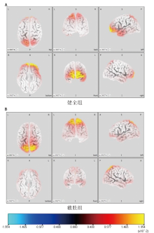
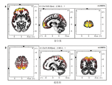
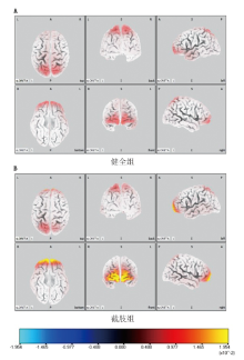
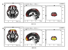
GUO Feng,HAO Ying,CHEN Yu. Brain sources characteristics during movement of residual limbs in forearm amputees based on standard low resolution brain electromagnetic tomography technology[J]. 《Chinese Journal of Rehabilitation Theory and Practice》, 2022, 28(8): 972-980.
"
| 组别 | n | 年龄/岁 | 身高/m | 体质量/kg | 残疾年限/年 |
|---|---|---|---|---|---|
| 健全组 | 15 | 22.6±2.7 | 1.78±0.1 | 71.2±8.5 | |
| 截肢组 | 9 | 24.7±8.6 | 1.73±0.1 | 64.2±16.4 | 13.4±5.4 |
| t值 | 0.887 | -1.186 | -1.385 | ||
| P值 | 0.495 | 0.229 | 0.260 |
"

"

"

"

"
| 脑叶 | 脑回 | BA | 残肢运动 | 健肢运动 | t值a | P值a | t值b | P值b | ||
|---|---|---|---|---|---|---|---|---|---|---|
| 健全组 | 截肢组 | 健全组 | 截肢组 | |||||||
| 额叶 | 中央前回 | BA4 | 36±10 | 41±12 | 44±15 | 44±11 | -1.101 | 0.283 | 0.001 | 0.999 |
| 额叶 | 额上回、额内侧回 | BA6 | 118±26 | 160±44 | 156±38 | 153±33 | -2.958 | 0.007 | 0.196 | 0.846 |
| 额叶 | 额上回、额中回 | BA8 | 63±12 | 49±12 | 66±10 | 48±12 | 2.767 | 0.011 | 3.964 | < 0.001 |
| 额叶 | 额上回、额中回 | BA9 | 90±12 | 26±7 | 59±20 | 68±29 | 14.509 | < 0.001 | -0.902 | 0.377 |
| 额叶 | 额上回、额中回 | BA10 | 178±36 | 74±14 | 134±32 | 143±38 | 8.240 | < 0.001 | -0.622 | 0.540 |
| 额叶 | 额上回、额中回、额下回 | BA11 | 143±37 | 57±12 | 94±45 | 132±46 | 6.712 | < 0.001 | -1.987 | 0.060 |
| 额叶 | 额中回、额下回 | BA45、46、47 | 161±53 | 63±15 | 95±34 | 150±45 | 5.376 | < 0.001 | -3.462 | 0.002 |
| 顶叶 | 中央后回 | BA1、2、3 | 43±11 | 54±13 | 61±16 | 64±15 | -2.217 | 0.037 | -0.455 | 0.654 |
| 顶叶 | 中央旁小叶 | BA5 | 63±16 | 60±15 | 68±17 | 58±12 | 0.455 | 0.654 | 1.670 | 0.074 |
| 顶叶 | 顶上小叶、中央后回 | BA7 | 348±59 | 308±72 | 332±87 | 265±61 | 1.482 | 0.153 | 2.023 | 0.055 |
| 顶叶 | 顶下小叶 | BA40 | 99±13 | 87±19 | 87±29 | 106±25 | 1.842 | 0.079 | -1.632 | 0.117 |
| 颞叶 | 颞中回、颞下回 | BA21 | 149±39 | 89±18 | 97±36 | 145±36 | 4.319 | < 0.001 | -3.162 | 0.005 |
| 颞叶 | 颞上回 | BA22 | 83±13 | 47±23 | 45±9 | 91±27 | 4.390 | < 0.001 | -6.132 | < 0.001 |
| 边缘叶 | 岛叶 | BA13 | 41±10 | 10±2 | 19±12 | 30±18 | 9.133 | < 0.001 | -1.992 | 0.082 |
| 边缘叶 | 扣带回后部 | BA31 | 25±7 | 36±7 | 17±8 | 16±6 | 3.292 | 0.002 | 0.323 | 0.750 |
| 枕叶 | 楔叶、舌回 | BA17 | 10±2 | 18±5 | 9±3 | 11±2 | -5.562 | < 0.001 | -1.770 | 0.091 |
| 枕叶 | 楔叶、楔前叶、枕上叶 | BA18 | 73±19 | 102±24 | 59±14 | 53±16 | -3.282 | 0.003 | 0.964 | 0.345 |
| 枕叶 | 楔叶、枕中回、梭状回 | BA19 | 137±41 | 185±43 | 112±22 | 101±18 | -2.782 | 0.012 | 1.264 | 0.219 |
| 枕叶 | 扣带回前部 | BA32 | 31±7 | 4±1 | 12±4 | 36±10 | 11.401 | < 0.001 | -8.433 | < 0.001 |
"
| 脑叶 | 脑回 | BA | 残肢运动 | 健肢运动 | t值a | P值a | t值b | P值b | ||
|---|---|---|---|---|---|---|---|---|---|---|
| 健全组 | 截肢组 | 健全组 | 截肢组 | |||||||
| 额叶 | 中央前回 | BA4 | 3.06±2.46 | 2.54±1.70 | 1.80±1.24 | 1.82±0.87 | 0.557 | 0.583 | -0.042 | 0.967 |
| 额叶 | 额上回、额中回 | BA6 | 1.45±0.72 | 1.77±1.07 | 1.89±0.62 | 1.44±0.55 | -0.869 | 0.394 | 2.096 | 0.056 |
| 额叶 | 额上回、额中回 | BA8 | 1.98±0.45 | 1.41±0.41 | 1.91±0.86 | 1.84±0.74 | 2.884 | 0.009 | 0.203 | 0.841 |
| 额叶 | 额上回、额中回 | BA9 | 2.16±1.35 | 0.96±0.48 | 1.41±0.79 | 2.38±1.02 | 2.552 | 0.018 | -2.612 | 0.016 |
| 额叶 | 额上回、额中回 | BA10 | 10.25±5.38 | 1.75±0.83 | 4.60±2.31 | 10.03±5.47 | 4.666 | < 0.001 | -3.408 | 0.003 |
| 额叶 | 额上回、额中回、额下回 | BA11 | 8.62±5.50 | 1.61±0.97 | 4.04±2.58 | 8.85±6.02 | 3.756 | 0.001 | -2.861 | 0.009 |
| 额叶 | 额中回、额下回 | BA45、46、47 | 4.78±2.88 | 1.38±0.82 | 2.01±1.01 | 2.98±1.21 | 4.278 | < 0.001 | -2.016 | 0.056 |
| 顶叶 | 中央后回 | BA1、2、3 | 1.45±0.97 | 1.59±1.06 | 1.53±0.96 | 1.73±0.81 | -0.331 | 0.744 | -0.444 | 0.661 |
| 顶叶 | 中央旁小叶 | BA5 | 4.21±1.87 | 3.80±1.46 | 3.46±1.39 | 3.42±1.46 | 0.561 | 0.580 | 0.067 | 0.947 |
| 顶叶 | 顶上小叶、中央后回 | BA7 | 5.99±1.71 | 4.32±1.04 | 4.49±2.09 | 4.43±2.09 | 2.638 | 0.015 | 0.068 | 0.946 |
| 顶叶 | 顶下小叶 | BA40 | 2.31±0.80 | 1.51±0.50 | 1.23±0.61 | 1.89±0.62 | 2.688 | 0.013 | -2.551 | 0.018 |
| 颞叶 | 颞中回、颞下回 | BA21 | 4.57±2.41 | 2.03±1.14 | 2.11±0.98 | 3.25±0.63 | 2.951 | 0.007 | -3.111 | 0.005 |
| 颞叶 | 颞上回 | BA22 | 2.91±1.73 | 1.54±0.52 | 1.57±0.78 | 2.56±0.79 | 2.296 | 0.032 | -2.996 | 0.007 |
| 边缘叶 | 岛叶 | BA13 | 1.47±1.20 | 0.65±0.22 | 0.87±0.55 | 1.11±0.43 | 2.012 | 0.050 | -2.013 | 0.052 |
| 边缘叶 | 扣带回前部 | BA24 | 0.71±0.39 | 0.75±0.35 | 0.70±0.30 | 0.82±0.36 | -0.252 | 0.803 | -0.881 | 0.388 |
| 边缘叶 | 扣带回后部 | BA31 | 1.20±0.39 | 1.20±0.37 | 0.73±0.32 | 1.12±0.26 | 0.061 | 0.951 | -3.088 | 0.005 |
| 枕叶 | 楔叶、舌回 | BA17 | 1.31±0.40 | 1.79±0.31 | 1.17±0.33 | 1.31±0.42 | -3.078 | 0.005 | -0.905 | 0.375 |
| 枕叶 | 楔叶、楔前叶、枕上回 | BA18 | 1.50±0.71 | 1.88±0.63 | 1.26±0.60 | 1.34±0.62 | -1.321 | 0.200 | -0.312 | 0.758 |
| 枕叶 | 楔叶、枕中回、梭状回 | BA19 | 2.03±1.01 | 2.97±1.02 | 1.62±0.87 | 1.76±0.85 | -2.199 | 0.039 | -0.385 | 0.704 |
| 枕叶 | 扣带回前部 | BA32 | 2.27±1.63 | 0.63±0.45 | 1.17±0.66 | 2.24±1.93 | 2.928 | 0.008 | -2.104 | 0.042 |
| [1] |
ZIEGLER-GRAHAM K, MACKENZIE E J, EPHRAIM P L, et al. Estimating the prevalence of limb loss in the United States: 2005 to 2050[J]. Arch Phys Med Rehabil, 2008, 89(3): 422-429.
doi: 10.1016/j.apmr.2007.11.005 |
| [2] | CUSACK W F, COPE M, NATHANSON S, et al. Neural activation differences in amputees during imitation of intact versus amputee movements[J]. Front Hum Neurosci, 2012, 6: 182. |
| [3] |
AZIZ-ZADEH L, SHENG T, LIEW S L, et al. Understanding otherness: the neural bases of action comprehension and pain empathy in a congenital amputee[J]. Cereb Cortex, 2012, 22(4): 811-819.
doi: 10.1093/cercor/bhr139 |
| [4] | BLANK A, OKAMURA A M, KUCHENBECKER K J. Identifying the role of proprioception in upper-limb prosthesis control: studies on targeted motion[J]. ACM Trans Applied Perc, 2010, 7(3): 1-19. |
| [5] |
MAKIN T R, FLOR H. Brain (re)organisation following amputation: implications for phantom limb pain[J]. Neuroimage, 2020, 218: 116943-116952.
doi: 10.1016/j.neuroimage.2020.116943 |
| [6] | 蒋光耀. 截肢后脑可塑性的磁共振成像研究[D]. 重庆: 第三军医大学,2016. |
| JIANG G Y. MRI study on brain plasticity after limb amputation[D]. Chongqing: Third Military Medical University, 2016. | |
| [7] | 吕元媛. 上肢截肢患者脑重塑的神经影像学研究[D]. 上海: 上海交通大学, 2019. |
| LÜ Y Y. Neuroimaging studies on brain reorganization in upper-limb amputees[D]. Shanghai: Shanghai Jiao Tong University, 2019. | |
| [8] |
GUO F, ZHANG T, HANSON N J, et al. Brain source imaging based on movement-related cortical potentials induced by fatigue during self-paced handgrip contractions[J]. Neuroreport, 2020, 31(4): 300-304.
doi: 10.1097/WNR.0000000000001395 |
| [9] | SADAT-NEJAD Y, BEHESHTI S. Higher resolution sLORETA (HR-sLORETA) in EEG source imaging[J]. Ann Int Conf IEEE Eng Med Biol Soc, 2019, 2019: 1690-1693. |
| [10] |
FERNANDEZ-CORAZZA M, FENG R, MA C, et al. Source localization of epileptic spikes using Multiple Sparse Priors[J]. Clin Neurophysiol, 2021, 132(2): 586-597.
doi: 10.1016/j.clinph.2020.10.030 |
| [11] |
KUO C C, TUCKER D M, LUU P, et al. EEG source imaging of epileptic activity at seizure onset[J]. Epilepsy Res, 2018, 146: 160-171.
doi: 10.1016/j.eplepsyres.2018.07.006 |
| [12] | 郝莹, 郭峰. 上肢截肢者大脑运动皮质区神经可塑性的研究进展[J]. 中国康复理论与实践, 2019, 25(7): 801-804. |
| HAO Y, GUO F. Advance in neural plasticity of cerebral motor cortex for upper-limb amputee[J]. Chin J Rehabil Theory Pract, 2019, 25(7): 801-804. | |
| [13] | 郭峰. 指屈肌次最大随意等长收缩诱发疲劳过程中中枢神经电生理学机制研究[D]. 长春: 吉林大学, 2014. |
| GUO F. The mechanisms on central electroneurophysiology during fatigue induced by submaximal voluntary contractions of flexor digitorum muscle[D]. Changchun: Jilin University, 2014. | |
| [14] |
MAZZIOTTA J, TOGA A, EVANS A, et al. A probabilistic atlas and reference system for the human brain: International Consortium for Brain Mapping (ICBM)[J]. Philos Trans R Soc Lond B Biol Sci, 2001, 356(1412): 1293-1322.
doi: 10.1098/rstb.2001.0915 |
| [15] | 吕墨竹, 郭峰. 基于 sLORETA 脑成像技术探究太极拳运动对中老年人安静状态下脑波影响的研究[J]. 沈阳体育学院学报, 2019, 38(2): 130-139. |
| LÜ M Z, GUO F. Effects of Tai Chi on resting state brainwave of middle-aged and elderly people based on sLORETA[J]. J Shenyang Sport Univ, 2019, 38(2): 130-139. | |
| [16] | NAKATA H, DOMOTO R, MIZUGUCHI N, et al. Negative BOLD responses during hand and foot movements: an fMRI study[J]. PLoS One, 2019, 14: e0215736. |
| [17] |
ANDERSSON P, RAGNI F, LINGNAU A. Visual imagery during real-time fMRI neurofeedback from occipital and superior parietal cortex[J]. Neuroimage, 2019, 200: 332-343.
doi: 10.1016/j.neuroimage.2019.06.057 |
| [18] |
BAI S, LIU W, GUAN Y. The Visuospatial and sensorimotor functions of posterior parietal cortex in drawing tasks: a review[J]. Front Aging Neurosci, 2021, 13: 717002.
doi: 10.3389/fnagi.2021.717002 |
| [19] |
ORBAN G A, SEPE A, BONINI L. Parietal maps of visual signals for bodily action planning[J]. Brain Struct Funct, 2021, 226(9): 2967-2988.
doi: 10.1007/s00429-021-02378-6 |
| [20] |
BAO B, WEI H, LUO P, et al. Parietal lobe reorganization and widespread functional connectivity integration in upper-limb amputees: a rs-fMRI study[J]. Front Neurosci, 2021, 15: 704079-704089.
doi: 10.3389/fnins.2021.704079 |
| [21] |
CUSACK W F, THACH S, PATTERSON R, et al. Enhanced neurobehavioral outcomes of action observation prosthesis training[J]. Neurorehabil Neural Repair, 2016, 30: 573-582.
doi: 10.1177/1545968315606992 |
| [22] | BRUURMIJN L C M, RAEMAEKERS M, BRANCO M P, et al. Decoding attempted phantom hand movements from ipsilateral sensorimotor areas after amputation[J]. J Neural Eng, 2021, 18(5): 121-126. |
| [23] |
BRAMATI I E, RODRIGUES E C, SIMÕES E L, et al. Lower limb amputees undergo long-distance plasticity in sensorimotor functional connectivity[J]. Sci Rep, 2019, 9(1): 2518-2528.
doi: 10.1038/s41598-019-39696-z |
| [24] |
BUCCINO G, LUI F, CANESSA N, et al. Neural circuits involved in the recognition of actions performed by nonconspecifics: an fMRI study[J]. Cogn Neurosci, 2004, 16: 114-126.
doi: 10.1162/089892904322755601 |
| [25] |
VAN OVERWALLE F, BAETENS K. Understanding others' actions and goals by mirror and mentalizing systems: a meta-analysis[J]. Neuroimage, 2009, 48: 564-584.
doi: 10.1016/j.neuroimage.2009.06.009 |
| [26] | 李坤, 褚蕾蕾, 朱世东, 等. 基于mu节律能量的运动意识分类研究[J]. 计算机技术与发展, 2006, 16(8): 157-159. |
| LI K, CHU L L, ZHU S D, et al. Study of classification of motor imageries based on energy of mu rhythm of EEG[J]. Comp Technol Devel, 2006, 16(8): 157-159. | |
| [27] | METZGER A J, DROMERICK A W, SCHABOWSKY C N, et al. Feed forward control strategies of subjects with transradial amputation in planar reaching[J]. Rehabil Res Dev, 2010, 47(3): 201-211. |
| [28] |
MIZELLE J C, OPARAH A, WHEATON L A. Reliability of visual and somatosensory feedback in skilled movement: the role of the cerebellum[J]. Brain Topogr, 2016, 29(1): 27-41.
doi: 10.1007/s10548-015-0446-2 |
| [29] |
BEAUCHAMP M S, LEE K E, HAXBY J V, et al. Parallel visual motion processing streams for manipulable objects and human movements[J]. Neuron, 2002, 34(1): 149-159.
doi: 10.1016/S0896-6273(02)00642-6 |
| [1] | CHEN Yu, GUO Feng, GUO Jianrui, DONG Tongtong, XIA Xuelian. Activation characteristics of motor cortex during mirror visual feedback based on electroencephalography [J]. 《Chinese Journal of Rehabilitation Theory and Practice》, 2023, 29(8): 967-976. |
| [2] | LIU Shaowen,WEI Conghui,SHAN Xinying,ZHANG Yan. Brain functional connectivity in lower limb amputees [J]. 《Chinese Journal of Rehabilitation Theory and Practice》, 2022, 28(1): 90-94. |
| [3] | DU Yu-peng,LI Xiao-dong,LIU Wen-bing,LIU Xue-yun. Intensities of Transcranial Direct Current Stimulation for Dysphagia after Cerebral Infarction [J]. 《Chinese Journal of Rehabilitation Theory and Practice》, 2020, 26(5): 583-587. |
| [4] | AN Wen-jun, LI Jin-hua, WANG He-ping. Advance in Electroencephalographic and Imaging Studies in Depression Children and Adolescents (review) [J]. 《Chinese Journal of Rehabilitation Theory and Practice》, 2019, 25(3): 314-318. |
| [5] | WEI Ze, XU Min-peng, MING Dong, MU Si-yu. Effects of Transcranial Alternating Current Stimulation on Serial Reaction Time Task [J]. 《Chinese Journal of Rehabilitation Theory and Practice》, 2019, 25(11): 1327-1331. |
| [6] | YAN Yun, LI Qing-ping, DONG Wen-bin, JIA Wen, GUO Lin, ZHAI Xue-song, KANG Lan. Effect of Mild Hypothermia Therapy on Neonatal Bilirubin Encephalopathy: Evaluated with 18F-fluorodeoxyglucose Positron Emission Tomography/CT and Amplitude Integrated Electroencephalogram [J]. 《Chinese Journal of Rehabilitation Theory and Practice》, 2017, 23(6): 690-695. |
| [7] | HU Meng;LIU Xin-you;FU Xiao-hui;et al.. Application of Benadrly in Test of Drug-induced Electroencephalography of Epilepsy [J]. 《Chinese Journal of Rehabilitation Theory and Practice》, 2010, 16(6): 587-588. |
| [8] | WANG Lei;HE Yi;ZHENG Hu;et al. Effects of Hyperbaric Oxygen Combined with Fastigial Nucleus Stimulation on Chronic Cerebral Circulation Insufficiency with Transcranial Doppler and Electroencephalography [J]. 《Chinese Journal of Rehabilitation Theory and Practice》, 2009, 15(12): 1130-1132. |
| Viewed | ||||||
|
Full text |
|
|||||
|
Abstract |
|
|||||
|
||