


《Chinese Journal of Rehabilitation Theory and Practice》 ›› 2020, Vol. 26 ›› Issue (10): 1152-1160.doi: 10.3969/j.issn.1006-9771.2020.10.006
Previous Articles Next Articles
PAN Lu1,TAN Bo-tao1,LUO Mei-ling1,LIU Yuan2,WU Ya-min2,YU Le-hua1,YIN Ying1( )
)
Received:2020-01-19
Revised:2020-02-05
Published:2020-10-25
Online:2020-10-29
Contact:
YIN Ying
E-mail:300735@cqmu.edu.cn
Supported by:CLC Number:
PAN Lu,TAN Bo-tao,LUO Mei-ling,LIU Yuan,WU Ya-min,YU Le-hua,YIN Ying. Effect of Task-based Rehabilitation Training on Neural Circuit Plasticity and Forelimb Motor Function post Spinal Cord Injury in Mice[J]. 《Chinese Journal of Rehabilitation Theory and Practice》, 2020, 26(10): 1152-1160.
"


"

"
| 组别 | n | P1 | N1 |
|---|---|---|---|
| 假手术组 | 7 | 5.300±0.265 | 6.343±0.658 |
| 模型组 | 7 | 7.986±1.404a | 9.386±1.826a |
| 训练组 | 7 | 5.886±0.471b | 6.943±0.820b |
| F值 | 18.508 | 12.285 | |
| P值 | < 0.001 | < 0.001 |

"

"
| 组别 | n | 术前 | 术后3 d | 术后2周 | 术后4周 | 术后6周 | 术后8周 |
|---|---|---|---|---|---|---|---|
| 假手术组 | 7 | 1.7±1.0 | 1.1±0.9 | 1.6±0.9 | 1.3±1.5 | 1.6±1.6 | 1.1±0.5 |
| 模型组 | 7 | 2.2±1.7 | 31.8±6.3a | 16.5±4.8a | 12.8±4.0a | 22.3±9.3a | 15.2±2.7a |
| 训练组 | 7 | 2.1±1.6 | 27.4±3.1a | 18.1±2.3a | 10.3±5.1a | 11.8±3.6a,b | 8.8±2.9a,b |
| F值 | 0.355 | 115.523 | 60.251 | 17.240 | 22.227 | 65.328 | |
| P值 | 0.706 | < 0.001 | < 0.001 | < 0.001 | < 0.001 | < 0.001 |
"
| 组别 | n | 术前 | 术后3 d | 术后2 周 | 术后4周 | 术后6周 | 术后8周 |
|---|---|---|---|---|---|---|---|
| 假手术组 | 7 | 8.6±6.9 | 10.0±5.8 | 12.1±3.9 | 12.8±4.9 | 10.8±8.4 | 10.8±4.5 |
| 模型组 | 7 | 11.4±6.9 | 1.4±3.8a | 3.6±6.2a | 5.0±5.0a | 4.3±4.5 | 3.6±4.8a |
| 训练组 | 7 | 10.0±7.1 | 2.9±4.9a | 2.9±4.9a | 4.1±5.3a | 8.6±6.9 | 14.3±5.3b |
| F值 | 0.265 | 6.200 | 7.136 | 6.138 | 1.629 | 8.750 | |
| P值 | 0.770 | 0.009 | 0.005 | 0.009 | 0.224 | 0.002 |

"

"
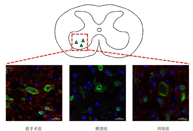
| 组别 | n | 轴突(a.u. /mm2) | NeuN/BDA(个/mm2) |
|---|---|---|---|
| 假手术组 | 7 | 267325.286±97575.678 | 17.143±4.451 |
| 模型组 | 7 | 768600.857±86511.326 | 19.143±4.811 |
| 训练组 | 7 | 1861362.762±771881.369a,b | 33.286±4.990a,b |
| F值 | 32.019 | 23.964 | |
| P值 | < 0.001 | < 0.001 |

"
"
| 组别 | n | Synapsin I(a.u. /mm2) |
|---|---|---|
| 假手术组 | 7 | 35.545±2.903 |
| 模型组 | 7 | 15.760±1.469a |
| 训练组 | 7 | 25.080±1.812a,b |
| F值 | 148.354 | |
| P值 | < 0.001 |
| [1] |
Kumar R, Lim J, Mekary R A, et al. Traumatic spinal injury: global epidemiology and worldwide volume[J]. World Neurosurg, 2018, 113:e345-e363.
doi: 10.1016/j.wneu.2018.02.033 |
| [2] |
Liu G, Keeler B E, Zhukareva V, et al. Cycling exercise affects the expression of apoptosis-associated microRNAs after spinal cord injury in rats[J]. Exp Neurol, 2010, 226(1):200-206.
doi: 10.1016/j.expneurol.2010.08.032 |
| [3] |
Keeler B E, Liu G, Siegfried R N, et al. Acute and prolonged hindlimb exercise elicits different gene expression in motoneurons than sensory neurons after spinal cord injury[J]. Brain Res, 2012, 1438:8-21.
doi: 10.1016/j.brainres.2011.12.015 |
| [4] | 李萌, 陈银海, 张慧, 等. 早期运动训练对脊髓损伤大鼠后肢运动功能影响及相关机制研究[J]. 中国康复医学杂志, 2015, 30(4):318-323. |
| Li M, Chen Y H, Zhang H, et al. Effects of early exercise training on hind limbs motor function in rats after spinal cord injury and the related mechanism[J]. Chin J Rehabil Med, 2015, 30(4):318-323. | |
| [5] |
Sachdeva R, Theisen C C, Ninan V, et al. Exercise dependent increase in axon regeneration into peripheral nerve grafts by propriospinal but not sensory neurons after spinal cord injury is associated with modulation of regeneration-associated genes[J]. Exp Neurol, 2016, 276:72-82.
doi: 10.1016/j.expneurol.2015.09.004 pmid: 26366525 |
| [6] |
Liu G, Detloff M R, Miller K N, et al. Exercise modulates microRNAs that affect the PTEN/mTOR pathway in rats after spinal cord injury[J]. Exp Neurol, 2012, 233(1):447-456.
doi: 10.1016/j.expneurol.2011.11.018 |
| [7] |
Wang H, Liu N K, Zhang Y P, et al. Treadmill training induced lumbar motoneuron dendritic plasticity and behavior recovery in adult rats after a thoracic contusive spinal cord injury[J]. Exp Neurol, 2015, 271:368-378.
doi: 10.1016/j.expneurol.2015.07.004 |
| [8] |
Sandrow-Feinberg H R, Houle J D. Exercise after spinal cord injury as an agent for neuroprotection, regeneration and rehabilitation[J]. Brain Res, 2015, 1619:12-21.
doi: 10.1016/j.brainres.2015.03.052 pmid: 25866284 |
| [9] | 刘鹏民, 李灵玲, 王良, 等. 督脉电针结合游泳训练对大鼠全横断脊髓损伤后GAP-43和Nogo-A表达的影响[J]. 中国康复医学杂志, 2016, 31(4):399-404. |
| Liu P M, Li L L, Wang L, et al. Effects of Du Meridian Electroacupuncture combined with swimming training on the expression of Nogo-A and GAP-43 aftert acute complete spinal cord injury in rats[J]. Chin J Rehabil Med, 2016, 31(4):399-404. | |
| [10] | 周治来, 俞洁, 黄子祥, 等. α-硫辛酸对大鼠全横断脊髓损伤后GAP-43和Caspase-3表达的影响[J]. 神经解剖学杂志, 2017, 33(4):397-402. |
| Zhou Z L, Yu J, Huang Z X, et al. Effects of α-lipoic acid on the expression of GAP-43 and Caspase-3 after spinal cord injury in rats[J]. Chin J Neuroanat, 2017, 33(4):397-402. | |
| [11] | 冯杰扬, 陈杨葭, 郭磊, 等. 减重步行训练对不完全脊髓损伤大鼠脚桥核可塑性影响的研究[J]. 中国康复医学杂志, 2017, 32(6):624-630. |
| Feng J Y, Chen Y J, Guo L, et al. Research on the plasticity of pedunculopontine tegmental nucleus after body weight supported treadmill training in incomplete spinal cord injury rats[J]. Chin J Rehabil Med, 2017, 32(6):624-630. | |
| [12] | 丁洁, 李向哲, 方露, 等. 阻断BDNF-Trk B信号通路后运动训练对脊髓损伤后大鼠痉挛状态及腰髓内GAD65表达的影响[J]. 中国康复医学杂志, 2019, 34(5):501-507. |
| Ding J, Li X Z, Fang L, et al. Effects of exercise training on the expression of GAD65 after blocking BDNF-TrkB pathway in spastic rats with spinal cord injury[J]. Chin J Rehabil Med, 2019, 34(5):501-507. | |
| [13] |
Torres-Espin A, Beaudry E, Fenrich K, et al. Rehabilitative training in animal models of spinal cord injury[J]. J Neurotrauma, 2018, 35(16):1970-1985.
doi: 10.1089/neu.2018.5906 |
| [14] |
Wahl A S, Omlor W, Rubio J C, et al. Asynchronous therapy restores motor control by rewiring of the rat corticospinal tract after stroke[J]. Science, 2014, 344(6189):1250-1255.
doi: 10.1126/science.1253050 |
| [15] |
Torres-Espin A, Forero J, Fenrich K K, et al. Eliciting inflammation enables successful rehabilitative training in chronic spinal cord injury[J]. Brain, 2018, 141(7):1946-1962.
doi: 10.1093/brain/awy128 pmid: 29860396 |
| [16] | 谭波涛, 刘捷, 虞乐华, 等. 成年小鼠颈5脊髓钳夹损伤模型的制备与评价[J]. 中国脊柱脊髓杂志, 2019, 29(2):164-169. |
| Tan B T, Liu J, Yu L H, et al. Establishment and evaluation of C5 dorsal spinal cord crush injury model in adult mice[J]. Chin J Spine Spinal Cord, 2019, 29(2):164-169. | |
| [17] |
Richards T M, Sharma P, Kuang A, et al. Novel speed-controlled automated ladder walking device reveals walking speed as a critical determinant of skilled locomotion after a spinal cord injury in adult rats[J]. J Neurotrauma, 2019, 36(18):2698-2721.
doi: 10.1089/neu.2018.6152 |
| [18] |
Hilton B J, Assinck P, Duncan G J, et al. Dorsolateral funiculus lesioning of the mouse cervical spinal cord at C4 but not at C6 results in sustained forelimb motor deficits[J]. J Neurotrauma, 2013, 30(12):1070-1083.
doi: 10.1089/neu.2012.2734 |
| [19] |
Jin D, Liu Y, Sun F, et al. Restoration of skilled locomotion by sprouting corticospinal axons induced by co-deletion of PTEN and SOCS3[J]. Nat Commun, 2015, 6:8074.
doi: 10.1038/ncomms9074 |
| [20] |
Zhang Q, Wu J F, Shi Q L, et al. The neuronal activation of deep cerebellar nuclei is essential for environmental enrichment-induced post-stroke motor recovery[J]. Aging Dis, 2019, 10(3):530-543.
doi: 10.14336/AD.2018.1220 pmid: 31164998 |
| [21] |
Weishaupt N, Li S, Di Pardo A, et al. Synergistic effects of BDNF and rehabilitative training on recovery after cervical spinal cord injury[J]. Behav Brain Res, 2013, 239:31-42.
doi: 10.1016/j.bbr.2012.10.047 pmid: 23131414 |
| [22] |
Wiersma A M, Fouad K, Winship I R. Enhancing spinal plasticity amplifies the benefits of rehabilitative training and improves recovery from stroke[J]. J Neurosci, 2017, 37(45):10983-10997.
doi: 10.1523/JNEUROSCI.0770-17.2017 |
| [23] |
Adkins D L, Jones T A. D-amphetamine enhances skilled reaching after ischemic cortical lesions in rats[J]. Neurosci Lett, 2005, 380(3):214-218.
doi: 10.1016/j.neulet.2005.01.036 |
| [24] |
MacLellan C L, Gyawali S, Colbourne F. Skilled reaching impairments follow intrastriatal hemorrhagic stroke in rats[J]. Behav Brain Res, 2006, 175(1):82-89.
pmid: 16956678 |
| [25] |
Vergara-Aragon P, Gonzalez C. A novel skilled-reaching impairment in paw supination on the "good" side of the hemi-parkinson rat improved with rehabilitation[J]. J Neurosci, 2003, 23(2):579-586.
doi: 10.1523/JNEUROSCI.23-02-00579.2003 |
| [26] |
Stackhouse S K, Murray M, Shumsky J S. Effect of cervical dorsolateral funiculotomy on reach-to-grasp function in the rat[J]. J Neurotrauma, 2008, 25(8):1039-1047.
doi: 10.1089/neu.2007.0419 |
| [27] |
Parmiani P, Lucchetti C, Bonifazzi C, et al. A kinematic study of skilled reaching movement in rat[J]. J Neurosci Methods, 2019, 328:108404.
doi: 10.1016/j.jneumeth.2019.108404 |
| [28] |
Maier I C, Baumann K, Thallmair M, et al. Constraint-induced movement therapy in the adult rat after unilateral corticospinal tract injury[J]. J Neurosci, 2008, 28(38):9386-9403.
doi: 10.1523/JNEUROSCI.1697-08.2008 |
| [29] |
Goldshmit Y, Lythgo N, Galea M P, et al. Treadmill training after spinal cord hemisection in mice promotes axonal sprouting and synapse formation and improves motor recovery[J]. J Neurotrauma, 2008, 25(5):449-465.
doi: 10.1089/neu.2007.0392 |
| [30] | Smith A C, Knikou M. A review on locomotor training after spinal cord injury: reorganization of spinal neuronal circuits and recovery of motor function[J]. Neural Plast, 2016, 2016:1216258. |
| [31] |
Ichiyama R M, Broman J, Roy R R, et al. Locomotor training maintains normal inhibitory influence on both alpha- and gamma-motoneurons after neonatal spinal cord transection[J]. J Neurosci, 2011, 31(1):26-33.
doi: 10.1523/JNEUROSCI.6433-09.2011 |
| [32] |
Ilha J, Centenaro L A, Broetto Cunha N, et al. The beneficial effects of treadmill step training on activity-dependent synaptic and cellular plasticity markers after complete spinal cord injury[J]. Neurochem Res, 2011, 36(6):1046-1055.
doi: 10.1007/s11064-011-0446-x |
| [33] |
Khalki L, Sadlaoud K, Lerond J, et al. Changes in innervation of lumbar motoneurons and organization of premotor network following training of transected adult rats[J]. Exp Neurol, 2018, 299(Pt A):1-14.
doi: 10.1016/j.expneurol.2017.09.002 |
| [1] | CHEN Junwen, CHEN Qian, CHEN Cheng, LI Shuyue, LIU Lingling, WU Cunshu, GONG Xiang, LU Jun, XU Guangxu. Effect of modified Baduanjin exercise on cardiopulmonary function, motor function and activities of daily living for stroke patients [J]. 《Chinese Journal of Rehabilitation Theory and Practice》, 2024, 30(1): 74-80. |
| [2] | LIU Dong, XU Zihan, LI Jiang, JU Ping. Effect of high-frequency repetitive transcranial magnetic stimulation in M1 region combined with dorsolateral prefrontal cortex on electroencephalogram θ frequency band amplitude of patients with neuropathic pain after spinal cord injury [J]. 《Chinese Journal of Rehabilitation Theory and Practice》, 2024, 30(1): 87-94. |
| [3] | SUN Tengfang, REN Mengting, YANG Lin, WANG Yaoting, WANG Hongyu, YAN Xingzhou. Effect of hyperbaric oxygen therapy combined with repetitive peripheral magnetic stimulation on ankle motor function and balance of stroke patients [J]. 《Chinese Journal of Rehabilitation Theory and Practice》, 2023, 29(8): 875-881. |
| [4] | WANG Ya'nan, LIU Xihua. Correlation and predictive effect of subjective and objective balance function measurements in stroke patients with hemiplegia [J]. 《Chinese Journal of Rehabilitation Theory and Practice》, 2023, 29(8): 890-895. |
| [5] | WANG Haiyun, WANG Yin, ZHOU Xinjie, HE Aiqun. Effect of transcranial direct current stimulation combined with acupuncture on central and upper limb function in stroke patients based on central-peripheral-central theory [J]. 《Chinese Journal of Rehabilitation Theory and Practice》, 2023, 29(8): 919-925. |
| [6] | CHEN Yiting, WANG Qian, CUI Shenhong, LI Yingcai, ZHANG Siyu, WEI Yanxu, REN Hui, LENG Jun, CHEN Bin. Effect of bilateral sequential repetitive transcranial magnetic stimulation on motor function of upper limbs in stroke patients [J]. 《Chinese Journal of Rehabilitation Theory and Practice》, 2023, 29(8): 926-932. |
| [7] | LI Fang, HUO Su, DU Jubao, LIU Xiuzhen, LI Xiaoshuang, SONG Weiqun. Effect of transcranial direct current stimulation combined with task-oriented rehabilitation training on forelimb motor dysfunction in rats with spinal cord injury [J]. 《Chinese Journal of Rehabilitation Theory and Practice》, 2023, 29(7): 777-781. |
| [8] | CUI Yao, CONG Fang, HUANG Fubiao, ZENG Ming, YAN Ruxiu. Brain and muscle activation under mirror neuron-based training strategies: a near-infrared spectroscopy and surface electromyography study [J]. 《Chinese Journal of Rehabilitation Theory and Practice》, 2023, 29(7): 782-790. |
| [9] | LIU Ning, LIU Yuquan, ZHU Bin, YU Lingjia, TAN Haining, YANG Yong, LI Xiang. Application of International Standards for Neurological Classification of Spinal Cord Injury in China: a bibliometrics re-analysis [J]. 《Chinese Journal of Rehabilitation Theory and Practice》, 2023, 29(7): 808-815. |
| [10] | LIU Hui, YIN Hang, JIA Shaohui, QIU Fubing. Structure, contents and psychometric properties of measurement of motor function and motor ability applicable to children and adolescents with disabilities: a systematic review [J]. 《Chinese Journal of Rehabilitation Theory and Practice》, 2023, 29(6): 630-638. |
| [11] | WANG Yiji, ZHOU Hongjun, HE Zejia, LIU Genlin, ZHENG Ying, HAO Chunxia, WEI Bo, KANG Haiqiong, ZHANG Ying, LU Xiaolei, YUAN Yuan, MENG Qianru. Relationship between symmetry of lower limb function and gait symmetry in patients with incomplete spinal cord injury [J]. 《Chinese Journal of Rehabilitation Theory and Practice》, 2023, 29(6): 639-645. |
| [12] | XU Miaomiao, LI Nan, YING Ying, YANG Kaixiang, YANG Jingrui, LI Jie, QIU Yanqun. Effect of repetitive peripheral magnetic stimulation on upper limb motor function of stroke patients after contralateral seventh cervical nerve transfer [J]. 《Chinese Journal of Rehabilitation Theory and Practice》, 2023, 29(6): 686-690. |
| [13] | ZHENG Li, BAO Zhicheng, ZHANG Qi, REN Xuyan, SU Min. Effect of transcutaneous auricular vagus nerve stimulation combined with robot-assisted therapy on upper limb function of stroke patients [J]. 《Chinese Journal of Rehabilitation Theory and Practice》, 2023, 29(6): 691-696. |
| [14] | ZHANG Qian, SUN Xinting. Effect of video-based mirror therapy on lower limb motor function of stroke patients at recovery stage [J]. 《Chinese Journal of Rehabilitation Theory and Practice》, 2023, 29(6): 703-707. |
| [15] | LIN Yufan, WEI Tianyuan, ZHANG Xiaoying, LI Chaojinzi, HE Jingjie, DU Xiaoxia. Effect of music therapy on post-stroke cognitive impairment [J]. 《Chinese Journal of Rehabilitation Theory and Practice》, 2023, 29(6): 714-719. |
| Viewed | ||||||
|
Full text |
|
|||||
|
Abstract |
|
|||||
|
||