


《Chinese Journal of Rehabilitation Theory and Practice》 ›› 2021, Vol. 27 ›› Issue (7): 785-790.doi: 10.3969/j.issn.1006-9771.2021.00.003
Previous Articles Next Articles
DAI Yan-hong1a,HUANG Jia-ying1b,LUO Hai-long1b,WANG Rui-qing1a,HUANG Ying-an1a,CHEN Zhuo-ming1a,WANG Hong1a,2( )
)
Received:2021-03-16
Revised:2021-05-12
Published:2021-07-25
Online:2021-07-28
Contact:
WANG Hong
E-mail:daxiaobaotwins@126.com
Supported by:CLC Number:
DAI Yan-hong, HUANG Jia-ying, LUO Hai-long, WANG Rui-qing, HUANG Ying-an, CHEN Zhuo-ming, WANG Hong. Changes in Gray Matter Volume in Chronic Nonfluent Aphasia after Cortical Cerebral Infarction[J]. 《Chinese Journal of Rehabilitation Theory and Practice》, 2021, 27(7): 785-790.
"
| 特征 | 对照组 (n = 28) | 患者组 (n = 19) | χ2/t/Z值 | P值 |
|---|---|---|---|---|
| 性别(男/女, n) | 15/13 | 12/7 | 0.426 | 0.514a |
| 年龄(岁) | 54.86±4.66 | 55.21±5.57 | 0.231 | 0.353b |
| 教育程度(年) | 9.46±2.36 | 9.55±2.68 | 0.242 | 0.821c |
| 病程(月) | – | 8.68±2.12 |
"
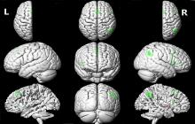
| 脑区 | 体素数 | MNI坐标 | t值 | ||
|---|---|---|---|---|---|
| X | Y | Z | |||
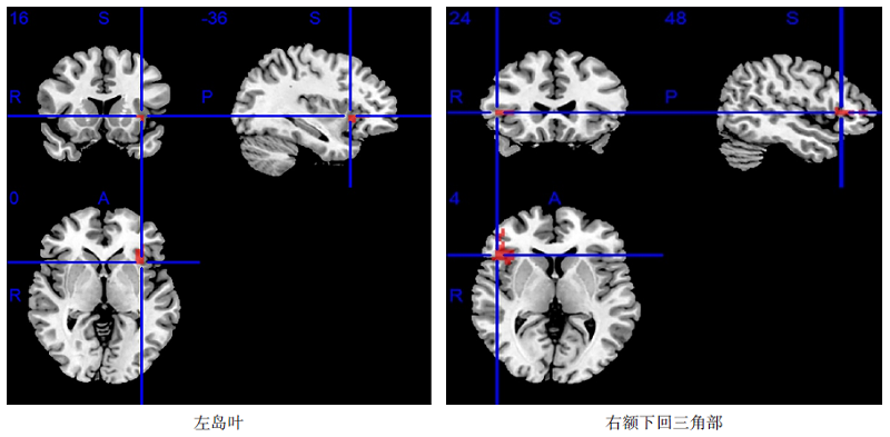
| 左岛叶 | 22 | -36 | 16 | 0 | -4.229 |
| 左内侧额上回 | 104 | -4 | 16 | 52 | -5.158 |
| 右额下回三角部 | 33 | 48 | 24 | 4 | -5.046 |
| 右岛盖部额下回 | 11 | 52 | 12 | 24 | -3.784 |
| 右角回 | 53 | 52 | -56 | 36 | -5.144 |
| 右内侧额上回 | 104 | 4 | 16 | 52 | -5.158 |
| 右侧苍白球 | 22 | 28 | -8 | -4 | 4.718 |
"
"
| [1] |
ARAS B, İNAL Ö, KESIKBURUN S, et al. Response to speech and language therapy according to artery involvement and lesion location in post-stroke aphasia[J]. J Stroke Cerebrovasc Dis, 2020, 29(10):105132.
doi: 10.1016/j.jstrokecerebrovasdis.2020.105132 |
| [2] |
KAPOOR A. Repetitive transcranial magnetic stimulation therapy for post-stroke non-fluent aphasia: a critical review[J]. Top Stroke Rehabil, 2017, 24(7):547-553.
doi: 10.1080/10749357.2017.1331417 |
| [3] |
STEFANIAK J D, HALAI A D, LAMBON RALPH M A. The neural and neurocomputational bases of recovery from post-stroke aphasia[J]. Nat Rev Neurol, 2020, 16(1):43-55.
doi: 10.1038/s41582-019-0282-1 |
| [4] |
BREINING B L, SEBASTIAN R. Neuromodulation in post-stroke aphasia treatment[J]. Curr Phys Med Rehabil Rep, 2020, 8(2):44-56.
doi: 10.1007/s40141-020-00257-5 |
| [5] |
NAKASHIMA A, MORIUCHI T, MITSUNAGA W, et al. Prediction of prognosis of upper extremity function following stroke-related paralysis using brain imaging[J]. J Phys Ther Sci, 2017, 29(8):1438-1443.
doi: 10.1589/jpts.29.1438 |
| [6] |
TORBEY M T, BOSEL J, RHONEY D H, et al. Evidence-based guidelines for the management of large hemispheric infarction: a statement for health care professionals from the Neurocritical Care Society and the German Society for Neuro intensive Care and Emergency Medicine[J]. Neurocrit Care, 2015, 22(1):146-164.
doi: 10.1007/s12028-014-0085-6 |
| [7] | 余晓凤, 陈芹, 高晴, 等. 颞叶癫痫患者语言网络基于Granger因果检验的功能磁共振特点[J]. 中华神经科杂志, 2014, 47(8):537-541. |
| YU X F, CHEN Q, GAO Q, et al. Evaluating the connectivity within the language network in temporal lobe epilepsy using Granger causality analysis: a functional magnetic resonance imaging study[J]. Chin J Neurol, 2014, 47(8):537-541. | |
| [8] |
JAKAB A, MOLNÁR P P, BOGNER P, et al. Connectivity-based parcellation reveals interhemispheric differences in the insula[J]. Brain Topogr, 2012, 25(3):264-271.
doi: 10.1007/s10548-011-0205-y |
| [9] |
OH A, DUERDEN E G, PANG E W. The role of the insula in speech and language processing[J]. Brain Lang, 2014, 135:96-103.
doi: 10.1016/j.bandl.2014.06.003 |
| [10] |
ARDILA A, BERNAL B, ROSSELLI M. Participation of the insula in language revisited: a meta-analytic connectivity study[J]. J Neurolinguistics, 2014, 29:31-41.
doi: 10.1016/j.jneuroling.2014.02.001 |
| [11] |
NORISE C, HAMILTON R H. Non-invasive brain stimulation in the treatment of post-stroke and neurodegenerative aphasia: parallels, differences, and lessons learned[J]. Front Hum Neurosci, 2016, 10:675.
doi: 10.3389/fpsyg.2019.00675 |
| [12] |
LOUGHNAN R, LORCA-PULS D L, GAJARDO-VIDAL A, et al. Generalizing post-stroke prognoses from research data to clinical data[J]. Neuroimage Clin, 2019, 24:102005.
doi: 10.1016/j.nicl.2019.102005 |
| [13] |
SREEDHARAN S, CHANDRAN A, YANAMALA V R, et al. Self-regulation of language areas using real-time functional MRI in stroke patients with expressive aphasia[J]. Brain Imaging Behav, 2020, 14(5):1714-1730.
doi: 10.1007/s11682-019-00106-7 |
| [14] |
HARTWIGSEN G, SAUR D, PRICE C J, et al. Perturbation of the left inferior frontal gyrus triggers adaptive plasticity in the right homologous area during speech production[J]. Proc Natl Acad Sci, 2013, 110(41):16402-16407.
doi: 10.1073/pnas.1310190110 |
| [15] |
TURKELTAUB P E. Brain stimulation and the role of the right hemisphere in aphasia recovery[J]. Curr Neurol Neurosci Rep, 2015, 15(11):72.
doi: 10.1007/s11910-015-0593-6 |
| [16] |
ROBSON H, ZAHN R, KEIDEL J L, et al. The anterior temporal lobes support residual comprehension in Wernicke's aphasia[J]. Brain, 2014, 137(Pt3):931-943.
doi: 10.1093/brain/awt373 |
| [17] |
JOHNSON J P, MEIER E L, PAN Y, et al. Treatment-related changes in neural activation vary according to treatment response and extent of spared tissue in patients with chronic aphasia[J]. Cortex, 2019, 121:147-168.
doi: 10.1016/j.cortex.2019.08.016 |
| [18] |
ABEL S, WEILLER C, HUBER W, et al. Therapy-induced brain reorganization patterns in aphasia[J]. Brain, 2015, 138(Pt 4):1097-1112.
doi: 10.1093/brain/awv022 |
| [19] | REN C, ZHANG G, XU X, et al. The effect of rTMS over the different targets on language recovery in stroke patients with global aphasia: a randomized sham-controlled study[J]. Biomed Res Int, 2019, 2019:4589056. |
| [20] |
HARVEY D Y, PODELL J, TURKELTAUB P E, et al. Functional reorganization of right prefrontal cortex underlies sustained naming improvements in chronic aphasia via repetitive transcranial magnetic stimulation[J]. Cogn Behav Neurol, 2017, 30(4):133-144.
doi: 10.1097/WNN.0000000000000141 |
| [21] |
ROSSETTI A, MALFITANO C, MALLOGGI C, et al. Phonemic fluency improved after inhibitory transcranial magnetic stimulation in a case of chronic aphasia[J]. Int J Rehabil Res, 2019, 42(1):92-95.
doi: 10.1097/MRR.0000000000000322 |
| [22] |
HAGHIGHI M, MAZDEH M, RANJBAR N, et al. Further evidence of the positive influence of repetitive transcranial magnetic stimulation on speech and language in patients with aphasia after stroke: results from a double-blind intervention with sham condition[J]. Neuropsychobiology, 2017, 75(4):185-192.
doi: 10.1159/000486144 |
| [23] | 邱国荣, 丘卫红, 邹艳, 等. 重复经颅磁刺激对卒中后失语语言功能重组的影响:基于功能磁共振的研究[J]. 中国康复理论与实践, 2018, 24(6):686-695. |
| QIU G R, QIU W H, ZOU Y, et al. Effect of repeated transcranial magnetic stimulation on reorganization of aphasia after stroke: a study based on functional magnetic resonance imaging[J]. Chin J Rehabil Theory Pract, 2018, 24(6):686-695. | |
| [24] |
HEIKKINEN P H, PULVERMULLER F, MÄKELÄ J P, et al. Combining rTMS with intensive language-action therapy in chronic aphasia: a randomized controlled trial[J]. Front Neurosci, 2019, 12:1036.
doi: 10.3389/fnins.2018.01036 |
| [25] | AL-JANABI S, NICKELS L A, SOWMAN P F, et al. Augmenting melodic intonation therapy with non-invasive brain stimulation to treat impaired left-hemisphere function: two case studies[J]. Front Psychol, 2014, 5:37. |
| [26] |
NARDO D, HOLLAND R, LEFF A P, et al. Less is more: neural mechanisms underlying anomia treatment in chronic aphasic patients[J]. Brain, 2017, 140(11):3039-3054.
doi: 10.1093/brain/awx234 |
| [27] |
CROSSON B, RODRIGUEZ A D, COPLAND D, et al. Neuroplasticity and aphasia treatments: new approaches for an old problem[J]. J Neurol Neurosurg Psychiatry, 2019, 90(10):1147-1155.
doi: 10.1136/jnnp-2018-319649 |
| [28] |
FORKEL S J, THIEBAUT DE SCHOTTEN M, DELL'ACQUA F, et al. Anatomical predictors of aphasia recovery: a tractography study of bilateral perisylvian language networks[J]. Brain, 2014, 137(Pt 7):2027-2039.
doi: 10.1093/brain/awu113 |
| [29] |
PANI E, ZHENG X, WANG J, et al. Right hemisphere structures predict poststroke speech fluency[J]. Neurology, 2016, 86(17):1574-1581.
doi: 10.1212/WNL.0000000000002613 |
| [30] |
HOPE T M H, LEFF A P, PREJAWA S, et al. Right hemisphere structural adaptation and changing language skills years after left hemisphere stroke[J]. Brain, 2017, 140(6):1718-1728.
doi: 10.1093/brain/awx086 |
| [31] | LUKIC S, BARBIERI E, WANG X, et al. Right hemisphere grey matter volume and language functions in stroke aphasia[J]. Neural Plast, 2017, 2017:1-14. |
| [32] |
XING S, LACEY E H, SKIPPER-KALLAL L M, et al. Right hemisphere grey matter structure and language outcomes in chronic left hemisphere stroke[J]. Brain, 2016, 139(Pt1):227-241.
doi: 10.1093/brain/awv323 |
| [33] |
BRODTMANN A, PARDOE H, LI Q, et al. Changes in regional brain volume three months after stroke[J]. J Neurol Sci, 2012, 322(1-2):122-128.
doi: 10.1016/j.jns.2012.07.019 |
| [34] |
GAUTHIER L V, TAUB E, MARK V W, et al. Atrophy of spared gray matter tissue predicts poorer motor recovery and rehabilitation response in chronic stroke[J]. Stroke, 2012, 43(2):453-457.
doi: 10.1161/STROKEAHA.111.633255 |
| [35] | KERR A L, CHENG S Y, JONES T A. Experience-dependent neural plasticity in the adult damaged brain[J]. J Commun Disord, 2011, 44(5):538-548. |
| [36] |
HARTWIGSEN G, SAUR D. Neuroimaging of stroke recovery from aphasia: insights into plasticity of the human language network[J]. Neuroimage, 2019, 190:14-31.
doi: 10.1016/j.neuroimage.2017.11.056 |
| [37] |
VOLZ L J, VOLLMER M, MICHELY J, et al. Time-dependent functional role of the contralesional motor cortex after stroke[J]. Neuroimage Clin, 2017, 16:165-174.
doi: 10.1016/j.nicl.2017.07.024 |
| [1] | JIANG Xiaocui, LIU Zhen, SU Qinglun, ZHAO Qin, XIA Xiaomei, LU Fei. Effect of intermittent theta burst transcranial magnetic stimulation on non-fluent aphasia after stroke [J]. 《Chinese Journal of Rehabilitation Theory and Practice》, 2023, 29(7): 839-843. |
| [2] | XU Minjie, WANG Bo, ZHOU Li, WANG Haifang, LEI Xiaojing, LI Ying, BAO Weiwei, MA Ya'nan, CHANG Jingling. Verbal and nonverbal cognitive function of aphasia after stroke based on Web of Science database: a visualized analysis [J]. 《Chinese Journal of Rehabilitation Theory and Practice》, 2023, 29(4): 452-464. |
| [3] | HU Xueyan, JIANG Xiaofeng, SHAN Lei, YANG Lingyu, CHEN Yudong, MA Lin, LIU Lixu, ZHANG Tong. Effect of low frequency or high frequency repetitive transcranial magnetic stimulation on stroke patients with nonfluent aphasia [J]. 《Chinese Journal of Rehabilitation Theory and Practice》, 2023, 29(3): 249-255. |
| [4] | CHEN Yating,ZHANG Jie,ZHANG Youmei,ZHANG Shuangshuang,YE Xiangming. Effects transcranial direct current stimulation on post-stroke aphasia: a systematic review [J]. 《Chinese Journal of Rehabilitation Theory and Practice》, 2022, 28(5): 534-543. |
| [5] | ZHOU Yufan,XU Minjie,TAN Yihai,MA Ya'nan,REN Qiaosheng,CHEN Jian,ZHANG Qingsu,WANG Bo,HE Yi,CHANG Jingling. Characteristics of post-stroke aphasia structural damage based on structural covariance network [J]. 《Chinese Journal of Rehabilitation Theory and Practice》, 2022, 28(10): 1198-1204. |
| [6] | Xiao-lin LI,Min-jie XU,Yun CAO,Dan-li ZHANG,Xin SHU,Chang-ming LI,Yu-fan ZHOU,Yi-hai TAN,Zhong-jian TAN,Jing-ling CHANG. Frequency-dependent Alterations in Amplitude of Low-frequency Fluctuations in Resting-state Functional Magnetic Resonance Imaging of Post Stroke Aphasia [J]. 《Chinese Journal of Rehabilitation Theory and Practice》, 2021, 27(5): 497-503. |
| [7] | Qiong-fen WANG,Feng-bo WANG,Ke WANG,Yong-qiang ZHONG,Jiao-jiao WANG. Effect of Electroacupuncture at Fengchi on Astrocytes and Neurons in Rats with Acute Cerebral Infarction [J]. 《Chinese Journal of Rehabilitation Theory and Practice》, 2021, 27(3): 302-309. |
| [8] | Xin-yu ZHANG,Xiao-zheng DU,Jin-hai WANG,Wen-jie HE,Na-na WANG,Ruo-zhou WANG,Wei-yao JING,Shang-wei YANG. Advance of Netrin-1 for Protection and Repairment of Injuries after Cerebral Infarction (review) [J]. 《Chinese Journal of Rehabilitation Theory and Practice》, 2021, 27(3): 316-319. |
| [9] | ZHANG Xiao-tong,LI Na,CHEN Zhao-cong,LIANG Jing-feng,YU Yong,WU Hui-xiang,KANG Zhuang,QIU Wei-hong. Potential Role of Right Cerebellum in Post-stroke Aphasia: A Preliminary Study Based on Granger Causality Analysis [J]. 《Chinese Journal of Rehabilitation Theory and Practice》, 2021, 27(12): 1458-1463. |
| [10] | YANG Fu-xia,HOU Dong-mei,GAO Jin-yun,GU Mei,ZHAO Bo-feng,ZENG Xu-mei,ZHAN Min-min,XIE Min-jiao,XIE Yao-qin. Effects of Acupuncture on Motor and Corticospinal Tract Impairment after Cerebral Infarction: Using Diffusion Tensor Imaging and Tractography [J]. 《Chinese Journal of Rehabilitation Theory and Practice》, 2021, 27(11): 1312-1317. |
| [11] | SHU Xin,DONG Xing-lu,HAO Xiao-hui,WEI Dong-jie,HUANG Xing,LI Xiao-lin,XU Min-jie,LI Chang-ming,KONG Qiao,HUANG Jia-qin,LIU Meng-yu,CHANG Jing-ling. Development of A Patient-reported Outcomes Scale for Post-stroke Aphasia Based on Traditional Chinese Medicine Holism [J]. 《Chinese Journal of Rehabilitation Theory and Practice》, 2021, 27(11): 1356-1364. |
| [12] | WANG Bo,ZHANG Qing-su,ZHANG Hao. Effect of Oral Reading Training on Post-stroke Alexia [J]. 《Chinese Journal of Rehabilitation Theory and Practice》, 2020, 26(7): 820-824. |
| [13] | LI Xiao-lin,ZHANG Bin-long,FAN Rui-wen,XU Min-jie,HUANG Xing,SHU Xin,LI Chang-ming,TAN Zhong-jian,CHANG Jing-ling. Stimulation Mode and Model of Word-picture Language Task in Functional Magnetic Resonance Imaging Test for Post-stroke Aphasia (review) [J]. 《Chinese Journal of Rehabilitation Theory and Practice》, 2020, 26(6): 668-672. |
| [14] | FAN Rui-wen,HUANG Xing,LI Xiao-lin,YAN He-ming,SHU Xin,KONG Qiao,LI Chang-ming,CHANG Jing-ling. Characteristics of Electroencephalogram Power Spectrum Network on Uninjured Side of Post-stroke Aphasia [J]. 《Chinese Journal of Rehabilitation Theory and Practice》, 2020, 26(6): 692-696. |
| [15] | GUO Lin-jia,CHEN Jing,GAO Feng-lian,ZHANG Yun,LIU Wen-hong. Clinical Features of Trousseau's Syndrome with Acute Multiple Cerebral Infarction as the First Manifestation [J]. 《Chinese Journal of Rehabilitation Theory and Practice》, 2020, 26(6): 730-737. |
| Viewed | ||||||||||||||||||||||||||||||||||||||||||||||||||
|
Full text 329
|
|
|||||||||||||||||||||||||||||||||||||||||||||||||
|
Abstract 598
|
|
|||||||||||||||||||||||||||||||||||||||||||||||||
|
||