


《Chinese Journal of Rehabilitation Theory and Practice》 ›› 2021, Vol. 27 ›› Issue (4): 436-444.doi: 10.3969/j.issn.1006-9771.2021.04.008
Previous Articles Next Articles
Hao-jie ZHANG1,2,3,Yun-lei WANG1,2,3,Ling-zhong FAN4,5,Fang LI1,2,3,Jing-ya LIU1,3,Shao-hong YU6,7,Yuan-yuan HOU1,2,3,Chen BAI1,2,3,Bing-jie LI1,3,Xiao-xia DU1,3,Tong ZHANG1,2,3( )
)
Received:2021-01-15
Revised:2021-03-10
Published:2021-04-25
Online:2021-04-20
Contact:
Tong ZHANG
E-mail:tom611@126.com
Supported by:CLC Number:
Hao-jie ZHANG,Yun-lei WANG,Ling-zhong FAN,Fang LI,Jing-ya LIU,Shao-hong YU,Yuan-yuan HOU,Chen BAI,Bing-jie LI,Xiao-xia DU,Tong ZHANG. Differences of Structural Plasticity between Hemispheres during Rehabilitation for Subacute Stroke[J]. 《Chinese Journal of Rehabilitation Theory and Practice》, 2021, 27(4): 436-444.
"
| 脑区 | 时间 | 时间×组间 | 组间 | T1 vs. T2 (P值) | T1 vs. T3 (P值) | T2 vs. T3 (P值) | |||
|---|---|---|---|---|---|---|---|---|---|
| F值 | P值 | F值 | P值 | F值 | P值 | ||||
| 皮质表面积 | |||||||||
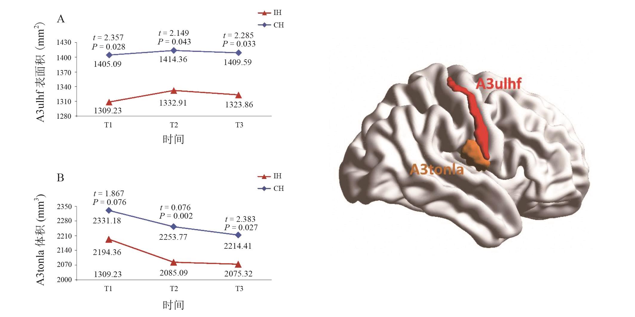
| dmPOS | 4.124 | 0.023 | 0.977 | 0.385 | 0.089 | 0.767 | 0.650 | 1 | 0.026 |
| A3ulhf | 2.382 | 0.105 | 0.347 | 0.709 | 5.397 | 0.025 | 0.391 | 1 | 0.462 |
| OPC | 6.717 | 0.003 | 1.524 | 0.230 | 0.336 | 0.565 | 1 | 0.029a | 0.003a |
| 皮质厚度 | |||||||||
| A10l | 4.330 | 0.020 | 0.058 | 0.944 | 0.311 | 0.580 | 0.357 | 0.024 | 0.094 |
| A44d | 13.968 | < 0.001 | 0.502 | 0.607 | 0.616 | 0.437 | 0.002 | < 0.001 | 0.640 |
| A45c | 10.217 | < 0.001 | 1.617 | 0.205 | 0.464 | 0.499 | 0.026 | < 0.001 | 0.373 |
| A6cvl | 14.513 | < 0.001 | 0.377 | 0.687 | 1.135 | 0.293 | < 0.001 | < 0.001 | 1 |
| TE1.2 | 13.671 | < 0.001 | 0.015 | 0.985 | 0.851 | 0.361 | < 0.001 | < 0.001 | 1 |
| A22r | 4.373 | 0.016 | 0.155 | 0.857 | 0.007 | 0.934 | 0.014 | 0.419 | 0.477 |
| A36r | 3.607 | 0.031 | 0.609 | 0.546 | 0.236 | 0.629 | 0.016a | 0.369 | 1 |
| A34 | 6.459 | 0.002 | 0.255 | 0.775 | 0.072 | 0.790 | 0.052 | 0.006 | 0.772 |
| A7c | 14.891 | < 0.001 | 0.035 | 0.966 | 3.806 | 0.058 | < 0.001 | < 0.001 | 1 |
| A39c | 10.531 | < 0.001 | 0.076 | 0.927 | < 0.001 | 0.983 | 0.004 | < 0.001 | 1 |
| A39rd | 13.161 | < 0.001 | 1.498 | 0.229 | 1.789 | 0.188 | 0.005 | < 0.001 | 0.809 |
| A40c | 10.628 | < 0.001 | 0.823 | 0.443 | 2.431 | 0.126 | 0.001 | 0.001 | 1 |
| A5m | 29.349 | < 0.001 | 2.975 | 0.056 | 1.801 | 0.187 | < 0.001 | < 0.001 | 0.023 |
| dmPOS | 12.465 | < 0.001 | 1.387 | 0.256 | 0.087 | 0.769 | 0.142 | < 0.001 | 0.028 |
| A3ulhf | 7.935 | 0.001 | 1.604 | 0.214 | 0.097 | 0.757 | 0.002 | 0.001 | 1 |
| A3tonIa | 14.043 | < 0.001 | 1.323 | 0.272 | 0.310 | 0.581 | < 0.001 | < 0.001 | 1 |
| A3tru | 7.408 | 0.002 | 0.726 | 0.490 | 0.363 | 0.550 | 0.002 | 0.004 | 0.923 |
| A32p | 6.226 | 0.003 | 0.309 | 0.735 | 0.086 | 0.770 | 0.003 | 0.057 | 0.821 |
| OPC | 8.132 | 0.001 | 0.629 | 0.536 | 0.002 | 0.968 | 0.139 | 0.001 | 0.144 |
| 皮质体积 | |||||||||
| A9m | 6.350 | 0.003 | 1.723 | 0.185 | 0.010 | 0.921 | 0.005 | 0.042 | 1 |
| A10l | 11.527 | < 0.001 | 0.685 | 0.507 | 1.963 | 0.169 | 0.028 | < 0.001 | 0.172 |
| A44d | 16.627 | < 0.001 | 0.064 | 0.938 | 0.630 | 0.432 | 0.001 | < 0.001 | 0.524 |
| A45c | 5.618 | 0.007 | 2.313 | 0.112 | < 0.001 | 0.997 | 0.157 | 0.005 | 0.693 |
| A6cvl | 7.769 | 0.001 | 0.677 | 0.511 | 2.869 | 0.098 | 0.040 | 0.001 | 0.783 |
| TE1.2 | 16.223 | < 0.001 | 0.729 | 0.485 | 0.059 | 0.810 | < 0.001 | < 0.001 | 1 |
| A22r | 3.638 | 0.031 | 0.902 | 0.410 | 0.115 | 0.736 | 0.044 | 0.119 | 1 |
| A36r | 5.805 | 0.004 | 0.177 | 0.838 | 0.001 | 0.980 | 0.007a | 0.947 | 0.06 |
| A34 | 7.155 | 0.001 | 2.087 | 0.130 | 0.110 | 0.742 | 0.290 | 0.001 | 0.148 |
| A7c | 7.16 | 0.001 | 0.578 | 0.563 | 0.108 | 0.744 | 0.001 | 0.054 | 0.963 |
| A39c | 10.815 | < 0.001 | 1.644 | 0.199 | 0.012 | 0.914 | 0.004 | 0.001 | 0.923 |
| A39rd | 8.368 | 0.001 | 0.201 | 0.819 | 0.807 | 0.374 | 0.072 | 0.001 | 0.910 |
| A40c | 7.080 | 0.001 | 0.588 | 0.557 | 0.066 | 0.798 | 0.041 | 0.003 | 1 |
| A5m | 20.528 | < 0.001 | 0.835 | 0.437 | 0.547 | 0.503 | 0.001 | < 0.001 | 0.115 |
| dmPOS | 8.295 | 0.001 | 0.030 | 0.971 | 0.011 | 0.915 | 0.131 | < 0.001 | 0.257 |
| A3ulhf | 9.662 | < 0.001 | 1.151 | 0.321 | 3.176 | 0.082 | 0.013 | 0.001 | 1 |
| A3tonIa | 13.709 | < 0.001 | 0.280 | 0.757 | 4.305 | 0.044 | 0.003 | < 0.001 | 0.886 |
| A3tru | 7.014 | 0.002 | 0.449 | 0.641 | 0.727 | 0.399 | 0.002 | 0.003 | 0.522 |
| A32p | 8.083 | 0.001 | 0.292 | 0.749 | 0.138 | 0.712 | 0.001 | 0.466 | 0.390 |
| OPC | 3.674 | 0.030 | 0.126 | 0.882 | 0.009 | 0.924 | 0.205 | 0.026 | 1 |

"
"
| 脑区 | 脑叶 | 脑回 | lh.MNI (X,Y,Z) | rh.MNI (X, Y, Z) |
|---|---|---|---|---|
| A9m | 额叶 | 额上回 | -5, 36, 38 | 6, 38, 35 |
| A10l | 额中回 | -26, 60, -6 | 25, 61, -4 | |
| A44d | 额下回 | -46, 13, 24 | 45, 16, 25 | |
| A45c | 额下回 | -53, 23, 11 | 54, 24, 12 | |
| A6cvl | 中央前回 | -49, 5, 30 | 51, 7, 30 | |
| TE1.2 | 颞叶 | 颞上回 | -50, -11, 1 | 51, -4, -1 |
| A22r | 颞上回 | -55, -3, -10 | 56, -12, -5 | |
| A36r | 海马旁回 | -27, -7, -34 | 28, -8, -33 | |
| A34 | 海马旁回 | -19, -12, -30 | 19, -10, -30 | |
| A7c | 顶叶 | 顶上小叶 | -15, -71, 52 | 19, -69, 54 |
| A39c | 顶下小叶 | -34, -80, 29 | 45, -71,20 | |
| A39rd | 顶下小叶 | -38, -61, 46 | 39, -65, 44 | |
| A40c | 顶下小叶 | -56, -49, 38 | 57, -44, 38 | |
| A5m | 楔前叶 | -8, -47, 57 | 7, -47, 58 | |
| dmPOS | 楔前叶 | -12, -67, 25 | 16, -64,25 | |
| A3ulhf | 中央后回 | -50, -16, 43 | 50, -14,44 | |
| A3tonIa | 中央后回 | -56, -14, 16 | 56, -10,15 | |
| A3tru | 中央后回 | -21, -35, 68 | 20, -33, 69 | |
| A32p | 边缘叶 | 扣带回 | -6, 34, 21 | 5, 28, 27 |
| OPC | 枕叶 | 枕叶外皮质 | -18, -99, 2 | 22, -97,4 |
"
| 1 | Brewer L, Horgan F, Hickey A, et al. Stroke rehabilitation: recent advances and future therapies [J]. QJM, 2013, 106(1): 11-25. |
| 2 | Wolf S L, Winstein C J, Miller J P, et al. Effect of constraint-induced movement therapy on upper extremity function 3 to 9 months after stroke: the EXCITE randomized clinical trial [J]. JAMA, 2006, 296(17): 2095-2104. |
| 3 | Lo AC, Guarino P D, Richards L G, et al. Robot-assisted therapy for long-term upper-limb impairment after stroke [J]. N Engl J Med, 2010, 362(19): 1772-1783. |
| 4 | McCabe J, Monkiewicz M, Holcomb J, et al. Comparison of robotics, functional electrical stimulation, and motor learning methods for treatment of persistent upper extremity dysfunction after stroke: a randomized controlled trial [J]. Arch Phys Med Rehabil, 2015, 96(6): 981-990. |
| 5 | Bach-y-Rita P. Brain plasticity as a basis for recovery of function in humans [J]. Neuropsychologia, 1990, 28(6): 547-554. |
| 6 | Johansson B B. Brain plasticity and stroke rehabilitation. The Willis lecture [J]. Stroke, 2000, 31(1): 223-230. |
| 7 | Johansen-Berg H, Dawes H, Guy C, et al. Correlation between motor improvements and altered fMRI activity after rehabilitative therapy [J]. Brain, 2002, 125(Pt 12): 2731-2742. |
| 8 | Luft A R, McCombe-Waller S, Whitall J, et al. Repetitive bilateral arm training and motor cortex activation in chronic stroke: a randomized controlled trial [J]. JAMA, 2004, 292(15): 1853-1861. |
| 9 | Pundik S, McCabe J P, Hrovat K, et al. Recovery of post stroke proximal arm function, driven by complex neuroplastic bilateral brain activation patterns and predicted by baseline motor dysfunction severity [J]. Front Hum Neurosci, 2015, 9: 394. |
| 10 | Kwakkel G, van Peppen R, Wagenaar R C, et al. Effects of augmented exercise therapy time after stroke: a meta-analysis [J]. Stroke, 2004, 35(11): 2529-2539. |
| 11 | Kwakkel G, Kollen B, Twisk J. Impact of time on improvement of outcome after stroke [J]. Stroke, 2006, 37(9): 2348-2353. |
| 12 | Dobkin B H, Carmichael S T. The specific requirements of neural repair trials for stroke [J]. Neurorehabil Neural Repair, 2016, 30(5): 470-478. |
| 13 | Cai J, Ji Q, Xin R, et al. Contralesional cortical structural reorganization contributes to motor recovery after sub-cortical stroke: a longitudinal voxel-based morphometry study [J]. Front Hum Neurosci, 2016, 10: 393. |
| 14 | Duering M, Righart R, Wollenweber F A, et al. Acute infarcts cause focal thinning in remote cortex via degeneration of connecting fiber tracts [J]. Neurology, 2015, 84(16): 1685-1692. |
| 15 | Fan F, Zhu C, Chen H, et al. Dynamic brain structural changes after left hemisphere subcortical stroke [J]. Hum Brain Mapp, 2013, 34(8): 1872-1881. |
| 16 | Milot M H, Cramer S C. Biomarkers of recovery after stroke [J]. Curr Opin Neurol, 2008, 21(6): 654-659. |
| 17 | Hosp J A, Luft A R. Cortical plasticity during motor learning and recovery after ischemic stroke [J]. Neural Plast, 2011: 871296. |
| 18 | Stinear C M, Barber P A, Petoe M, et al. The PREP algorithm predicts potential for upper limb recovery after stroke [J]. Brain, 2012, 135(Pt 8): 2527-2535. |
| 19 | Burke E, Cramer S C. Biomarkers and predictors of restorative therapy effects after stroke [J]. Curr Neurol Neurosci Rep, 2013, 13(2): 329. |
| 20 | Byblow W D, Stinear C M, Barber P A, et al. Proportional recovery after stroke depends on corticomotor integrity [J]. Ann Neurol, 2015, 78(6): 848-859. |
| 21 | Zeiler S R, Krakauer J W. The interaction between training and plasticity in the poststroke brain [J]. Curr Opin Neurol, 2013, 26(6): 609-616. |
| 22 | Lin D J, Cloutier A M, Erler K S, et al. Corticospinal tract injury estimated from acute stroke imaging predicts upper extremity motor recovery after stroke [J]. Stroke, 2019, 50(12): 3569-3577. |
| 23 | Cassidy J M, Tran G, Quinlan E B, et al. Neuroimaging identifies patients most likely to respond to a restorative stroke therapy [J]. Stroke, 2018, 49(2): 433-438. |
| 24 | Lam T K, Binns M A, Honjo K, et al. Variability in stroke motor outcome is explained by structural and functional integrity of the motor system [J]. Sci Rep, 2018, 8(1): 9480. |
| 25 | Kolb B, Whishaw I Q. Brain plasticity and behavior [J]. Annu Rev Psychol, 1998, 49: 43-64. |
| 26 | Sampaio-Baptista C, Sanders Z B, Johansen-Berg H. Structural plasticity in adulthood with motor learning and stroke rehabilitation [J]. Annu Rev Neurosci, 2018, 41: 25-40. |
| 27 | Jones P W, Borich M R, Vavsour I, et al. Cortical thickness and metabolite concentration in chronic stroke and the relationship with motor function [J]. Restor Neurol Neurosci, 2016, 34(5): 733-746. |
| 28 | Lotan E, Tavor I, Barazany D, et al. Selective atrophy of the connected deepest cortical layers following small subcortical infarct [J]. Neurology, 2019, 92(6): e567-e575. |
| 29 | Carmichael S T. Rodent models of focal stroke: size, mechanism, and purpose [J]. NeuroRx, 2005, 2(3): 396-409. |
| 30 | Gauthier L V, Taub E, Mark V W, et al. Atrophy of spared gray matter tissue predicts poorer motor recovery and rehabilitation response in chronic stroke [J]. Stroke, 2012, 43(2): 453-457. |
| 31 | Dang C, Liu G, Xing S, et al. Longitudinal cortical volume changes correlate with motor recovery in patients after acute local subcortical infarction [J]. Stroke, 2013, 44(10): 2795-2801. |
| 32 | Tardif C L, Gauthier C J, Steele C J, et al. Advanced MRI techniques to improve our understanding of experience-induced neuroplasticity [J]. Neuroimage, 2016, 131: 55-72. |
| 33 | Brodtmann A, Pardoe H, Li Q, et al. Changes in regional brain volume three months after stroke [J]. J Neurol Sci, 2012, 322(1-2): 122-128. |
| 34 | Aho K, Harmsen P, Hatano S, et al. Cerebrovascular disease in the community: results of a WHO collaborative study [J]. Bull World Health Organ, 1980, 58(1): 113-130. |
| 35 | Nasreddine Z S, Phillips N A, Bédirian V, et al. The Montreal Cognitive Assessment, MoCA: a brief screening tool for mild cognitive impairment [J]. J Am Geriatr Soc, 2005,53(4):695-699. |
| 36 | Fugl-Meyer A R, Jaasko L, Leyman I, et al. The post-stroke hemiplegic patient. 1. a method for evaluation of physical performance [J]. Scand J Rehabil Med, 1975, 7(1): 13-31. |
| 37 | Winkler A M, Sabuncu M R, Yeo B T, et al. Measuring and comparing brain cortical surface area and other areal quantities[J]. Neuroimage, 2012, 61(4): 1428-1443. |
| 38 | Fan L, Li H, Zhuo J, et al. The human brainnetome atlas: a new brain atlas based on connectional architecture [J]. Cereb Cortex, 2016, 26(8): 3508-3526. |
| 39 | Fischl B, Dale A M. Measuring the thickness of the human cerebral cortex from magnetic resonance images [J]. Proc Natl Acad Sci U S A, 2000, 97(20): 11050-11055. |
| 40 | Greve D N, Fischl B. False positive rates in surface-based anatomical analysis [J]. Neuroimage, 2018, 171: 6-14. |
| 41 | Winkler A M, Greve D N, Bjuland K J, et al. Joint analysis of cortical area and thickness as a replacement for the analysis of the volume of the cerebral cortex [J]. Cereb Cortex, 2018, 28(2): 738-749. |
| 42 | Bethe A, Woitas E. Studien über die Plastizität des Nervensystems [J]. Pflüger's Archiv für die gesamte Physiologie des Menschen und der Tiere, 1930, 224(1): 821-835. |
| 43 | Vaquero L, Hartmann K, Ripollés P, et al. Structural neuroplasticity in expert pianists depends on the age of musical training onset [J]. Neuroimage, 2016, 126: 106-119. |
| 44 | Pundik S, Scoco A, Skelly M, et al. Greater cortical thickness is associated with enhanced sensory function after arm rehabilitation in chronic stroke [J]. Neurorehabil Neural Repair, 2018,32(6-7): 590-601. |
| 45 | Cheng B, Schulz R, Bönstrup M, et al. Structural plasticity of remote cortical brain regions is determined by connectivity to the primary lesion in subcortical stroke [J]. J Cereb Blood Flow Metab, 2015, 35(9): 1507-1514. |
| 46 | Li Q, Pardoe H, Lichter R, et al. Cortical thickness estimation in longitudinal stroke studies: a comparison of 3 measurement methods [J]. Neuroimage Clin, 2015, 8: 526-535. |
| 47 | Sterr A, Dean P J, Vieira G, et al. Cortical thickness changes in the non-lesioned hemisphere associated with non-paretic arm immobilization in modified CI therapy [J]. Neuroimage Clin, 2013, 2: 797-803. |
| 48 | Leung A W, Cheng S K, Mak A K, et al. Functional gain in hemorrhagic stroke patients is predicted by functional level and cognitive abilities measured at hospital admission [J]. NeuroRehabilitation, 2010, 27(4): 351-358. |
| [1] | LUO Lihua, WANG Yusheng, LI Jianfeng, DONG Jige. Effect of early postoperative comprehensive rehabilitation on children and youth with supracondylar fracture of humerus complicated with ulnar nerve injury [J]. 《Chinese Journal of Rehabilitation Theory and Practice》, 2024, 30(1): 105-110. |
| [2] | WANG Zihao, LI Xinhua, JIANG Huiping, GUO Sainan, LIANG Qiuman, SHI Tingqi. Short-term knee function after total knee arthroplasty and related factors [J]. 《Chinese Journal of Rehabilitation Theory and Practice》, 2024, 30(1): 111-118. |
| [3] | LIN Na, GAO Hanlu, LU Huiping, CHEN Yanqing, ZHENG Junfan, CHEN Shurong. Effect of virtual reality on upper limb function after stroke: a study of diffusion tensor imaging [J]. 《Chinese Journal of Rehabilitation Theory and Practice》, 2024, 30(1): 61-67. |
| [4] | WANG Haoyi, SHI Yawei, LU Jun, XU Guangxu. Impact of subjective vertical perception impairment on function in stroke patients: a retrospective study [J]. 《Chinese Journal of Rehabilitation Theory and Practice》, 2024, 30(1): 68-73. |
| [5] | CHEN Junwen, CHEN Qian, CHEN Cheng, LI Shuyue, LIU Lingling, WU Cunshu, GONG Xiang, LU Jun, XU Guangxu. Effect of modified Baduanjin exercise on cardiopulmonary function, motor function and activities of daily living for stroke patients [J]. 《Chinese Journal of Rehabilitation Theory and Practice》, 2024, 30(1): 74-80. |
| [6] | HU Yonglin, MA Ying, DOU Chao, LU Anmin, JIANG Xiaoge, SONG Xinjian, XIAO Yuhua. Effect of neural mobilization based on shoulder control training on shoulder pain and upper limb function in stroke patients with hemiplegia [J]. 《Chinese Journal of Rehabilitation Theory and Practice》, 2024, 30(1): 81-86. |
| [7] | WANG He, HAN Liang, KAN Mengfan, YU Shaohong. Efficacy of electrical stimulation on shoulder-hand syndrome after stroke: a systematic review and meta-analysis [J]. 《Chinese Journal of Rehabilitation Theory and Practice》, 2023, 29(9): 1048-1056. |
| [8] | SHI Jiawei, LI Lingyu, YANG Haojie, WANG Qinlu, ZOU Haiou. Effect of preoperative prerehabilitation training on total knee arthroplasty: a systematic review of systematic reviews [J]. 《Chinese Journal of Rehabilitation Theory and Practice》, 2023, 29(9): 1057-1064. |
| [9] | CAI Huanian, FEI Sixian, ZHANG Yichen, SUN Qing, GUO Shuai, SONG Tao. Motion assistance analysis for robot-assisted tele-rehabilitation based on bilateral admittance control [J]. 《Chinese Journal of Rehabilitation Theory and Practice》, 2023, 29(9): 1104-1109. |
| [10] | SUN Tengfang, REN Mengting, YANG Lin, WANG Yaoting, WANG Hongyu, YAN Xingzhou. Effect of hyperbaric oxygen therapy combined with repetitive peripheral magnetic stimulation on ankle motor function and balance of stroke patients [J]. 《Chinese Journal of Rehabilitation Theory and Practice》, 2023, 29(8): 875-881. |
| [11] | WANG Ya'nan, LIU Xihua. Correlation and predictive effect of subjective and objective balance function measurements in stroke patients with hemiplegia [J]. 《Chinese Journal of Rehabilitation Theory and Practice》, 2023, 29(8): 890-895. |
| [12] | WANG Haiyun, WANG Yin, ZHOU Xinjie, HE Aiqun. Effect of transcranial direct current stimulation combined with acupuncture on central and upper limb function in stroke patients based on central-peripheral-central theory [J]. 《Chinese Journal of Rehabilitation Theory and Practice》, 2023, 29(8): 919-925. |
| [13] | CHEN Yiting, WANG Qian, CUI Shenhong, LI Yingcai, ZHANG Siyu, WEI Yanxu, REN Hui, LENG Jun, CHEN Bin. Effect of bilateral sequential repetitive transcranial magnetic stimulation on motor function of upper limbs in stroke patients [J]. 《Chinese Journal of Rehabilitation Theory and Practice》, 2023, 29(8): 926-932. |
| [14] | LI Zhenya, SUN Jie, GUO Pengfei, WANG Guangming. Correlation between changes of swallowing function in oral and pharyngeal phases, and aspiration in stroke patients based on videofluroscopic swallowing study [J]. 《Chinese Journal of Rehabilitation Theory and Practice》, 2023, 29(8): 933-939. |
| [15] | LIU Yang, ZHANG Peng, HUANG Ying, CHEN Han, XU Chen, LI Min. Path analysis of mediating effect of perceived stress affecting impact of event in rehabilitation patients with traumatic injury [J]. 《Chinese Journal of Rehabilitation Theory and Practice》, 2023, 29(8): 954-960. |
| Viewed | ||||||
|
Full text |
|
|||||
|
Abstract |
|
|||||
|
||