


《Chinese Journal of Rehabilitation Theory and Practice》 ›› 2021, Vol. 27 ›› Issue (4): 466-471.doi: 10.3969/j.issn.1006-9771.2021.04.012
Previous Articles Next Articles
Xiao-qian YING1,2,Li-min LIAO1,2,3,4( )
)
Received:2021-02-22
Revised:2021-03-08
Published:2021-04-25
Online:2021-04-20
Contact:
Li-min LIAO
E-mail:lmliao@263.net
Supported by:CLC Number:
Xiao-qian YING,Li-min LIAO. Changes of Brain Functional Connections in Patients with Overactive Bladder[J]. 《Chinese Journal of Rehabilitation Theory and Practice》, 2021, 27(4): 466-471.
"

"
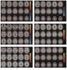
| 大脑区域 | 左/右 | 体素数 | X | Y | Z | t值 |
|---|---|---|---|---|---|---|
| ROI 1(额上回) | 右 | |||||
| 楔叶 | 右 | 72 | 6 | -54 | 33 | -8.75 |
| 额中回 | 右 | 55 | 24 | 39 | 45 | 10.48 |
| ROI 2(前扣带回) | 右 | |||||
| 颞上回 | 右 | 27 | 48 | 12 | -24 | 10.53 |
| 后扣带回 | 左 | 37 | -9 | -57 | 30 | -10.98 |
| ROI 3(中央后回) | 左 | |||||
| 楔叶 | 右 | 106 | 6 | -75 | 27 | -12.75 |
| ROI 4(辅助运动区) | 右 | |||||
| 楔叶 | 左 | 30 | -9 | -75 | 30 | 8.12 |
| ROI 5(脑岛) | 左 | |||||
| 内侧额上回 | 右 | 93 | 24 | 57 | 6 | 31.26 |
| ROI 6(脑岛) | 右 | |||||
| 顶下小叶 | 左 | 44 | -33 | -60 | 42 | 20.55 |
| 1 | Abrams P, Cardozo L, Fall M, et al. The standardisation of terminology of lower urinary tract function: report from the Standardisation Sub-committee of the International Continence Society [J]. Neurourol Urodyn, 2002, 21 (2): 167-178. |
| 2 | Griffiths D, Derbyshire S, Stenger A, et al. Brain control of normal and overactive bladder [J]. J Urol, 2005, 174(5): 1862-1867. |
| 3 | Hulls C M, Lentle R G, King Q M, et al. Spatiotemporal analysis of spontaneous myogenic contractions in the urinary bladder of the rabbit: timing and patterns reflect reported electrophysiology [J]. Am J Physiol Renal Physiol, 2017, 313(3): F687-F698. |
| 4 | Hubeaux K, Deffieux X, Desseaux K, et al. Stand up urgency: is this symptom related to a urethral mechanism? [J]. Prog Urol, 2012, 22(8): 475-481. |
| 5 | Kushida N, Fry C H. On the origin of spontaneous activity in the bladder [J]. BJU Int, 2016,117(6): 982-992. |
| 6 | Deruyver Y, Hakim L, Franken J, et al. The use of imaging techniques in understanding lower urinary tract (dys)function [J]. Auton Neurosci, 2016, 200: 11-20. |
| 7 | Mehnert U, Michels L, Zempleni M Z, et al. The supraspinal neural correlate of bladder cold sensation: an fMRI study [J]. Human Brain Mapp, 2011, 32(6): 835-845. |
| 8 | Jarrahi B, Mantini D, Balsters J H, et al. Differential functional brain network connectivity during visceral interoception as revealed by independent component analysis of fMRI TIME‐series [J]. Hum Brain Mapp, 2015, 36(11): 4438‐4468. |
| 9 | Pievani M, Filippini N, van den Heuvel M P, et al. Brain connectivity in neurodegenerative diseases: from phenotype to proteinopathy [J]. Nat Rev Neurol, 2014, 10(11): 620-633. |
| 10 | Tadic S D, Griffiths D, Schaefer W, et al. Brain activity underlying impaired continence control in older women with overactive bladder [J]. Neurourol Urodyn, 2012, 31(5): 652-658. |
| 11 | Fowler C J, Griffiths D, de Groat W C. The neural control of micturition [J]. Nat Rev Neurosci, 2008, 9(6): 453-466. |
| 12 | Lei D, Ma J, Du X, et al. Spontaneous brain activity changes in children with primary monosymptomatic nocturnal enuresis: a resting-state fMRI study [J]. Neurourol Urodyn, 2012, 31(1): 99-104. |
| 13 | Blok B F. Central pathways controlling micturition and urinary continence [J]. Urology, 2002, 59(5): 13‐17. |
| 14 | Vancea R, Simonyan K, Petracca M, et al. Cognitive performance in mid‐stage Parkinson's disease: functional connectivity under chronic antiparkinson treatment [J]. Brain Imaging Behav, 2019,13(1): 200‐209. |
| 15 | Kitta T, Chancellor M B, de Groat W C, et al. Role of the anterior cingulate cortex in the control of micturition reflex in a rat model of Parkinson's disease [J]. J Urol, 2016, 195(5): 1613-1620. |
| 16 | Kuhtz-Buschbeck J P, van der Horst C, Pott C, et al. Cortical representation of the urge to void: a functional magnetic resonance imaging study [J]. J Urol, 2005, 174(4Pt 1): 1477-1481. |
| 17 | Fuchs T A, Ziccardi S, Benedict R H B, et al. Functional connectivity and structural disruption in the default-mode network predicts cognitive rehabilitation outcomes in multiple sclerosis [J]. J Neuroimaging, 2020, 304(4): 523-530. |
| 18 | Müller N C J, Dresler M, Janzen G, et al. Medial prefrontal decoupling from the default mode network benefits memory [J]. Neuroimage, 2020, 210: 116543. |
| 19 | Watanabe K, Hirano S, Kojima K, et al. Altered cerebral blood flow in the anterior cingulate cortex is associated with neuropathic pain [J]. J Neurol Neurosurg Psychiatry, 2018, 89(10): 1082-1087. |
| 20 | Godlewska B R, Browning M, Norbury R, et al. Predicting treatment response in depression: the role of anterior cingulate cortex [J]. Int J Neuropsychopharmacol, 2018, 21(11): 988-996. |
| 21 | Griffiths D. Neural control of micturition in humans: a working model [J]. Nat Rev Urol, 2015,12(12): 695-705. |
| 22 | Kuhtz-Buschbeck J P, van der Horst C, Wolff S, et al. Activation of the supplementary motor area (SMA) during voluntary pelvic floor muscle contractions: an fMRI study [J]. Neuroimage, 2007, 35(2): 449-457. |
| 23 | Gjone R. Excitatory and inhibitory bladder responses to stimulation of 'limbic', diencephalic and mesencephalic structures in the cat [J]. Acta Physiol Scand, 1966, 66(1): 91‐102. |
| 24 | Gao Y, Liao L, Blok B F. A resting-state functional MRI study on central control of storage: brain response provoked by strong desire to void [J]. Int Urol Nephrol, 2015, 47(6): 927-935. |
| 25 | Yao J, Zhang Q, Liao X, et al. A corticopontine circuit for initiation of urination [J]. Nat Neurosci, 2018, 21(11): 1541-1550. |
| 26 | Berron D, van Westen D, Ossenkoppele R, et al. Medial temporal lobe connectivity and its associations with cognition in early Alzheimer's disease [J]. Brain, 2020,143(4): 1233-1248. |
| 27 | Papagno C, Mattavelli G, Casarotti A, et al. Defective recognition and naming of famous people from voice in patients with unilateral temporal lobe tumours [J]. Neuropsychologia, 2018, 116(Pt B): 194-204. |
| 28 | Barbeau E B, Chai X J, Chen J K, et al. The role of the left inferior parietal lobule in second language learning: An intensive language training fMRI study [J]. Neuropsychologia, 2017, 98: 169-176. |
| 29 | Thakral P P, Madore K P, Schacter D L. A role for the left angular gyrus in episodic simulation and memory [J]. J Neurosci, 2017, 37(34): 8142-8149. |
| 30 | Bonnici H M, Cheke L G, Green D A E, et al. Specifying a causal role for angular gyrus in autobiographical memory [J]. J Neurosci, 2018, 38(49): 10438‐10443. |
| 31 | Nardos R, Karstens L, Carpenter S, et al. Abnormal functional connectivity in women with urgency urinary incontinence: can we predict disease presence and severity in individual women using rs‐fMRI [J]. Neurourol Urodyn, 2016, 35(5): 564‐573. |
| 32 | Pang D, GaoY, Liao L, et al. Brain functional network alterations caused by a strong desire to void in healthy adults: a graph theory analysis study [J]. Neurourol Urodyn, 2020, 39(7): 1966-1976. |
| 33 | Astafiev S V, Stanley C M, Shulman G L, et al. Extrastriate body area in human occipital cortex responds to the performance of motor actions [J]. Nat Neurosci, 2004, 7(5): 542-548. |
| 34 | Heo W, Kim J S, Chung C K, et al. Relationship between cortical resection and visual function after occipital lobe epilepsy surgery [J]. J Neurosurg, 2018, 129(2): 524-532. |
| [1] | YUAN Yuan, ZHOU Hongjun, CONG Xinying, LIU Genlin, WEI Bo, ZHENG Ying, HAO Chunxia, ZHANG Ying, WANG Yiji, KANG Haiqiong, LU Xiaolei, MENG Qianru. Relationship between impairment and magnetic resonance imaging finding in patients with traumatic cervical spinal cord injury after surgery [J]. 《Chinese Journal of Rehabilitation Theory and Practice》, 2023, 29(6): 725-730. |
| [2] | YANG Yanhui, WANG Haochong, DONG Yuanyuan, SHI Gaige, LI Qiuxia, ZHANG Jie, SHI Xiu'e. Effect of visual motion-induced brain computer interface technology on upper limb motor and cognitive function of patients with stroke [J]. 《Chinese Journal of Rehabilitation Theory and Practice》, 2023, 29(4): 472-478. |
| [3] | LIU Mingyue, FAN Yalei, ZHANG Meng, SONG Xueyi, LI Zhe. Brain-computer interface technology for stroke in the past decade: a visualized analysis [J]. 《Chinese Journal of Rehabilitation Theory and Practice》, 2023, 29(2): 223-230. |
| [4] | LUO Qihang, WU Yuxi, ZHANG Jiaxuan, LI Wanying, OU Haining, LIN Qiang, LIANG Junjie. Brain network during balance in older adults: a functional near-infrared spectroscopy study [J]. 《Chinese Journal of Rehabilitation Theory and Practice》, 2023, 29(2): 238-242. |
| [5] | FU Wenjuan, HOU Jianhua, YU Xiaonan, CHEN Tianyong. Relationship of common dyadic coping to marital satisfaction and quality of life for patients with brain injury and their spouses in a rehabilitation facility: using common fate model [J]. 《Chinese Journal of Rehabilitation Theory and Practice》, 2023, 29(12): 1446-1453. |
| [6] | LIU Mingyue, LI Zhe, CAO Yongsheng, HAO Daojian, SONG Xueyi. Effect of brain-computer interface training based on motor imagery on hand function for subacute stroke patients [J]. 《Chinese Journal of Rehabilitation Theory and Practice》, 2023, 29(1): 71-76. |
| [7] | LIU Junming, HUANG Fubiao, LIU Jingya, YANG Xu. Effects of cathodic transcranial direct current stimulation on motor function of upper limbs and fingers in patients with right brain injury [J]. 《Chinese Journal of Rehabilitation Theory and Practice》, 2023, 29(1): 82-87. |
| [8] | ZHANG Xiaoyu,YANG Fan,WEN Jianzhong,YU Weiyong. Application of resting-state functional magnetic resonance imaging in acute mild traumatic brain injury [J]. 《Chinese Journal of Rehabilitation Theory and Practice》, 2022, 28(9): 1084-1088. |
| [9] | GUO Feng,HAO Ying,CHEN Yu. Brain sources characteristics during movement of residual limbs in forearm amputees based on standard low resolution brain electromagnetic tomography technology [J]. 《Chinese Journal of Rehabilitation Theory and Practice》, 2022, 28(8): 972-980. |
| [10] | LIANG Yanan,HE Panqi,LIAO Limin. A novel wearable transcutaneous tibial nerve stimulator inhibits bladder reflexes in cats [J]. 《Chinese Journal of Rehabilitation Theory and Practice》, 2022, 28(7): 797-802. |
| [11] | LIU Haoqiang,CHEN Hanzhe,YANG Yaru. Effect of resilience intervention on rehabilitation of traumatic brain injury: a systematic review [J]. 《Chinese Journal of Rehabilitation Theory and Practice》, 2022, 28(6): 670-677. |
| [12] | WANG Chen,LENG Wenwu,WANG Zhipeng,CHEN Hanzhe,ZHANG Ji,XU Mingchao,ZHONG Xiaoke,JIANG Changhao. Effect of twelve-week aerobic exercise on inhibitory control abilities in overweight children [J]. 《Chinese Journal of Rehabilitation Theory and Practice》, 2022, 28(6): 684-689. |
| [13] | TIAN Jing,LIU Jue,HE Zhijie,FAN Chenyu,LI Haozheng,YANG Qing,WU Yi,YU Kewei. Brain network functional connectivity as unilateral or bilateral upper limb training for patients with upper limb motor dysfunction after stroke: study with functional near-infrared spectroscopy [J]. 《Chinese Journal of Rehabilitation Theory and Practice》, 2022, 28(5): 497-501. |
| [14] | XUAN Wenru,SHEN Yuqing,ZHOU Miao,FENG Shiwen. Bilingual training for cognition of older adults: a systematic review [J]. 《Chinese Journal of Rehabilitation Theory and Practice》, 2022, 28(5): 578-584. |
| [15] | HUANG Feifei,WANG Wansong,DONG Xiaoyang,FENG Zhen. Repair of blood-brain barrier by hyperbaric oxygen via autophagy in rats with cerebral ischemia-reperfusion injury [J]. 《Chinese Journal of Rehabilitation Theory and Practice》, 2022, 28(4): 415-420. |
| Viewed | ||||||
|
Full text |
|
|||||
|
Abstract |
|
|||||
|
||