


《Chinese Journal of Rehabilitation Theory and Practice》 ›› 2021, Vol. 27 ›› Issue (5): 522-529.doi: 10.3969/j.issn.1006-9771.2021.05.005
Previous Articles Next Articles
Wen-mei ZHOU1,2,Tao TAO2,3( ),Shuang WU1,Ting-long WANG4,Zheng-yi YANG4,Ying ZHANG1,2
Received:2020-06-09
Revised:2020-11-27
Published:2021-05-25
Online:2021-05-26
Contact:
Tao TAO
E-mail:835707237@qq.com
Supported by:CLC Number:
Wen-mei ZHOU,Tao TAO,Shuang WU,Ting-long WANG,Zheng-yi YANG,Ying ZHANG. Effects of Enriched Environment on Neurological Function and Glucose Metabolism in Ischemic Penumbra in Cerebral Ischemia-reperfusion Injury Rats[J]. 《Chinese Journal of Rehabilitation Theory and Practice》, 2021, 27(5): 522-529.
"
| 组别 | n | 0 d | 1 d | 7 d | 14 d | 21 d | 28 d |
|---|---|---|---|---|---|---|---|
| 假手术组 | 8 | 0 | 0 | 0 | 0 | 0 | 0 |
| 模型组 | 8 | 0 | 10.38±0.92a | 9.25±0.71a | 8.63±0.74a | 7.75±1.04 | 7.13±0.64a |
| 丰富环境组 | 8 | 0 | 10.50±0.93a | 9.13±0.99a | 7.88±0.83a,b | 6.88±0.64a,b | 5.63±0.92a,b |
| F值 | 513.800 | 455.675 | 438.300 | 291.723 | 270.900 | ||
| P值 | < 0.001 | < 0.001 | < 0.001 | < 0.001 | < 0.001 |
"

"
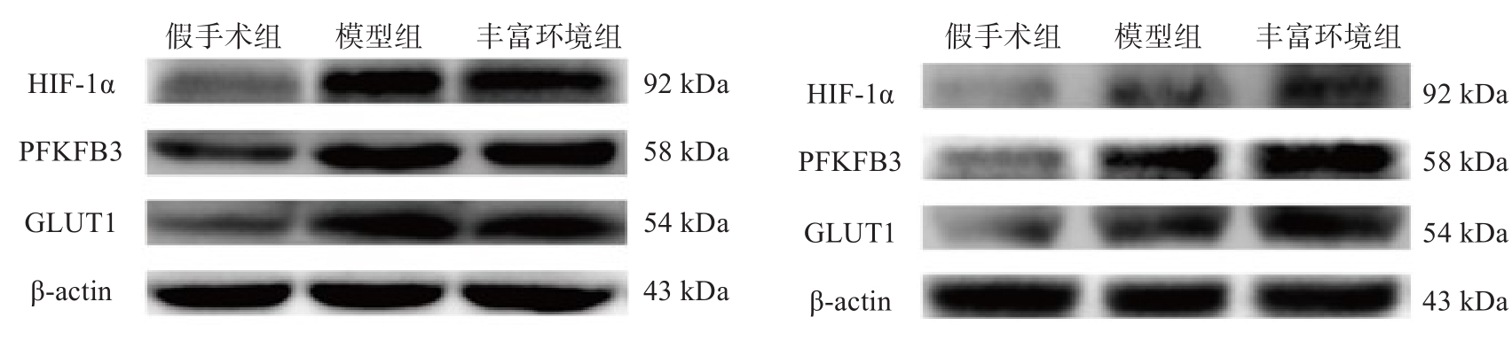
| 组别 | n | HIF-1α | GLUT1 | PFKFB3 | |||||
|---|---|---|---|---|---|---|---|---|---|
| 1 d | 28 d | 1 d | 28 d | 1 d | 28 d | ||||
| 假手术组 | 6 | 0.02±0.02 | 0.02±0.03 | 0.01±0.02 | 0.01±0.02 | 0.01±0.01 | 0.03±0.04 | ||
| 模型组 | 6 | 0.79±0.19a | 0.10±0.01a | 0.64±0.04a | 0.14±0.06a | 2.25±0.29a | 0.28±0.03a | ||
| 丰富环境组 | 6 | 0.81±0.18a | 0.24±0.04a,b | 0.62±0.09a | 0.23±0.04a,b | 2.10±0.24a | 0.87±0.14a,b | ||
| F值 | 27.281 | 46.799 | 119.965 | 18.401 | 127.415 | 73.408 | |||
| P值 | 0.001 | < 0.001 | < 0.001 | 0.003 | < 0.001 | < 0.001 | |||
"
"
| 组别 | n | HIF-1α | GLUT1 | PFKFB3 | |||||
|---|---|---|---|---|---|---|---|---|---|
| 1 d | 28 d | 1 d | 28 d | 1 d | 28 d | ||||
| 假手术组 | 6 | 0.38±0.04 | 0.43±0.03 | 0.41±0.08 | 0.48±0.08 | 0.51±0.05 | 0.41±0.06 | ||
| 模型组 | 6 | 1.22±0.11a | 0.75±0.08a | 1.24±0.08a | 0.75±0.03a | 1.58±0.10a | 1.04±0.12a | ||
| 丰富环境组 | 6 | 1.27±0.09a | 0.93±0.07a,b | 1.18±0.06a | 0.88±0.06a,b | 1.56±0.05a | 1.31±0.06a,b | ||
| F值 | 105.196 | 44.379 | 121.472 | 41.537 | 236.893 | 91.819 | |||
| P值 | < 0.001 | < 0.001 | < 0.001 | < 0.001 | < 0.001 | < 0.001 | |||
"

"
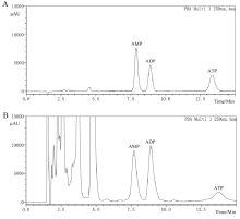
| 组别 | n | ATP(μg/g) | ADP(μg/g) | AMP(μg/g) | EC | |||||||
|---|---|---|---|---|---|---|---|---|---|---|---|---|
| 1 d | 28 d | 1 d | 28 d | 1 d | 28 d | 1 d | 28 d | |||||
| 假手术组 | 3 | 16.15±1.31 | 17.40±0.56 | 215.09±4.31 | 167.91±1.15 | 49.32±1.67 | 49.10±0.60 | 0.44±0.00 | 0.43±0.00 | |||
| 模型组 | 3 | 4.91±0.88a | 9.95±0.38a | 71.97±2.24a | 67.81±0.03a | 105.93±1.27a | 38.13±0.21a | 0.22±0.01a | 0.38±0.01a | |||
| 丰富环境组 | 3 | 3.83±1.65a | 13.82±1.86a,b | 67.15±3.64a | 158.70±1.58a | 87.23±1.35a | 56.24±2.96 | 0.24±0.02a | 0.41±0.00a,b | |||
| F值 | 179.228 | 193.752 | 2994.473 | 13650.446 | 3508.858 | 134.914 | 333.841 | 196.000 | ||||
| P值 | < 0.001 | < 0.001 | < 0.001 | < 0.001 | < 0.001 | < 0.001 | < 0.001 | < 0.001 | ||||
| 1 | PANISELLO-ROSELLÓ A, ROSELLÓ-CATAFAU J. Molecular mechanisms and pathophysiology of ischemia-reperfusion injury [J]. Int J Mol Sci, 2018, 19(12): 4093. |
| 2 | ASTRUP J, SIESJÖ B K, SYMON L. Thresholds in cerebral ischemia-the ischemic penumbra [J]. Stroke, 1981, 12(6): 723-725. |
| 3 | ZHANG Y J, XU D, QI H, et al. Enriched environment promotes post-stroke neurogenesis through NF-κB-mediated secretion of IL-17A from astrocytes [J]. Brain Res, 2018, 1687: 20-31. |
| 4 | BALAMURUGAN K. HIF-1 at the crossroads of hypoxia, inflammation, and cancer [J]. In J Cancer, 2016, 138(5): 1058-1066. |
| 5 | MASOUD G N, LI W. HIF-1α pathway: role, regulation and intervention for cancer therapy [J]. Acta Pharm Sin B, 2015, 5(5): 378-389. |
| 6 | HU L, ZENG Z, XIA Q, et al. Metformin attenuates hepatoma cell proliferation by dcreasing glycolytic flux through the HIF-1α/PFKFB3/PFK1 pathway [J]. Life Sci, 2019, 239: 116966. |
| 7 | 邓敏,章军建,沈军,等. 丰富环境对慢性脑低灌注大鼠低氧诱导因子血管内皮生长因子的表达及其认知功能的影响[J]. 中华老年医学, 2016, 35(1): 79-84. |
| DENG M, ZHANG J J, SHEN J, et al. Effect of environmental enrichment on cognitive function and the expressions of hypoxia inducible factor 1α and vascular endothelial growth factor in rats with chronic cerebral hypoperfusion [J]. Chin J Geriatr, 2016, 35(1): 79-84. | |
| 8 | WU X, LIU S, HU Z, et al. Enriched housing promotes post-stroke neurogenesis through calpain 1-STAT3/HIF-1α/VEGF signaling [J]. Brain Res Bull, 2018, 139: 133-143. |
| 9 | YAN R Y, WANG S J, YAO G T, et al. The protective effect and its mechanism of 3-n-butylphthalide pretreatment on cerebral ischemia reperfusion injury in rats [J]. Eur Rev Med Pharmacol Sci, 2017, 21(22): 5275-5282. |
| 10 | ZHU Y, DENG L, TANG H J, et al. Electroacupuncture improves neurobehavioral function and brain injury in rat model of intracerebral hemorrhage [J]. Brain Res Bull, 2017, 131: 123-132. |
| 11 | XIE H, YU K, ZHOU N, et al. Enriched environment elicits proangiogenic mechanisms after focal cerebral ischemia [J]. Transl Stroke Res, 2019, 10(2): 150-159. |
| 12 | THIRUGNANACHANDRAN T, MA H, SINGHAL S, et al. Refining the ischemic penumbra with topography [J]. Int J Stroke, 2018, 13(3): 277-284. |
| 13 | ZHAN C, YANG J, ZHAN L. RP-HPLC determination of effects of isoliquiritigenin on brain energy metabolism in repeated cerebral ischemia-reperfusion mice [J]. Chin J Pharm Anal, 2005, 25: 639-642. |
| 14 | PANISELLO-ROSELLÓ A, ROSELLÓ-CATAFAU J. Molecular mechanisms and pathophysiology of ischemia-reperfusion injury [J]. Int J Mol Sci, 2018, 19 (12): 4093. |
| 15 | MALÁ H, RASMUSSEN C P. The effect of combined therapies on recovery after acquired brain injury: systematic review of preclinical studies combining enriched environment, exercise, or task-specific training with other therapies [J]. Restor Neurol Neuros, 2017, 35(1): 25-64. |
| 16 | 吕富岩,张雷红,宫兆帅,等. 丰富环境促进缺氧缺血性脑损伤神经可塑性的研究进展[J]. 中国康复理论与实践, 2018, 24(5): 509-512. |
| LÜ F Y, ZHANG L H, GONG Z S, et al. Advance of enriched environment in neural plasticity post hypoxic-ischemic brain damage (review) [J]. Chin J Rehabil Theory Pract, 2018, 24(5): 509-512. | |
| 17 | 李敏,陶陶,张继荣. 丰富环境对脑缺血再灌注损伤后相关凋亡基因的研究进展[J]. 中国康复医学杂志, 2018, 33(10): 1238-1241. |
| LI M, TAO T, ZHANG J R. Chin J Rehabil Med, 2018, 33(10): 1238-1241. | |
| 18 | WANG D, YUAN X, LIU T, et al. Neuroprotective activity of lavender oil on transient focal cerebral ischemia in mice [J]. Molecules, 2012, 17(8): 9803-9817. |
| 19 | KANG H, CHOI D H, KIM S K, et al. Alteration of energy metabolism and antioxidative processing in the hippocampus of rats reared in long-term environmental enrichment [J]. Dev Neurosci, 2016, 38(3): 186-194. |
| 20 | BRIONES T L, WOODS J, ROGOZINSKA M. Decreased neuroinflammation and increased brain energy homeostasis following environmental enrichment after mild traumatic brain injury is associated with improvement in cognitive function [J]. Acta Neuropathol Com, 2013, 1: 57. |
| 21 | UZDENSKY A B. Regulation of apoptosis in the ischemic penumbra in the first day post-stroke [J]. Neural Regen Res, 2020, 15(2): 253-254. |
| 22 | AMARAL A I, TEIXEIRA A P, MARTENS S, et al. Metabolic alterations induced by ischemia in primary cultures of astrocytes:merging 13C NMR spectroscopy and metabolic flux analysis [J]. Neurochem, 2010, 113(3): 735-748. |
| 23 | MARIN-VALENCIA I, GOOD L B, MA Q, et al. Glut1 deficiency (G1D): epilepsy and metabolic dysfunction in a mouse model of the most common human phenotype [J]. Neurobiol Dis, 2012, 48(1): 92-101. |
| 24 | WU X, WANG C, WANG J, et al. Hypoxia preconditioning protects neuronal cells against traumatic brain injury through stimulation of glucose transport mediated by HIF-1α/GLUTs signaling pathway in rat [J]. Neurosurg Rev, 2021, 44(1): 411-422. |
| 25 | LV Y, ZHANG B, ZHAI C, et al. PFKFB3-mediated glycolysis is involved in reactive astrocyte proliferation after oxygen-glucose deprivation/reperfusion and is regulated by Cdh1 [J]. Neurochem Int, 2015, 91: 26-33. |
| 26 | LI C, ZHANG B, ZHU Y, et al. Post-stroke constraint-induced movement therapy increases functional recovery, angiogenesis, and neurogenesis with enhanced expression of HIF-1α and VEGF [J]. Curr Neurovasc Res, 2017, 14(4): 368-377. |
| 27 | ZHANG Z, YAN J, SHI H. Role of hypoxia inducible factor 1 in hyperglycemia-exacerbated blood-brain barrier disruption in ischemic stroke [J]. Neurobiol Dis, 2016, 95: 82-92. |
| 28 | SADLECKI P, BODNAR M, GRABIEC M, et al. The role of hypoxia-inducible factor-1α, glucose transporter-1 (GLUT-1) and carbon anhydrase IX in endometrial cancer patients [J]. Biomed Res Int, 2014, 2014: 616850. |
| 29 | FUKASAWA M, TSUCHIYA T, TAKAYAMA E, et al. Identification and characterization of the hypoxia-responsive element of the human placental 6-phosphofructo-2-kinase/fructose-2, 6-bisphosphatase gene [J]. J Biochem, 2004, 136(3): 273-277. |
| 30 | VRIEND J, REITER R J. Melatonin and the von Hippel-Lindau/HIF-1 oxygen sensing mechanism: a review [J]. Biochim Biophys Acta, 2016, 1865(2): 176-183. |
| 31 | 金信浩,郑兵,吴雪莲,等. 丰富环境对脑卒中患者神经功能康复的效果[J]. 中国康复理论与实践, 2017, 23(3): 323-325. |
| JIN X H, ZHENG B, WU X L, et al. Effects of environmental enrichment on neurological functions of patients with stroke [J]. Chin J Rehabil Theory Pract, 2017, 23(3): 323-325. |
| [1] | LI Fang, HUO Su, DU Jubao, LIU Xiuzhen, LI Xiaoshuang, SONG Weiqun. Effect of transcranial direct current stimulation combined with task-oriented rehabilitation training on forelimb motor dysfunction in rats with spinal cord injury [J]. 《Chinese Journal of Rehabilitation Theory and Practice》, 2023, 29(7): 777-781. |
| [2] | KANG Xiaoyu, LIU Lixu, WANG Wenzhu, WANG Yunlei. Effects of pramipexole combined with levodopa on cognitive and mitochondrial function of rats after global cerebral ischemia-reperfusion injury [J]. 《Chinese Journal of Rehabilitation Theory and Practice》, 2023, 29(5): 533-540. |
| [3] | WEI Tianqi, LUO Jiaqi, LI Zijuan, WU Xueliang, XU Panpan, ZHANG Yanmei, ZHAO Xiaomeng, WU Qinfeng. Effect of augmented reality training based on enriched environment on walking function after stroke [J]. 《Chinese Journal of Rehabilitation Theory and Practice》, 2023, 29(12): 1439-1445. |
| [4] | HUANG Zhilin, XU Fashao, SHI Jing, HUANG Gan, LIU Meifang, ZHANG Xiahui. Establishment of rat model of dysphagia after stroke by thread embolism [J]. 《Chinese Journal of Rehabilitation Theory and Practice》, 2023, 29(10): 1147-1153. |
| [5] | QIN Yanqiang, DONG Hao, SUN Yingchun, CHENG Xiankuan, YAO Haijiang. Effects of different acupuncture schemes on neurotransmitters and related inflammatory factors in rats with post-stroke depression [J]. 《Chinese Journal of Rehabilitation Theory and Practice》, 2023, 29(1): 30-37. |
| [6] | MIAO Pei,ZHANG Tong,MI Haixia,ZHANG Weidong. Learning and memory ability and its mechanism in rats with focal cerebral ischemia induced by two filament-occluded methods [J]. 《Chinese Journal of Rehabilitation Theory and Practice》, 2022, 28(7): 789-796. |
| [7] | ZHOU Xiaojue,FENG Jing,PANG Rizhao,LIU Jie,ZHANG Anren. Every-other-day fasting attenuated inflammation in rats after spinal cord injury via the aryl hydrocarbon receptor/suppressor of cytokine signaling 2/nuclear transcription factor-κB signaling pathway [J]. 《Chinese Journal of Rehabilitation Theory and Practice》, 2022, 28(5): 544-551. |
| [8] | SONG Shaofei,HOU Yuanyuan,WANG Yunlei,ZHANG Tong. Effects of circadian misalignment induced by abnormal photoperiod on expression rhythm of clock genes and glucose uptake related genes in gastrocnemius in rats [J]. 《Chinese Journal of Rehabilitation Theory and Practice》, 2022, 28(5): 552-558. |
| [9] | LI Tong,FANG Zhipeng,SHAO Yuping,WANG Ping. Effect of aerobic exercise on learning and memory, and synaptic plasticity of hippocampal neurons for sleep-deprived rats [J]. 《Chinese Journal of Rehabilitation Theory and Practice》, 2022, 28(11): 1270-1277. |
| [10] | Jing-yi WANG,Jie YIN,Jian-cheng LIU,Ri-zhao PANG,Wen-chun WANG. Effect of Iridoid-rich Fraction from Valeriana Jatamansi Jones on Neuron Pyroptosis in Rats with Acute Spinal Cord Injury [J]. 《Chinese Journal of Rehabilitation Theory and Practice》, 2021, 27(6): 653-660. |
| [11] | Qiong-fen WANG,Feng-bo WANG,Ke WANG,Yong-qiang ZHONG,Jiao-jiao WANG. Effect of Electroacupuncture at Fengchi on Astrocytes and Neurons in Rats with Acute Cerebral Infarction [J]. 《Chinese Journal of Rehabilitation Theory and Practice》, 2021, 27(3): 302-309. |
| [12] | Xiang-zhe LI,Jie DING,Qing-hua WANG,Chuan-ming DONG,Tong WANG,Qin-feng WU. Effects of Body Weight-supported Treadmill Training on Neuropathic Pain and Expression of Glutamate Decarboxylase (GAD)-65/67 in Spinal Dorsal Horn of Rats with Spinal Cord Injury [J]. 《Chinese Journal of Rehabilitation Theory and Practice》, 2021, 27(2): 131-136. |
| [13] | Kun AI,Ming XU,Qiong LIU,Shi-feng DENG,Ji-sheng LIU,Fang QI,Xi-qin YI,Qi-rui QU,Hong ZHANG. Effect of Electroacupuncture on Content of Cyclic Adenosine Monophosphate and Protein Kinase A, and Phosphorylation of Myosin Light Chain Kinase in Detrusor of Rats with Detrusor Hyperreflex after Suprasacral Spinal Cord Injury [J]. 《Chinese Journal of Rehabilitation Theory and Practice》, 2021, 27(2): 137-144. |
| [14] | LING Meng-yu,YANG Yi-zhuo,LIU Shuai,YE Chao-qun. Effect of Exercise on Expression of NeuN and SynapsinI in Cortex and Cognitive Function in Rats after Cerebral Ischemia-reperfusion [J]. 《Chinese Journal of Rehabilitation Theory and Practice》, 2021, 27(11): 1272-1281. |
| [15] | PENG Zhi-feng, ZHANG Yi-ping, ZHANG Ji-hong. Effect of Early Exercise Combined with Pentylenetetrazol on Neurologic Function after Cerebral Ischemia-reperfusion in Rats and Its Mechanism [J]. 《Chinese Journal of Rehabilitation Theory and Practice》, 2021, 27(1): 27-30. |
| Viewed | ||||||
|
Full text |
|
|||||
|
Abstract |
|
|||||
|
||