


《Chinese Journal of Rehabilitation Theory and Practice》 ›› 2021, Vol. 27 ›› Issue (7): 791-796.doi: 10.3969/j.issn.1006-9771.2021.07.009
Previous Articles Next Articles
CHEN Ming-zhen1a,JIANG Fan1a( ),SHAN Yong1a,HONG Yong-feng1b,LIU Xue1a,XIAO Hong-bo2,CHEN Rui-quan2
Received:2020-11-16
Revised:2021-06-02
Published:2021-07-25
Online:2021-07-28
Contact:
JIANG Fan
E-mail:ahultrasound2005@126.com
CLC Number:
CHEN Ming-zhen,JIANG Fan,SHAN Yong,HONG Yong-feng,LIU Xue,XIAO Hong-bo,CHEN Rui-quan. Measurement of Structure and Stiffness of Gastrocnemius Muscle for Stroke Patients with Multimodal Ultrasound Imaging[J]. 《Chinese Journal of Rehabilitation Theory and Practice》, 2021, 27(7): 791-796.
"
| 组别 | n | 性别(男/女, n) | 年龄(岁) | 类型(出血/梗死, n) | 偏瘫侧(左/右,n) |
|---|---|---|---|---|---|
| 对照组 | 46 | 32/14 | 53.83±10.25 | ||
| 试验组 | 44 | 33/11 | 53.43±13.54 | 29/15 | 25/19 |
| χ2/t值 | 0.156 | 0.331 | |||
| P值 | 0.876 | 0.565 |
"
"

"
"
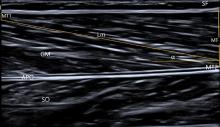
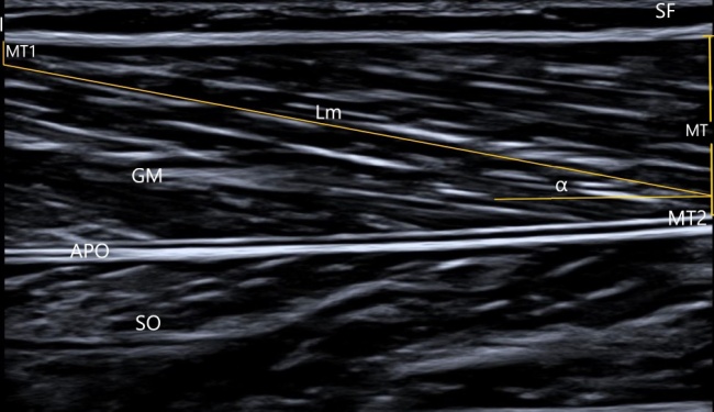
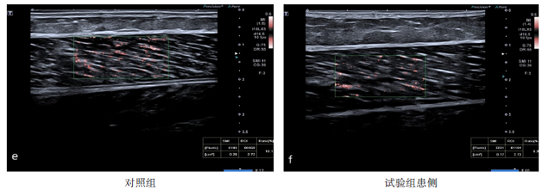
| 组别 | n | FL(mm) | MT(mm) | PA(°) | SWV(m/s) | SMI(%) |
|---|---|---|---|---|---|---|
| 对照组 | 46 | 48.05±2.15 | 13.75±2.68 | 13.07±3.23 | 2.69±0.40 | 7.49±2.11 |
| 试验组健侧 | 44 | 47.69±3.31 | 11.14±2.55 | 12.02±2.93 | 2.72±0.56 | |
| 试验组患侧 | 44 | 43.22±3.40 | 12.25±2.21 | 14.64±3.18 | 3.70±0.85 | 6.28±2.31 |
| t1值 | -6.235 | 2.198 | 4.006 | 6.346 | ||
| P1值 | < 0.001 | 0.031 | <0.001 | < 0.001 | ||
| t2值 | -8.073 | -2.881 | 2.326 | 7.119 | 2.604 | |
| P2值 | < 0.001 | 0.005 | 0.022 | < 0.001 | 0.011 |
"
| 组别 | n | 差值 |
|---|---|---|
| 对照组 | 46 | 10.91±4.23 |
| 试验组健侧 | 44 | 9.10±4.06 |
| 试验组患侧 | 44 | 3.52±1.94 |
| Z1值 | 2.055 | |
| P1值 | 0.040 | |
| Z2值 | 6.703 | |
| P2值 | < 0.001 | |
| Z3值 | 7.318 | |
| P3值 | < 0.001 |
| [1] |
FEIGIN V L, FOROUZANFAR M H, KRISHNAMURTHI R, et al. Global and regional burden of stroke during 1990-2010: findings from the global burden of disease study 2010[J]. Lancet, 2014, 383(9913):245-254.
doi: 10.1016/S0140-6736(13)61953-4 |
| [2] |
GAO J, RUBIN J M, CHEN J, et al. Ultrasound elastography to assess Botulinum toxin A treatment for post-stroke spasticity: a feasibility study[J]. Ultrasound Med Biol, 2019, 45(5):1094-1102.
doi: 10.1016/j.ultrasmedbio.2018.10.034 |
| [3] |
EBY S, ZHAO H, SONG P, et al. Quantitative evaluation of passive muscle stiffness in chronic stroke[J]. Am J Phys Med Rehabil, 2016, 95(12):899-910.
doi: 10.1097/PHM.0000000000000516 |
| [4] |
PRADINES M, GHEDIRA M, PORTERO R, et al. Ultrasound structural changes in triceps surae after a 1-year daily self-stretch program: a prospective randomized controlled trial in chronic hemiparesis[J]. Neurorehabil Neural Repair, 2019, 33(4):245-259.
doi: 10.1177/1545968319829455 |
| [5] |
GHASEMI E, KHADEMI-KALANTARI K, KHALKHALI-ZAVIEH M, et al. The effect of functional stretching exercises on neural and mechanical properties of the spastic medial gastrocnemius muscle in patients with chronic stroke: a randomized controlled trial[J]. J Stroke Cerebrovasc Dis, 2018, 27(7):1733-1742.
doi: 10.1016/j.jstrokecerebrovasdis.2018.01.024 |
| [6] | 秦鹍, 冯亚男, 李亚鹏, 等. 剪切波弹性成像技术量化评估肌腱肌肉弹性模量的信度[J]. 中国康复理论与实践, 2018, 24(10):1201-1205. |
| QIN K, FENG Y N, LI Y P, et al. Intra- and inter-rater reliability of shear wave elastic imaging technique for elastic modulus measurements of muscle and tendon[J]. Chin Rehabil Theory Pract, 2018, 24(10):1201-1205. | |
| [7] |
LEHOUX M C, SOBCZAK S, CLOUTIER F, et al. Shear wave elastography potential to characterize spastic muscles in stroke survivors: literature review[J]. Clin Biomech (Bristol, Avon), 2020, 72:84-93.
doi: 10.1016/j.clinbiomech.2019.11.025 |
| [8] | 中华医学会神经病学分会, 中华医学会神经病学分会脑血管病学组. 中国各类主要脑血管病诊断要点2019[J]. 中华神经科杂志, 2019, 52(9):710-715. |
| Chinese Society of Neurology, Chinese Stroke Society. Diagnosis of Cerebrovascular Diseases in China (version 2019)[J]. Chin J Neurol, 2019, 52(9):710-715. | |
| [9] |
IKEZOE T, MORI N, NAKAMURA M, et al. Atrophy of the lower limbs in elderly women: is it related to walking ability?[J]. Eur J Appl Physiol, 2011, 111(6):989-995.
doi: 10.1007/s00421-010-1728-8 |
| [10] | 甄希成, 陈新, 张辉. 持续被动运动治疗脑卒中患者下肢肌痉挛的效果[J]. 中国老年学杂志, 2017, 37(4):880-882. |
| ZHEN X C, CHEN X, ZHANG H. Effect of continuous passive exercise on treatment of lower extremity muscle spasm in patients with stroke[J]. Chin J Gerontol, 2017, 37(4):880-882. | |
| [11] |
KUO C, HU G. Post-stroke spasticity: a review of epidemiology, pathophysiology, and treatments[J]. Int J Gerontol, 2018, 12(4):280-284.
doi: 10.1016/j.ijge.2018.05.005 |
| [12] |
JAKUBOWSKI K L, TERMAN A, SANTANA R V C, et al. Passive material properties of stroke-impaired plantarflexor and dorsiflexor muscles[J]. Clin Biomech (Bristol, Avon), 2017, 49:48-55.
doi: 10.1016/j.clinbiomech.2017.08.009 |
| [13] | 樊留博, 韩文胜, 江莹莹, 等. 超声弹性成像评价脑卒中偏瘫肢体硬度对痉挛性偏瘫患者预后的影响[J]. 中华全科医学, 2017, 15(8):1323-1325. |
| FAN L B, HAN W S, JIANG Y Y, et al. Role of elastosonography in evaluating hemiplegic limb hardness and improving the prognosis of spastic hemiplegia patients[J]. Chin General Pract, 2017, 15(8):1323-1325. | |
| [14] |
HONG M J, PARK J B, LEE Y J, et al. Quantitative evaluation of post-stroke spasticity using neurophysiological and radiological tools: a pilot study[J]. Ann Rehabil Med, 2018, 42(3):384-395.
doi: 10.5535/arm.2018.42.3.384 |
| [15] | 邓思宇, 卢茜, 郄淑燕, 等. 等速测试指标与改良Ashworth量表用于踝痉挛评定的相关性研究[J]. 中国康复理论与实践, 2016, 22(2):178-183. |
| DENG S Y, LU Q, QIE S Y, et al. Correlation of isokinetic parameter and Modified Ashworth Scale applied in evaluation of ankle spasticity[J]. Chin Rehabil Theory Pract, 2016, 22(2):178-183. | |
| [16] |
DOS SANTOS A N, ROCHA N A C F. Immediate effect of kinesio taping on knee extensor torque of children with cerebral palsy: three case reports[J]. NeuroRehabilitation, 2018, 43(4):519-523.
doi: 10.3233/NRE-161921 |
| [17] |
MATHEVON L, MICHEL F, AUBRY S, et al. Two-dimensional and shear wave elastography ultrasound: a reliable method to analyse spastic muscles?[J]. Muscle Nerve, 2018, 57(2):222-228.
doi: 10.1002/mus.25716 |
| [18] |
DIAS C P, FREIRE B, GOULART N B, et al. Muscle architecture and torque production in stroke survivors: an observational study[J]. Top Stroke Rehabil, 2017, 24(3):206-213.
doi: 10.1080/10749357.2016.1210873 |
| [19] |
SON J, RYMER W Z, LEE S S M. Limited fascicle shortening and fascicle rotation may be associated with impaired voluntary force-generating capacity in pennate muscles of chronic stroke survivors[J]. Clin Biomech (Bristol, Avon), 2020, 75:105007.
doi: 10.1016/j.clinbiomech.2020.105007 |
| [20] | YANG Y B, ZHANG J, LENG Z P, et al. Evaluation of spasticity after stroke by using ultrasound to measure the muscle architecture parameters: a clinical study[J]. Int J Clin Exp Med, 2014, 7(9):2712-2717. |
| [21] |
D'SOUZA A, BOLSTERLEE B, HERBERT R D. Architecture of the medial gastrocnemius muscle in people who have had a stroke: a diffusion tensor imaging investigation[J]. Clin Biomech (Bristol, Avon), 2020, 74:27-33.
doi: 10.1016/j.clinbiomech.2020.02.004 |
| [22] |
THIELMAN G, YOUREY L. Ultrasound imaging of upper extremity spastic muscle post-stroke and the correlation with function: a pilot study[J]. NeuroRehabilitation, 2019, 45(2):213-220.
doi: 10.3233/NRE-192742 |
| [23] | 刘美快, 徐乐义, 李海燕, 等. 脑卒中患者小腿肌肉形态结构变化的定量超声研究[J]. 中国康复医学杂志, 2018, 33(10):1183-1187. |
| LIU M K, XU L Y, LI H Y, et al. A study on quantitative ultrasound on evaluating the architectural parameters of lower leg muscles in stroke survivors[J]. Chin J Rehabil Med, 2018, 33(10):1183-1187. | |
| [24] |
LEE S S M, SPEAR S, RYMER W Z. Quantifying changes in material properties of stroke-impaired muscle[J]. Clin Biomech (Bristol, Avon), 2015, 30(3):269-275.
doi: 10.1016/j.clinbiomech.2015.01.004 |
| [25] |
LECHARTE T, GROSS R, NORDEZ A, et al. Effect of chronic stretching interventions on the mechanical properties of muscles in patients with stroke: a systematic review[J]. Ann Phys Rehabil Med, 2020, 63(3):222-229.
doi: 10.1016/j.rehab.2019.12.003 |
| [26] |
LIEBER R L, ROBERTS T J, BLEMKER S S, et al. Skeletal muscle mechanics, energetics and plasticity[J]. J Neuroeng Rehabil, 2017, 14(1):108.
doi: 10.1186/s12984-017-0318-y |
| [27] |
FRIDÉN J, LIEBER R L. Spastic muscle cells are shorter and stiffer than normal cells[J]. Muscle Nerve, 2003, 27(2):157-164.
doi: 10.1002/mus.v27:2 |
| [28] |
LEE S S M, JAKUBOWSKI K L, SPEAR S C, et al. Muscle material properties in passive and active stroke-impaired muscle[J]. J Biomech, 2019, 83:197-204.
doi: 10.1016/j.jbiomech.2018.11.043 |
| [29] | LIU J, PAN H, BAO Y, et al. The value of real-time shear wave elastography before and after rehabilitation of upper limb spasm in stroke patients[J]. Biomed Res Int, 2020, 2020:1-7. |
| [30] |
VOLA E A, ALBANO M, DI LUISE C, et al. Use of ultrasound shear wave to measure muscle stiffness in children with cerebral palsy[J]. J Ultrasound, 2018, 21(3):241-247.
doi: 10.1007/s40477-018-0313-6 |
| [31] |
COSGROVE D, PISCAGLIA F, BAMBER J, et al. EFSUMB Guidelines and Recommendations on the Clinical Use of Ultrasound Elastography. Part 2: Clinical Applications[J]. Ultraschall Med, 2013, 34(3):238-253.
doi: 10.1055/s-00000089 |
| [32] |
LENG Y, WANG Z, BIAN R, et al. Alterations of elastic property of spastic muscle with its joint resistance evaluated from shear wave elastography and biomechanical model[J]. Front Neurol, 2019, 10:736.
doi: 10.3389/fneur.2019.00736 |
| [33] |
WU C, HO Y, HSIAO M Y, et al. Evaluation of post-stroke spastic muscle stiffness using shear wave ultrasound elastography[J]. Ultrasound Med Biol, 2017, 43(6):1105-1111.
doi: 10.1016/j.ultrasmedbio.2016.12.008 |
| [34] |
EBY S F, ZHAO H, SONG P, et al. Quantifying spasticity in individual muscles using shear wave elastography[J]. Radiol Case Rep, 2017, 12(2):348-352.
doi: 10.1016/j.radcr.2017.01.004 |
| [35] |
KWON D R, PARK G Y, LEE S U, et al. Spastic cerebral palsy in children: dynamic sonoelastographic findings of medial gastrocnemius[J]. Radiology, 2012, 263(3):794-801.
doi: 10.1148/radiol.12102478 |
| [36] |
LE SANT G, NORDEZ A, HUG F, et al. Effects of stroke injury on the shear modulus of the lower leg muscle during passive dorsiflexion[J]. J Appl Physiol (1985), 2019, 126(1):11-22.
doi: 10.1152/japplphysiol.00968.2017 |
| [37] |
ASKIN A, KALAYCI O T, BAYRAM K B, et al. Strain sonoelastographic evaluation of biceps muscle intrinsic stiffness after botulinum toxin-A injection[J]. Top Stroke Rehabil, 2017, 24(1):12-17.
doi: 10.1080/10749357.2016.1183865 |
| [38] |
HUANG M, MILLER T, YING M, et al. Whole-body vibration modulates leg muscle reflex and blood perfusion among people with chronic stroke: a randomized controlled crossover trial[J]. Sci Rep, 2020, 10(1):1473.
doi: 10.1038/s41598-020-58479-5 |
| [39] | 蓝晓锋, 姜凡, 彭梅, 等. SMI技术对血管性阴茎勃起功能障碍的诊断价值[J]. 中国超声医学杂志, 2018, 34(11):1028-1031. |
| LAN X F, JIANG F, PENG M, et al. The diagnosis value of SMI technology in vascular penile erectile dysfunction[J]. Chin J Ultrasound Med, 2018, 34(11):1028-1031. | |
| [40] |
JIANG Z Z, HUANG Y H, SHEN H L, et al. Clinical applications of superb microvascular imaging in the liver, breast, thyroid, skeletal muscle, and carotid plaques[J]. J Ultrasound Med, 2019, 38(11):2811-2820.
doi: 10.1002/jum.v38.11 |
| [41] |
CALISKAN E, AKKOC O, BAYRAMOGLU Z, et al. Effects of static stretching duration on muscle stiffness and blood flow in the rectus femoris in adolescents[J]. Med Ultrason, 2019, 21(2):136-143.
doi: 10.11152/mu-1859 |
| [1] | LIN Na, GAO Hanlu, LU Huiping, CHEN Yanqing, ZHENG Junfan, CHEN Shurong. Effect of virtual reality on upper limb function after stroke: a study of diffusion tensor imaging [J]. 《Chinese Journal of Rehabilitation Theory and Practice》, 2024, 30(1): 61-67. |
| [2] | WANG Haoyi, SHI Yawei, LU Jun, XU Guangxu. Impact of subjective vertical perception impairment on function in stroke patients: a retrospective study [J]. 《Chinese Journal of Rehabilitation Theory and Practice》, 2024, 30(1): 68-73. |
| [3] | CHEN Junwen, CHEN Qian, CHEN Cheng, LI Shuyue, LIU Lingling, WU Cunshu, GONG Xiang, LU Jun, XU Guangxu. Effect of modified Baduanjin exercise on cardiopulmonary function, motor function and activities of daily living for stroke patients [J]. 《Chinese Journal of Rehabilitation Theory and Practice》, 2024, 30(1): 74-80. |
| [4] | HU Yonglin, MA Ying, DOU Chao, LU Anmin, JIANG Xiaoge, SONG Xinjian, XIAO Yuhua. Effect of neural mobilization based on shoulder control training on shoulder pain and upper limb function in stroke patients with hemiplegia [J]. 《Chinese Journal of Rehabilitation Theory and Practice》, 2024, 30(1): 81-86. |
| [5] | WANG He, HAN Liang, KAN Mengfan, YU Shaohong. Efficacy of electrical stimulation on shoulder-hand syndrome after stroke: a systematic review and meta-analysis [J]. 《Chinese Journal of Rehabilitation Theory and Practice》, 2023, 29(9): 1048-1056. |
| [6] | SUN Tengfang, REN Mengting, YANG Lin, WANG Yaoting, WANG Hongyu, YAN Xingzhou. Effect of hyperbaric oxygen therapy combined with repetitive peripheral magnetic stimulation on ankle motor function and balance of stroke patients [J]. 《Chinese Journal of Rehabilitation Theory and Practice》, 2023, 29(8): 875-881. |
| [7] | WANG Ya'nan, LIU Xihua. Correlation and predictive effect of subjective and objective balance function measurements in stroke patients with hemiplegia [J]. 《Chinese Journal of Rehabilitation Theory and Practice》, 2023, 29(8): 890-895. |
| [8] | WANG Haiyun, WANG Yin, ZHOU Xinjie, HE Aiqun. Effect of transcranial direct current stimulation combined with acupuncture on central and upper limb function in stroke patients based on central-peripheral-central theory [J]. 《Chinese Journal of Rehabilitation Theory and Practice》, 2023, 29(8): 919-925. |
| [9] | CHEN Yiting, WANG Qian, CUI Shenhong, LI Yingcai, ZHANG Siyu, WEI Yanxu, REN Hui, LENG Jun, CHEN Bin. Effect of bilateral sequential repetitive transcranial magnetic stimulation on motor function of upper limbs in stroke patients [J]. 《Chinese Journal of Rehabilitation Theory and Practice》, 2023, 29(8): 926-932. |
| [10] | LI Zhenya, SUN Jie, GUO Pengfei, WANG Guangming. Correlation between changes of swallowing function in oral and pharyngeal phases, and aspiration in stroke patients based on videofluroscopic swallowing study [J]. 《Chinese Journal of Rehabilitation Theory and Practice》, 2023, 29(8): 933-939. |
| [11] | HUA Ling, ZHANG Yi'nan, ZHENG Yu, SUN Qiaoyi, FANG Hui, SONG Da. Effect of hand controlled rhythm music therapy on unilateral spatial neglect after stroke [J]. 《Chinese Journal of Rehabilitation Theory and Practice》, 2023, 29(7): 833-838. |
| [12] | JIANG Xiaocui, LIU Zhen, SU Qinglun, ZHAO Qin, XIA Xiaomei, LU Fei. Effect of intermittent theta burst transcranial magnetic stimulation on non-fluent aphasia after stroke [J]. 《Chinese Journal of Rehabilitation Theory and Practice》, 2023, 29(7): 839-843. |
| [13] | XU Miaomiao, LI Nan, YING Ying, YANG Kaixiang, YANG Jingrui, LI Jie, QIU Yanqun. Effect of repetitive peripheral magnetic stimulation on upper limb motor function of stroke patients after contralateral seventh cervical nerve transfer [J]. 《Chinese Journal of Rehabilitation Theory and Practice》, 2023, 29(6): 686-690. |
| [14] | ZHENG Li, BAO Zhicheng, ZHANG Qi, REN Xuyan, SU Min. Effect of transcutaneous auricular vagus nerve stimulation combined with robot-assisted therapy on upper limb function of stroke patients [J]. 《Chinese Journal of Rehabilitation Theory and Practice》, 2023, 29(6): 691-696. |
| [15] | GU Bin, ZHANG Jinqin, XIA Yuanhao, HU Jingran, NAOKI Morohashi, HUANG Fubiao. Effect of repetitive facilitative exercise on hand function of stroke patients with hemiplegic during recovery period [J]. 《Chinese Journal of Rehabilitation Theory and Practice》, 2023, 29(6): 697-702. |
| Viewed | ||||||
|
Full text |
|
|||||
|
Abstract |
|
|||||
|
||