


《Chinese Journal of Rehabilitation Theory and Practice》 ›› 2022, Vol. 28 ›› Issue (4): 399-406.doi: 10.3969/j.issn.1006-9771.2022.04.005
Previous Articles Next Articles
ZHANG Shiwen1,ZHI Jincao1,HUANG Meiling1,PEI Fei2,WANG Yan2( )
)
Received:2021-12-22
Revised:2022-02-14
Published:2022-04-25
Online:2022-05-05
Contact:
WANG Yan
E-mail:swallow-1113@163.com
Supported by:ZHANG Shiwen,ZHI Jincao,HUANG Meiling,PEI Fei,WANG Yan. Visualized analysis of researches on denervated muscle atrophy in the past five years[J]. 《Chinese Journal of Rehabilitation Theory and Practice》, 2022, 28(4): 399-406.

"

"
"

"

"
| 序号 | 发文量 | 作者 | 被引频次 | 作者 |
|---|---|---|---|---|
| 1 | 12 | 孙华林 | 119 | Bodine S C |
| 2 | 9 | Sandra Zampieri | 99 | Sandri M |
| 3 | 8 | 朱建伟 | 73 | Schiaffino S |
| 4 | 8 | Holly Van Remmen | 52 | Cohen S |
| 5 | 8 | 丁飞 | 45 | Bonaldo P |
"

"
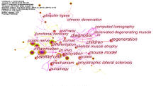
| 序号 | 频次 | 关键词 | 中心性 | 关键词 |
|---|---|---|---|---|
| 1 | 167 | skeletal muscle | 0.33 | activation |
| 2 | 87 | atrophy | 0.3 | mouse model |
| 3 | 77 | expression | 0.28 | functional recovery |
| 4 | 77 | denervation | 0.26 | regeneration |
| 5 | 72 | muscle atrophy | 0.25 | mechanism |
| 6 | 49 | neuromuscular junction | 0.25 | children |
| 7 | 41 | regeneration | 0.22 | protein |
| 8 | 38 | mechanism | 0.22 | in vivo |
| 9 | 38 | oxidative stress | 0.21 | autophagy |
| 10 | 36 | activation | 0.21 | inflammation |
"

"
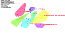
| 分类号 | 大小 | 轮廓值 | 关键词 |
|---|---|---|---|
| 0 | 19 | 0.939 | 肌肉萎缩;蛋白质合成;泛素连接酶;骨骼肌;泛素-蛋白酶体系统 |
| 1 | 19 | 0.795 | 肌萎缩侧索硬化症;基因治疗;小鼠模型;机制;儿童 |
| 2 | 18 | 0.814 | 吸烟;雷帕霉素的哺乳动物靶点;骨骼肌;线粒体;肌萎缩侧索硬化症 |
| 3 | 16 | 0.885 | 人类;神经肌肉接头;脊髓性肌萎缩;失神经萎缩;突变 |
| 4 | 15 | 0.788 | 氧化应激;年龄;肥胖;运动锻炼;运动神经 |
| 5 | 15 | 0.954 | 脊髓损伤;失神经肌肉退行性变;皮肤活检;纤维;家庭功能性电刺激 |
| 6 | 12 | 0.951 | 骨骼肌萎缩;分子机制;微阵列;细胞;萎缩素第一型基因 |
"

"
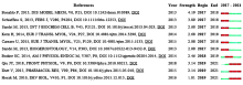
| 序号 | 频次 | 作者 | 期刊 | 参考文献 |
|---|---|---|---|---|
| 1 | 27 | Cohen S | Nature Reviews Drug Discovery | Muscle wasting in disease: molecular mechanisms and promising therapies |
| 2 | 26 | Bodine S C | American Journal of Physiology-Endocrinology and Metabolism | Skeletal muscle atrophy and the E3 ubiquitin ligases MuRF1 and MAFbx/atrogin-1 |
| 3 | 20 | Milan G | Nature Communications | Regulation of autophagy and the ubiquitin-proteasome system by the FoxO transcriptional network during muscle atrophy |
"
| 序号 | 中心性 | 作者 | 期刊 | 参考文献 |
|---|---|---|---|---|
| 1 | 0.31 | He Q R | Experimental and Therapeutic Medicine | MicroRNA-351 inhibits denervation-induced muscle atrophy by targeting TRAF6 |
| 2 | 0.18 | Carnio S | Cell Reports | Autophagy Impairment in Muscle Induces Neuromuscular Junction Degeneration and Precocious Aging |
| 3 | 0.16 | Karam C | Skeletal Muscle | Absence of physiological Ca2+ transients is an initial trigger for mitochondrial dysfunction in skeletal muscle following denervation |
"

| [1] |
PASSIPIERI J A, DIENES J, FRANK J, et al. Adipose stem cells enhance nerve regeneration and muscle function in a peroneal nerve ablation model[J]. Tissue Eng Part A, 2021, 27(5-6):297-310.
doi: 10.1089/ten.tea.2018.0244 |
| [2] | 宋凯凯, 张锴, 贾龙. 周围神经系统损伤的微环境与修复方式[J]. 中国组织工程研究, 2021, 25(4):651-656. |
| SONG K K, ZHANG K, JIA L. Microenvironment and repair methods of peripheral nervous system injury[J]. Chin J Tissue Engineer Res, 2021, 25(4):651-656. | |
| [3] | 王世杨, 孙慧哲, 颜南, 等. 被动训练促进失神经肌萎缩模型大鼠骨骼肌结构和功能的恢复[J]. 中国组织工程研究, 2020, 24(32):5138-5144. |
| WANG S Y, SUN H Z, YAN N, et al. Passive training improves the recovery of skeletal muscle structure and function in rats with denervated muscle atrophy[J]. Chin J Tissue Engineer Res, 2020, 24(32):5138-5144. | |
| [4] |
AGÜERA E, CASTILLA S, LUQUE E, et al. Denervated muscle extract promotes recovery of muscle atrophy through activation of satellite cells. An experimental study[J]. J Sport Health Sci, 2019, 8(1):23-31.
doi: 10.1016/j.jshs.2017.05.007 |
| [5] | MA W, CAI Y, SHEN Y, et al. HDAC4 knockdown alleviates denervation-induced muscle atrophy by inhibiting myogenin-dependent atrogene activation[J]. Front Cell Neurosci, 2021, 15:663384. |
| [6] |
SHIRAKAWA T, MIYAWAKI A, KAWAMOTO T, et al. Natural compounds attenuate denervation-induced skeletal muscle atrophy[J]. Int J Mol Sci, 2021, 22(15):8310.
doi: 10.3390/ijms22158310 |
| [7] | 陈悦, 陈超美, 刘则渊, 等. CiteSpace知识图谱的方法论功能[J]. 科学学研究, 2015, 33(2):242-253. |
| CHEN Y, CHEN C M, LIU Z Y, et al. The methodology function of CiteSpace mapping knowledge domains[J]. Stud Sci Sci, 2015, 33(2):242-253. | |
| [8] | SUN H, SUN J, LI M, et al. Transcriptome analysis of immune receptor activation and energy metabolism reduction as the underlying mechanisms in interleukin-6-induced skeletal muscle atrophy[J]. Front Immunol, 2021, 12:730070. |
| [9] |
HUGHES D C, TURNER D C, BAEHR L M, et al. Knockdown of the E3 ubiquitin ligase UBR5 and its role in skeletal muscle anabolism[J]. Am J Physiol Cell Physiol, 2021, 320(1):C45-C56.
doi: 10.1152/ajpcell.00432.2020 |
| [10] |
HUGHES D C, BAEHR L M, DRISCOLL J R, et al. Identification and characterization of Fbxl22, a novel skeletal muscle atrophy-promoting E3 ubiquitin ligase[J]. Am J Physiol Cell Physiol, 2020, 319(4):C700-C719.
doi: 10.1152/ajpcell.00253.2020 |
| [11] | FRANCO-ROMERO A, SANDRI M. Role of autophagy in muscle disease[J]. Mol Aspects Med, 2021, 82:101041. |
| [12] |
CHEN J, REN S, DUSCHER D, et al. Exosomes from human adipose-derived stem cells promote sciatic nerve regeneration via optimizing Schwann cell function[J]. J Cell Physiol, 2019, 234(12):23097-23110.
doi: 10.1002/jcp.28873 |
| [13] |
SHEHATA A S, AL-GHONEMY N M, AHMED S M, et al. Effect of mesenchymal stem cells on induced skeletal muscle chemodenervation atrophy in adult male albino rats[J]. Int J Biochem Cell Biol, 2017, 85:135-148.
doi: 10.1016/j.biocel.2017.01.016 |
| [14] |
MCKENNA C F, FRY C S. Altered satellite cell dynamics accompany skeletal muscle atrophy during chronic illness, disuse, and aging[J]. Curr Opin Clin Nutr Metab Care, 2017, 20(6):447-452.
doi: 10.1097/MCO.0000000000000409 |
| [15] |
WONG A, GARCIA S M, TAMAKI S, et al. Satellite cell activation and retention of muscle regenerative potential after long-term denervation[J]. Stem Cells, 2021, 39(3):331-344.
doi: 10.1002/stem.3316 |
| [16] |
COHEN S, NATHAN J A, GOLDBERG A L. Muscle wasting in disease: molecular mechanisms and promising therapies[J]. Nat Rev Drug Discov, 2015, 14(1):58-74.
doi: 10.1038/nrd4467 |
| [17] |
HE Q, QIU J, DAI M, et al. MicroRNA-351 inhibits denervation-induced muscle atrophy by targeting TRAF6[J]. Exp Ther Med, 2016, 12(6):4029-4034.
doi: 10.3892/etm.2016.3856 |
| [18] |
BONALDO P, SANDRI M. Cellular and molecular mechanisms of muscle atrophy[J]. Dis Model Mech, 2013, 6(1):25-39.
doi: 10.1242/dmm.010389 |
| [19] |
QIU J, FANG Q, XU T, et al. Mechanistic role of reactive oxygen species and therapeutic potential of antioxidants in denervation- or fasting-induced skeletal muscle atrophy[J]. Front Physiol, 2018, 9:215.
doi: 10.3389/fphys.2018.00215 |
| [20] |
DUTT V, GUPTA S, DABUR R, et al. Skeletal muscle atrophy: potential therapeutic agents and their mechanisms of action[J]. Pharmacol Res, 2015, 99:86-100.
doi: 10.1016/j.phrs.2015.05.010 |
| [21] |
DULAC M, LEDUC-GAUDET J P, REYNAUD O, et al. Drp1 knockdown induces severe muscle atrophy and remodelling, mitochondrial dysfunction, autophagy impairment and denervation[J]. J Physiol, 2020, 598(17):3691-3710.
doi: 10.1113/JP279802 |
| [22] |
SOUTHERN W M, NICHENKO A S, TEHRANI K F, et al. PGC-1α overexpression partially rescues impaired oxidative and contractile pathophysiology following volumetric muscle loss injury[J]. Sci Rep, 2019, 9(1):4079.
doi: 10.1038/s41598-019-40606-6 |
| [23] |
SALUCCI S, FALCIERI E. Polyphenols and their potential role in preventing skeletal muscle atrophy[J]. Nutr Res, 2020, 74:10-22.
doi: 10.1016/j.nutres.2019.11.004 |
| [24] |
YANG X, XUE P, YUAN M, et al. SESN2 protects against denervated muscle atrophy through unfolded protein response and mitophagy[J]. Cell Death Dis, 2021, 12(9):805.
doi: 10.1038/s41419-021-04094-9 |
| [25] |
WADA A, NISHIO N, YOKOI S, et al. Safety and feasibility of fat injection therapy with adipose-derived stem cells in a rabbit hypoglossal nerve paralysis model: a pilot study[J]. Auris Nasus Larynx, 2021, 48(2):274-280.
doi: 10.1016/j.anl.2020.08.003 |
| [26] |
TABBAA M, RUZ G T, CAMPELJ D G, et al. The regulation of polyamine pathway proteins in models of skeletal muscle hypertrophy and atrophy: a potential role for mTORC1[J]. Am J Physiol Cell Physiol, 2021, 320(6):C987-C999.
doi: 10.1152/ajpcell.00078.2021 |
| [27] | COSTA D M, CRUZ-FILHO J D, VASCONCELOS A, et al. Oxytocin induces anti-catabolic and anabolic effects on protein metabolism in the female rat oxidative skeletal muscle[J]. Life Sci, 2021, 279:119665. |
| [28] | CUI W, LIU C X, ZHANG Y C, et al. A novel oleanolic acid derivative HA-19 ameliorates muscle atrophy via promoting protein synjournal and preventing protein degradation[J]. Toxicol Appl Pharmacol, 2019, 378:114625. |
| [29] |
LANGER H T, SENDEN J, GIJSEN A P, et al. Muscle atrophy due to nerve damage is accompanied by elevated myofibrillar protein synjournal rates[J]. Front Physiol, 2018, 9:1220.
doi: 10.3389/fphys.2018.01220 |
| [30] |
FERLIN A, DE TONI L, AGOULNIK A I, et al. Protective role of testicular hormone INSL3 From atrophy and weakness in skeletal muscle[J]. Front Endocrinol (Lausanne), 2018, 9:562.
doi: 10.3389/fendo.2018.00562 |
| [31] |
XU Z, FENG X, DONG J, et al. Cardiac troponin T and fast skeletal muscle denervation in ageing[J]. J Cachexia Sarcopenia Muscle, 2017, 8(5):808-823.
doi: 10.1002/jcsm.12204 |
| [32] |
WILSON R J, DRAKE J C, CUI D, et al. Mitochondrial protein S-nitrosation protects against ischemia reperfusion-induced denervation at neuromuscular junction in skeletal muscle[J]. Free Radic Biol Med, 2018, 117:180-190.
doi: 10.1016/j.freeradbiomed.2018.02.006 |
| [33] |
MACHADO J, SILVEIRA W A, GONÇALVES D A, et al. α-Calcitonin gene-related peptide inhibits autophagy and calpain systems and maintains the stability of neuromuscular junction in denervated muscles[J]. Mol Metab, 2019, 28:91-106.
doi: 10.1016/j.molmet.2019.06.024 |
| [34] |
LI L, YOKOYAMA H, KABURAGI H, et al. Remnant neuromuscular junctions in denervated muscles contribute to functional recovery in delayed peripheral nerve repair[J]. Neural Regen Res, 2020, 15(4):731-738.
doi: 10.4103/1673-5374.266925 |
| [35] |
MONTI E, REGGIANI C, FRANCHI M V, et al. Neuromuscular junction instability and altered intracellular calcium handling as early determinants of force loss during unloading in humans[J]. J Physiol, 2021, 599(12):3037-3061.
doi: 10.1113/JP281365 |
| [36] |
CARRARO U, KERN H, GAVA P, et al. Recovery from muscle weakness by exercise and FES: lessons from masters, active or sedentary seniors and SCI patients[J]. Aging Clin Exp Res, 2017, 29(4):579-590.
doi: 10.1007/s40520-016-0619-1 |
| [37] | HARBO T, MARKVARDSEN L K, HELLFRITZSCH M B, et al. Neuromuscular electrical stimulation in early rehabilitation of Guillain-Barré syndrome: a pilot study[J]. Muscle Nerve, 2019, 59(4):481-484. |
| [38] |
ALBERTIN G, HOFER C, ZAMPIERI S, et al. In complete SCI patients, long-term functional electrical stimulation of permanent denervated muscles increases epidermis thickness[J]. Neurol Res, 2018, 40(4):277-282.
doi: 10.1080/01616412.2018.1436877 |
| [39] |
PINHEIRO-DARDIS C M, RUSSO T L. Electrical stimulation based on chronaxie increases fibrosis and modulates TWEAK/Fn14, TGF-β/Myostatin, and MMP pathways in denervated muscles[J]. Am J Phys Med Rehabil, 2017, 96(4):260-267.
doi: 10.1097/PHM.0000000000000601 |
| [40] |
OTZEL D M, KOK H J, GRAHAM Z A, et al. Pharmacologic approaches to prevent skeletal muscle atrophy after spinal cord injury[J]. Curr Opin Pharmacol, 2021, 60:193-199.
doi: 10.1016/j.coph.2021.07.023 |
| [41] |
RODRÍGUEZ-CUETO C, GARCÍA-TOSCANO L, SANTOS-GARCÍA I, et al. Targeting the CB(2) receptor and other endocannabinoid elements to delay disease progression in amyotrophic lateral sclerosis[J]. Br J Pharmacol, 2021, 178(6):1373-1387.
doi: 10.1111/bph.15386 |
| [42] |
YIN M Z, KIM H J, SUH E Y, et al. Endurance exercise training restores atrophy-induced decreases of myogenic response and ionic currents in rat skeletal muscle artery[J]. J Appl Physiol (1985), 2019, 126(6):1713-1724.
doi: 10.1152/japplphysiol.00962.2018 |
| [43] |
ZHAO J, TIAN Z, KADOMATSU T, et al. Age-dependent increase in angiopoietin-like protein 2 accelerates skeletal muscle loss in mice[J]. J Biol Chem, 2018, 293(5):1596-1609.
doi: 10.1074/jbc.M117.814996 |
| [44] |
CAMPOS J C, BAEHR L M, GOMES K, et al. Exercise prevents impaired autophagy and proteostasis in a model of neurogenic myopathy[J]. Sci Rep, 2018, 8(1):11818.
doi: 10.1038/s41598-018-30365-1 |
| [45] |
REZA M M, SUBRAMANIYAM N, SIM C M, et al. Irisin is a pro-myogenic factor that induces skeletal muscle hypertrophy and rescues denervation-induced atrophy[J]. Nat Commun, 2017, 8(1):1104.
doi: 10.1038/s41467-017-01131-0 |
| [1] | ZOU Yucong, ZHOU Jing, LIN Weiming, LI Dongxia, WANG Juan, WANG Yuqi, WANG Yulong. Treatments for prolonged disorder of consciousness in recent five years: a visualized analysis [J]. 《Chinese Journal of Rehabilitation Theory and Practice》, 2023, 29(9): 1065-1071. |
| [2] | MA Tiantian, YU Zifu, QIN Fang, LENG Xiaoxuan, LIU Xihua. Application of constraint-induced movement therapy in the field of rehabilitation: a visualized analysis [J]. 《Chinese Journal of Rehabilitation Theory and Practice》, 2023, 29(7): 822-832. |
| [3] | WU Qianhao, HOU Rongjie, FU Liyuan. Pelvic floor rehabilitation domestic and abroad in the last decade: a visualized analysis [J]. 《Chinese Journal of Rehabilitation Theory and Practice》, 2023, 29(6): 673-685. |
| [4] | XU Minjie, WANG Bo, ZHOU Li, WANG Haifang, LEI Xiaojing, LI Ying, BAO Weiwei, MA Ya'nan, CHANG Jingling. Verbal and nonverbal cognitive function of aphasia after stroke based on Web of Science database: a visualized analysis [J]. 《Chinese Journal of Rehabilitation Theory and Practice》, 2023, 29(4): 452-464. |
| [5] | TANG Zhiqing, LIU Tianhao, HAN Kaiyue, LIU Ying, ZHAO Jingdu, ZHANG Hao. Transcranial magnetic stimulation for stroke in recent five years: a visualized analysis [J]. 《Chinese Journal of Rehabilitation Theory and Practice》, 2023, 29(3): 294-301. |
| [6] | YUAN Yuan, ZHANG Guohui, ZHANG Hong. Exercise-induced muscle damage and physiotherapy: a visualized analysis [J]. 《Chinese Journal of Rehabilitation Theory and Practice》, 2023, 29(3): 312-319. |
| [7] | LIU Mingyue, FAN Yalei, ZHANG Meng, SONG Xueyi, LI Zhe. Brain-computer interface technology for stroke in the past decade: a visualized analysis [J]. 《Chinese Journal of Rehabilitation Theory and Practice》, 2023, 29(2): 223-230. |
| [8] | HU Jiaquan, ZHU Liling, JIANG Zhimei. Early screening tools for autism spectrum disorder in the past two decades: a visualized analysis [J]. 《Chinese Journal of Rehabilitation Theory and Practice》, 2023, 29(11): 1304-1315. |
| [9] | ZHANG Ning, YANG Yuanbin, TIAN Haolin, WAN Mengying. Application of functional near-infrared spectroscopy in rehabilitation: a visualized analysis [J]. 《Chinese Journal of Rehabilitation Theory and Practice》, 2023, 29(10): 1171-1178. |
| [10] | XU Hongli, ZHANG Qian, XUE Qing, YANG Yulin, MA Lihong, QIU Zhengang. Domestic and international researches related to postpartum rehabilitation in the last decade: a visualized analysis [J]. 《Chinese Journal of Rehabilitation Theory and Practice》, 2023, 29(10): 1179-1188. |
| [11] | SHI Manxinyu,MENG Detao,FANG Boyan. Advance in Parkinson's disease rehabilitation: a visualization analysis [J]. 《Chinese Journal of Rehabilitation Theory and Practice》, 2022, 28(9): 1060-1064. |
| [12] | ZHENG Wei,SUN Libing,LIU Chunhua,HAO Chuanping. Physical fitness for children and adolescents with intellectual disabilities: a visualized analysis [J]. 《Chinese Journal of Rehabilitation Theory and Practice》, 2022, 28(12): 1435-1443. |
| [13] | JIANG Jiarui,NIU Zhendong. Deep learning for diagnosis of Alzheimer's disease in the past five years: a visualized analysis [J]. 《Chinese Journal of Rehabilitation Theory and Practice》, 2022, 28(11): 1318-1324. |
| [14] | LAN Wanting,ZHONG Jiugen,SHEN Yingying,GONG Jiaheng,ZOU Zhi,HOU Xiaohui. Mechanism of exercise reducing neuroinflammation in autism spectrum disorder: a visualized analysis [J]. 《Chinese Journal of Rehabilitation Theory and Practice》, 2022, 28(10): 1190-1197. |
| [15] | LIU Jun,LI Jian-jun,GAO Feng,ZHANG Wen-hao,LIU Chang-bin,LI Jun,YANG De-gang,ZHANG Xin,ZHANG Chao,QIN Chuan. Advance in Nutritional Status for Spinal Cord Injury [J]. 《Chinese Journal of Rehabilitation Theory and Practice》, 2020, 26(4): 388-392. |
| Viewed | ||||||
|
Full text |
|
|||||
|
Abstract |
|
|||||
|
||