


《Chinese Journal of Rehabilitation Theory and Practice》 ›› 2022, Vol. 28 ›› Issue (4): 415-420.doi: 10.3969/j.issn.1006-9771.2022.04.007
Previous Articles Next Articles
HUANG Feifei1,WANG Wansong2,DONG Xiaoyang2,FENG Zhen2( )
)
Received:2022-01-11
Revised:2022-03-04
Published:2022-04-25
Online:2022-05-05
Contact:
FENG Zhen
E-mail:fengzhenly@sina.com
Supported by:HUANG Feifei,WANG Wansong,DONG Xiaoyang,FENG Zhen. Repair of blood-brain barrier by hyperbaric oxygen via autophagy in rats with cerebral ischemia-reperfusion injury[J]. 《Chinese Journal of Rehabilitation Theory and Practice》, 2022, 28(4): 415-420.
"
| 组别 | n | EB含量 |
|---|---|---|
| 假手术照组 | 6 | 0.887±0.191 |
| 模型组 | 6 | 2.423±0.223a |
| 高压氧组 | 6 | 1.989±0.163b |
| 抑制剂组 | 6 | 2.249±0.174c |
| F值 | 79.726 | |
| P值 | < 0.001 |
"

"
"
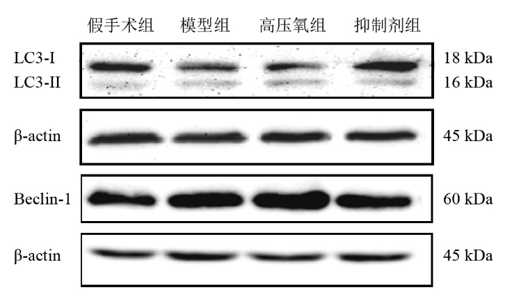
| 组别 | n | LC3-Ⅱ/LC3-Ⅰ | Beclin-1/β-actin |
|---|---|---|---|
| 假手术组 | 6 | 0.255±0.041 | 0.873±0.174 |
| 模型组 | 6 | 0.385±0.064a | 1.235±0.090a |
| 高压氧组 | 6 | 0.487±0.066b | 1.549±0.660b |
| 抑制剂组 | 6 | 0.268±0.082c,d | 0.934±0.048c,d |
| F值 | 16.927 | 51.700 | |
| P值 | < 0.001 | < 0.001 |
| [1] | 殷鹏, 齐金蕾, 刘韫宁, 等. 2005~2017年中国疾病负担研究报告[J]. 中国循环杂志, 2019, 34(12):1145-1154. |
| YIN P, QI J L, LIU Y N, et al. Burden of disease in the Chinese population from 2005 to 2017[J]. Chin Circul J, 2019, 34(12):1145-1154. | |
| [2] | WANG Y, DUAN K, ZHANG A, et al. Clinical and imaging analysis of cerebral infarction caused by spontaneous cerebral artery dissection based on augmented reality technology[J]. J Healthc Eng, 2021, 2021(7):6671121. |
| [3] | JIANG X, ANDJELKOVIC A V, ZHU L, et al. Blood-brain barrier dysfunction and recovery after ischemic stroke[J]. Prog Neurobiol, 2018, 163- 164:144-171. |
| [4] | LI H Y, GAO A J, FENG D X, et al. Evaluation of the protective potential of brain microvascular endothelial cell autophagy on blood-brain barrier integrity during experimental cerebral ischemia-reperfusion injury[J]. Stroke Res, 2014, 5(5):618-626. |
| [5] | 胡慧军, 范丹峰. 2018年版高压氧治疗适应证与禁忌证解读[J]. 中华航海医学与高气压医学杂志, 2020, 27(1):127-128. |
| HU H J, FAN D F. Interpretation of indications and contraindications of hyperbaric oxygen therapy in 2018[J]. Chin J Naut Med Hyperbar Med, 2020, 27(1):127-128. | |
| [6] |
GOTTFRIED I, SCHOTTLENDER N, ASHERY U. Hyperbaric oxygen treatment: from mechanisms to cognitive improvement[J]. Biomolecules, 2021, 11(10):1520.
doi: 10.3390/biom11101520 |
| [7] |
CHEN C Y, WU R W, NAI W T, et al. Increased circulating endothelial progenitor cells and improved short-term outcomes in acute non-cardioembolic stroke after hyperbaric oxygen therapy[J]. J Transl Med, 2018, 16(1):255.
doi: 10.1186/s12967-018-1629-x |
| [8] |
YAN D, SHAN J, ZE Y, et al. The effects of combined hyperbaric oxygen therapy on patients with post-stroke depression[J]. J Phys Therapy Sci, 2015, 27(5):1295-1297.
doi: 10.1589/jpts.27.1295 |
| [9] |
ABDULLAHI W, TRIPATHI D, RONALDSON P T. Blood‐brain barrier dysfunction in ischemic stroke: targeting tight junctions and transporters for vascular protection[J]. Am J Physiol Cell Physiol, 2018, 315(3):C343-C356.
doi: 10.1152/ajpcell.00095.2018 |
| [10] |
FANG Z P, FENG Y, LI Y H, et al. Neuroprotective autophagic flux induced by hyperbaric oxygen preconditioning is mediated by cystatin C[J]. Neurosci Bull, 2019, 35(2):336-346.
doi: 10.1007/s12264-018-0313-8 |
| [11] |
CHEN Z, HU Q, XIE Q, et al. Effects of treadmill exercise on motor and cognitive function recovery of MCAO mice through the caveolin-1/VEGF signaling pathway in ischemic penumbra[J]. Neurochem Res, 2019, 44(4):930-946.
doi: 10.1007/s11064-019-02728-1 |
| [12] |
SUGAWARA T, AYER R, JADHAV V, et al. A new grading system evaluating bleeding scale in filament perforation subarachnoid hemorrhage rat model[J]. J Neurosci Methods, 2008, 167(2):327-334.
doi: 10.1016/j.jneumeth.2007.08.004 |
| [13] | 赵龙, 李红玲, 赵艳颖, 等. 不同吸氧时间高压氧治疗对脑出血大鼠血肿周围AQP4和SOD表达的影响[J]. 中国康复医学杂志, 2016, 31(11):1213-1218. |
| ZHAO L, LI H L, ZHAO Y Y, et al. Effects of different oxygen uptake time of hyperbaric oxygen on perihematomal edema, expression of aquaporin-4 and superoxide dismutase in rats with intracerebral hemorrhage[J]. Chin J Rehabil Med, 2016, 31(11):1213-1218. | |
| [14] | 梁碧莹, 朱路文, 唐强, 等. 运动预处理对大鼠脑缺血再灌注损伤后血脑屏障通透性的影响[J]. 中国康复理论与实践, 2019, 25(3):302-306. |
| LIANG B Y, ZHU L W, TANG Q. Effects of exercise preconditioning on blood-brain barrier permeability in rats after cerebral ischemia-reperfusion injury[J]. Chin J Rehabil Theory Pract, 2019, 25(3):302-306. | |
| [15] |
YUN H R, YONG H J, KIM J, et al. Roles of autophagy in oxidative stress[J]. Int J Mol Sci, 2020, 21(9):3289.
doi: 10.3390/ijms21093289 |
| [16] |
LI J, YANG F, GUO J, et al. 17-AAG post-treatment ameliorates memory impairment and hippocampal CA1 neuronal autophagic death induced by transient global cerebral ischemia[J]. Brain Res, 2015, 1610:80-88.
doi: 10.1016/j.brainres.2015.03.051 |
| [17] |
IZUSHIMA N. Methods for monitoring autophagy[J]. Int J Biochem Cell Biol, 2004, 36(12):2491-2502.
doi: 10.1016/j.biocel.2004.02.005 |
| [18] |
SUN Q, FAN W, ZHONG Q. Regulation of Beclin 1 in autophagy[J]. Autophagy, 2009, 5(5):713-716.
doi: 10.4161/auto.5.5.8524 |
| [19] | GAO Q. Oxidative stress and autophagy[J]. Adv Exp Med Biol, 2019, 1206:179-198. |
| [20] |
WU M Y, YIANG G T, LIAO W T, et al. Current mechanistic concepts in ischemia and reperfusion injury[J]. Cell Physiol Biochem, 2018, 46(4):1650-1667.
doi: 10.1159/000489241 |
| [21] | 李丹丹, 肖学进, 秦书俭, 等. 脑缺血再灌注损伤程度与自噬相关基因LC3、mTOR表达的相关性研究[J]. 中国临床解剖学杂志, 2018, 36(2):182-186. |
| LI D D, XIAO X J, QIN S J, et al. Correlation between the degree of cerebral ischemia-reperfusion injury and the expression of LC3 and mTOR[J]. Chin J Clin Anatome, 2018, 36(2):182-186. | |
| [22] | DEL ZOPPO G J, HALLENBECK J M. Advances in the vascular pathophysiology of ischemic stroke[J]. Thromb Res, 2000, 98(3):73-81. |
| [23] |
YANG C, HAWKINS K E, DORE S, et al. Neuroinflammatory mechanisms of blood-brain barrier damage in ischemic stroke[J]. AJP Cell Physiology, 2019, 316(2):C135-C153.
doi: 10.1152/ajpcell.00136.2018 |
| [24] |
LIU J Y, THOM M, CATARINO C B, et al. Neuropathology of the blood-brain barrier and pharmaco-resistance in human epilepsy[J]. Brain, 2012, 135(10):3115-3133.
doi: 10.1093/brain/aws147 |
| [25] |
FANG L, LI X, ZHONG Y, et al. Autophagy protects human brain microvascular endothelial cells against methylglyoxal-induced injuries, reproducible in a cerebral ischemic model in diabetic rats[J]. J Neurochem, 2015, 135(2):431-440.
doi: 10.1111/jnc.13277 |
| [26] | CHEN C H, CHEN S Y, WANG V, et al. Effects of repetitive hyperbaric oxygen treatment in patients with acute cerebral infarction: a pilot study[J]. Sci World J, 2012, 2012(7):1-5. |
| [27] |
HU Q, MANAENKO A, BIAN H, et al. Hyperbaric oxygen reduces infarction volume and hemorrhagic transformation through ATP/NAD+/Sirt1 pathway in hyperglycemic middle cerebral artery occlusion rats[J]. Stroke, 2017, 48(6):1655-1664.
doi: 10.1161/STROKEAHA.116.015753 |
| [28] | 张爱民, 蒋宗滨. 高压氧预处理通过HIF-1α/VEGF通路减轻大脑缺血再灌注损伤[J]. 中国病理生理杂志, 2018, 34(11):2048-2053. |
| ZHANG A M, JIANG Z B. Role of HIF-1α/VEGF pathway in treatment of cerebral ischemia-reperfusion injury by hyperbaric oxygen pretreatment[J]. Chin J Pathophysiol, 2018, 34(11):2048-2053. | |
| [29] |
LI H Z, CHEN J F, LIU M, et al. Effect of hyperbaric oxygen on the permeability of the blood-brain barrier in rats with global cerebral ischemia/reperfusion injury[J]. Biomed Pharmacother, 2018, 108:1725-1730.
doi: 10.1016/j.biopha.2018.10.025 |
| [30] |
GAO Z X, RAO J, LI Y H. Hyperbaric oxygen preconditioning improves postoperative cognitive dysfunction by reducing oxidant stress and inflammation[J]. Neural Regen Res, 2017, 12(2):329-336.
doi: 10.4103/1673-5374.200816 |
| [31] |
WANG S D, FU Y Y, HAN X Y, et al. Hyperbaric oxygen preconditioning protects against cerebral ischemia/reperfusion injury by inhibiting mitochondrial apoptosis and energy metabolism disturbance[J]. Neurochem Res, 2021, 46(4):866-877.
doi: 10.1007/s11064-020-03219-4 |
| [32] |
XING P C, MA K, LI L J, et al. The protection effect and mechanism of hyperbaric oxygen therapy in rat brain with traumatic injury[J]. Acta Cir Bras, 2018, 33(4):341-353.
doi: 10.1590/s0102-865020180040000006 |
| [33] |
DONG H L, LU Y, BAI X G, et al. Autophagy activation is involved in neuroprotection induced by hyperbaric oxygen preconditioning against focal cerebral ischemia in rats[J]. Brain Res, 2011, 1402:109-121.
doi: 10.1016/j.brainres.2011.05.049 |
| [34] | 于敏, 梁维娣, 张玉鹏, 等. 超早期高压氧处理对持续脑缺血大鼠血脑屏障通透性的影响[J]. 中华航海医学与高气压医学杂志, 2018, 25(6):378-381. |
| YU M, LIANG W D, ZHANG Y P, et al. Effect of ultra early hyperbaric oxygen treatment on the permeability of blood-brain barrier in persistent cerebral ischemic rats[J]. Chin J Nautic Med Hyperbaric Med, 2018, 25(6):378-381. | |
| [35] |
CHEN L F, TIAN Y F, LIN C H. Repetitive hyperbaric oxygen therapy provides better effects on brain inflammation and oxidative damage in rats with focal cerebral ischemia[J]. J Formos Med Assoc, 2014, 113(9):620-628.
doi: 10.1016/j.jfma.2014.03.012 |
| [1] | SUN Tengfang, REN Mengting, YANG Lin, WANG Yaoting, WANG Hongyu, YAN Xingzhou. Effect of hyperbaric oxygen therapy combined with repetitive peripheral magnetic stimulation on ankle motor function and balance of stroke patients [J]. 《Chinese Journal of Rehabilitation Theory and Practice》, 2023, 29(8): 875-881. |
| [2] | KANG Xiaoyu, LIU Lixu, WANG Wenzhu, WANG Yunlei. Effects of pramipexole combined with levodopa on cognitive and mitochondrial function of rats after global cerebral ischemia-reperfusion injury [J]. 《Chinese Journal of Rehabilitation Theory and Practice》, 2023, 29(5): 533-540. |
| [3] | YUAN Huiping, FENG Xiaojun, JIANG Dongsheng, CHE Xingwang, FAN Lianbin. Effect of hyperbaric oxygen combined with dynamic scalp acupuncture on motor function for stroke patients with hemiplegia [J]. 《Chinese Journal of Rehabilitation Theory and Practice》, 2023, 29(10): 1208-1213. |
| [4] | TAO Miao-miao,XU Ming-shu,ZHANG Ying-jie,CHENG Ai-fang,DENG Yun-yi. Advance in Role of Mitochondria in Remote Ischemic Postconditioning for Cerebral Ischemia-reperfusion Injury (review) [J]. 《Chinese Journal of Rehabilitation Theory and Practice》, 2020, 26(9): 1061-1065. |
| [5] | LI Hong-yu,ZHANG Ji-yao,ZHANG Yu,ZHANG Shi-qiang,HUANG Hui-lin,LI Jia-shuai,TANG Qiang. Effects of Exercise Preconditioning on Expression of Autophagy-related Proteins and Apoptosis after Myocardial Ischemia-reperfusion Injury in Rats [J]. 《Chinese Journal of Rehabilitation Theory and Practice》, 2020, 26(8): 903-907. |
| [6] | WEI Hai-ping,GUO Jia,WANG Huan,GE Zhao-ming. Effects of Vagus Nerve Stimulation on Adenosine Monophosphate Activated Protein Kinase-Silent Mating Type Information Regulation 2 Homolog 1 Pathway in Ischemia-reperfusion Rats [J]. 《Chinese Journal of Rehabilitation Theory and Practice》, 2020, 26(7): 775-779. |
| [7] | HUANG Qin,ZHOU Li-zhi,TAN Cai-ling,ZHENG Su,PENG Li. Effects of Acupoint Catgut Embedding on Motor Function in Cerebral Ischemia-reperfusion Injury in Rats [J]. 《Chinese Journal of Rehabilitation Theory and Practice》, 2020, 26(10): 1167-1175. |
| [8] | LI Dan-dan, MA Rui, HUANG Min-ying, ZHAO Hong-yi, HUANG Yong-hua. Influence of Sleep Deprivation on Blood-brain Barrier in Rats [J]. 《Chinese Journal of Rehabilitation Theory and Practice》, 2019, 25(9): 1020-1025. |
| [9] | LIANG Bi-ying, ZHU Lu-wen, TANG Qiang, YE Tao, LI Hong-yu, LI Bao-long, RUAN Ye. Effects of Exercise Preconditioning on Blood-brain Barrier Permeability in Rats after Cerebral Ischemia-reperfusion Injury [J]. 《Chinese Journal of Rehabilitation Theory and Practice》, 2019, 25(3): 302-306. |
| [10] | LIANG Ji-ling, XIE Jin-feng, WANG Cen-yi, CHEN Ning. Regulatory Role of Exercise-induced Autophagy in Rehabilitation of Sarcopenia (review) [J]. 《Chinese Journal of Rehabilitation Theory and Practice》, 2019, 25(3): 334-337. |
| [11] | YANG Zhi-xue, TANG Cheng-lin, LI Xiao-hong, ZHU Zheng-wei, HUANG Si-qin, DAI Ni, WU Meng-jia, QIU Li, AN Hui-yu. Effect of Electroacupuncture on Blood-brain Barrier in SAMP8 Mice [J]. 《Chinese Journal of Rehabilitation Theory and Practice》, 2019, 25(2): 156-161. |
| [12] | LIU Wei-lin, ZHENG Yi, JIN Ting-ting, ZHANG Yu-hao, SHI Dan, CHEN Li-dian, TAO Jing. Effect of Electroacupuncture on Neural Stem Cell Differentiation via Regulating MicroRNA-34a in Rats with Cerebral Ischemia-reperfusion Injury [J]. 《Chinese Journal of Rehabilitation Theory and Practice》, 2019, 25(2): 162-171. |
| [13] | AN Hui-yu, TANG Cheng-lin, HUANG Si-qin, ZHAO Dan-dan, LUO Ao, WU Meng-jia, TAN Cheng-fang, QIU Li, WAN Xiao-feng, MA Xiang. Effect of Tuina on Autophagy-related Factor Beclin-1, Vacuolar Protein Sorting and Microtubule-associated Protein Light Chain 3 in Rats with Denervated Skeletal Muscle Atrophy [J]. 《Chinese Journal of Rehabilitation Theory and Practice》, 2019, 25(2): 184-191. |
| [14] | TIAN Chao, MANA Lu-lu, YUAN Meng-chen, WANG Li-qin, AN Na, ZHANG Han-lai, XING Yan-wei, SUN Yi-kun, GAO Yong-hong. Effect of Qingkailing on Expression of Toll-like Receptor 4, gp91phox and Zonula Occludens-1 in Cerebrovascular Endothelial Cells Induced by Hypoxia Activating Microglias [J]. 《Chinese Journal of Rehabilitation Theory and Practice》, 2019, 25(11): 1303-1308. |
| [15] | PAN Song-bin, WAN Lin, SHAO Wei, TANG Kun, YAO Han-yun. Effect of Huangjiao Granule on Inflammatory Factors and Apoptosis-related Proteins in Rats with Cerebral Ischemia-reperfusion Injury [J]. 《Chinese Journal of Rehabilitation Theory and Practice》, 2018, 24(8): 893-899. |
| Viewed | ||||||
|
Full text |
|
|||||
|
Abstract |
|
|||||
|
||