


Chinese Journal of Rehabilitation Theory and Practice ›› 2024, Vol. 30 ›› Issue (2): 223-231.doi: 10.3969/j.issn.1006-9771.2024.02.013
Previous Articles Next Articles
ZHANG Minglan1a, ZHANG Lingling1b, WANG Lisha1a, LIU Li1b, GAO Run1b, RAO Jiang1b, LIU Wan1b, XIA Zi'an1b, ZHANG Chuanwen1b, CHENG Xinxin1b( )
)
Received:2023-04-17
Revised:2023-12-12
Published:2024-02-25
Online:2024-03-01
Contact:
CHENG Xinxin
E-mail:417735101@qq.com
Supported by:CLC Number:
ZHANG Minglan, ZHANG Lingling, WANG Lisha, LIU Li, GAO Run, RAO Jiang, LIU Wan, XIA Zi'an, ZHANG Chuanwen, CHENG Xinxin. Impact of autonomic nerve function on motor function in patients with post-stroke depression[J]. Chinese Journal of Rehabilitation Theory and Practice, 2024, 30(2): 223-231.
Table 1
Comparison of baseline data between two groups"
| 组别 | n | 性别/n | 年龄/岁 | 病程/周 | 受损半球/n | ||
|---|---|---|---|---|---|---|---|
| 男 | 女 | 左侧 | 右侧 | ||||
| 对照组 | 30 | 16 | 14 | 61.72±14.36 | 14.46±3.11 | 15 | 15 |
| PSD组 | 30 | 17 | 13 | 63.80±11.90 | 15.23±4.38 | 14 | 16 |
| χ2/t值 | 0.211 | 0.289 | -0.376 | 0.443 | |||
| P值 | 0.786 | 0.598 | 0.710 | 0.561 | |||
Table 2
Comparison of parameters of HRV between two groups"
| 指标 | 对照组(n = 30) | PSD组(n = 30) | t值 | P值 |
|---|---|---|---|---|
| SDNN/ms | 30.43±11.97 | 20.73±12.12 | -3.065 | 0.003 |
| RMSSD/ms | 16.50±4.87 | 15.47±2.92 | -2.224 | 0.030 |
| PNN50/% | 15.03±4.18 | 14.37±2.59 | -2.121 | 0.039 |
| TP/(ms2·Hz-1) | 1047.67±516.58 | 513.47±89.56 | -5.487 | < 0.001 |
| VLF/(ms2·Hz-1) | 517.43±90.76 | 256.21±78.06 | -11.751 | < 0.001 |
| LF/(ms2·Hz-1) | 215.41±84.72 | 176.93±51.34 | -2.092 | 0.041 |
| HF/(ms2·Hz-1) | 163.75±62.10 | 130.82±25.93 | -2.635 | 0.011 |
| LF/HF | 1.44±0.80 | 1.39±0.42 | -2.240 | 0.031 |
Table 3
Comparison of scores of HAMD, FMA and MBI between two groups"
| 组别 | n | HAMD | FMA | MBI |
|---|---|---|---|---|
| 对照组 | 30 | 4.60±1.81 | 70.17±12.84 | 67.57±9.06 |
| PSD组 | 30 | 16.47±5.39 | 21.96±9.91 | 30.19±9.95 |
| t值 | 11.524 | -16.276 | -14.797 | |
| P值 | < 0.001 | < 0.001 | < 0.001 |
Table 4
Correlation of FMA score to HRV parameters and HAMD in PSD group"
| 项目 | FMA | |
|---|---|---|
| r值 | P值 | |
| SDNN | 0.905 | < 0.001 |
| RMSSD | 0.957 | < 0.001 |
| PNN50 | 0.982 | < 0.001 |
| TP | 0.940 | < 0.001 |
| VLF | 0.870 | < 0.001 |
| LF | 0.394 | 0.034 |
| HF | 0.223 | 0.246 |
| LF/HF | 0.248 | 0.195 |
| HAMD | -0.952 | < 0.001 |
Table 5
Correlation of MBI score to HRV parameters and HAMD in PSD group"
| 项目 | MBI | |
|---|---|---|
| r值 | P值 | |
| SDNN | 0.934 | < 0.001 |
| RMSSD | 0.954 | < 0.001 |
| PNN50 | 0.952 | < 0.001 |
| TP | 0.961 | < 0.001 |
| VLF | 0.919 | < 0.001 |
| LF | 0.409 | 0.027 |
| HF | 0.350 | 0.063 |
| LF/HF | 0.179 | 0.352 |
| HAMD | -0.919 | < 0.001 |
Table 6
Correlation of HAMD score to HRV parameters in PSD group"
| 项目 | HAMD | |
|---|---|---|
| r值 | P值 | |
| SDNN | -0.810 | < 0.001 |
| RMSSD | -0.966 | < 0.001 |
| PNN50 | -0.954 | < 0.001 |
| TP | -0.883 | < 0.001 |
| VLF | -0.769 | < 0.001 |
| LF | -0.355 | 0.054 |
| HF | -0.247 | 0.188 |
| LF/HF | -0.211 | 0.263 |
Table 7
Brain regions with increased ReHo in PSD group"
| 脑区 | MNI坐标 | 体素 | t值 | ||
|---|---|---|---|---|---|
| X | Y | Z | |||
| 右侧直回 | 9 | 57 | -18 | 142 | 6.575 |
| 左侧内侧和旁扣带脑回 | -9 | -42 | 33 | 204 | 4.925 |
Table 8
Brain regions with decreased ReHo in PSD group"
| 脑区 | MNI坐标 | 体素 | t值 | ||
|---|---|---|---|---|---|
| X | Y | Z | |||
| 右侧小脑 | 27 | -51 | -24 | 191 | -6.487 |
| 左侧颞中回 | -51 | -72 | 18 | 140 | -5.516 |
| 左侧中央前回 | -36 | -12 | 51 | 119 | -4.764 |
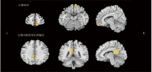
Figure 1
Brain regions with increased ReHo in PSD group"

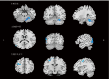
Figure 2
Brain regions with reduced ReHo in PSD group"

| [1] | PAFFIANOWICZ D, ESPIRIDION E D. Chronic post-stroke psychosis with left cortical and bilateral inferior cerebellar involvement[J]. Cureus, 2019, 11(12): e6437. |
| [2] | 郑书恭. 缺血性脑卒中患者卒中后抑郁的相关因素分析[J]. 中国现代药物应用, 2021, 15(12): 105-107. |
| ZHENG S G. Analysis of factors associated with post-stroke depression in ischaemic stroke patients[J]. Chin J Mod Drug Appl, 2021, 15(12): 105-107. | |
| [3] | CAI W, MUELLER C, LI Y J, et al. Post stroke depression and risk of stroke recurrence and mortality: a systematic review and meta-analysis[J]. Ageing Res Rev, 2019(50): 102-109. |
| [4] |
房翠, 王秀丽, 李玲钰, 等. 缺血性脑卒中后抑郁的发病机制及新生物学指标[J]. 医学研究与教育, 2023, 40(2): 18-24.
doi: 10.3969/j.issn.1674-490X.2023.02.003 |
| FANG C, WANG X L, LI L Y, et al. Pathogenesis and new biological markers of post-ischemic stroke depression[J]. Med Res Educ, 2023, 40(2): 18-24. | |
| [5] |
TAY J, MORRIS R G, MARKUS H S. Apathy after stroke: diagnosis, mechanisms, consequences, and treatment[J]. Int J Stroke, 2021, 16(5): 510-518.
doi: 10.1177/1747493021990906 pmid: 33527880 |
| [6] | 袁淑雅, 邸聪冉, 席子明, 等. 脑卒中后抑郁患者的临床治疗[J]. 中国医药科学, 2021, 11(21): 45-48, 78. |
| YUAN S Y, DI C R, XI Z M, et al. Clinical treatment of depressed patients after stroke[J]. Chin Med Pharm, 2021, 11 (21): 45-48, 78. | |
| [7] | PAN C, LI G, JING P, et al. Structural disconnection-based prediction of poststroke depression[J]. Transl Psych, 2022, 12(1): 461. |
| [8] |
HAYANO J, YUDA E. Pitfalls of assessment of autonomic function by heart rate variability[J]. J Physiol Anthropol, 2019, 38(1): 3.
doi: 10.1186/s40101-019-0193-2 pmid: 30867063 |
| [9] |
BOISSONEAULT J, LETZEN J, ROBINSON M, et al. Cerebral blood flow and heart rate variability predict fatigue severity in patients with chronic fatigue syndrome[J]. Brain Imaging Behav, 2019, 13(3): 789-797.
doi: 10.1007/s11682-018-9897-x pmid: 29855991 |
| [10] |
KORPELAINEN J T, SOTANIEMI K A, M YLLYLÄ V V. Autonomic nervous system disorders in stroke[J]. Clin Auton Res, 1999, 9(6): 325-333.
pmid: 10638806 |
| [11] | LUO X, FANG W, JI J, et al. Association of lesion location with post-stroke depression in China: a systematic review and meta-analysis[J]. EC Psychol Psychiatr, 2023, 12(3): 34-45. |
| [12] | 中华医学会神经病学分会,中华医学会神经病学分会脑血管病学组. 中国各类主要脑血管病诊断要点2019[J]. 中华神经科杂志, 2019, 52(9): 710-715. |
| Chinese Society of Neurology, Chinese Stroke Society. Diagnostic criteria of cerebrovascular diseases in China (version 2019)[J]. Chin J Neurol, 2019, 52(9): 710-715. | |
| [13] | 中华医学会精神病学分会. 中国精神障碍分类与诊断标准第三版(精神障碍分类)[J]. 中华精神科杂志, 2001, 34(3): 184-188. |
| [14] |
LIMAMPAI P, WONGSRITHEP W, KUPTNIRATSAIKUL V. Depression after stroke at 12-month follow-up: a multicenter study[J]. Int J Neurosci, 2017, 127(10): 887-892.
doi: 10.1080/00207454.2016.1277344 pmid: 28052710 |
| [15] | 陶希, 王佳, 刘楚娟, 等. 缺血性卒中后抑郁动物模型构建及核心症状评定研究进展[J]. 中国康复医学杂志, 2020, 35(10): 1264-1269. |
| [16] |
ZHANG X F, HE X, WU L, et al. Altered functional connectivity of amygdala with the fronto-limbic-striatal circuit in temporal lobe lesion as a proposed mechanism for poststroke depression[J]. Am J Phys Med Rehabil, 2019, 98(4): 303-310.
doi: 10.1097/PHM.0000000000001081 |
| [17] |
ZHANG C, JING H, YAN H, et al. Disrupted interhemispheric coordination of sensory-motor networks and insula in major depressive disorder[J]. Front Neurosci, 2023, 17: 1135337.
doi: 10.3389/fnins.2023.1135337 |
| [18] |
Y-HASSAN S. Acute cardiac sympathetic disruption and left ventricular wall motion abnormality in takotsubo syndrome[J]. Acute Card Care, 2015, 17(1): 24-25.
doi: 10.3109/17482941.2014.989858 pmid: 25535745 |
| [19] | 李长清, 董为伟. 大脑半球卒中患者的心脏自律神经活性与心电图改变[J]. 中国神经精神疾病杂志, 1996, 22(3): 141-144. |
| LI C Q, DONG W W. Cardiac autonomic neural activity and ECG changes in patients with cerebral hemisphere stroke[J]. Chin J Nervous Mental Dis, 1996, 22(3): 141-144. | |
| [20] | 胡梦叶. 动态心电图心率变异性指标在评估急性脑卒中患者心脏自主神经功能及预后中的价值[J]. 医学理论与实践, 2019, 32(11): 1746-1747. |
| HU M Y. The value of Holter heart rate variability in evaluating cardiac autonomic function and prognosis in patients with acute stroke[J]. J Med Theor Prac, 2019, 32(11): 1746-1747. | |
| [21] | ZHANG J, DU L, LI J, et al. Association between circadian variation of heart rate and mortality among critically ill patients: a retrospective cohort study[J]. BMC Anesthesiol, 2022, 12, 22(1): 45. |
| [22] | 杨法, 苏明兰, 李小珠, 等. 心率变异性指标评估急性脑卒中患者心脏自主神经功能的应用价值[J]. 深圳中西医结合杂志, 2016, 26(11): 37-38. |
| YANG F, SU M L, LI X Z, et al. Application of heart rate variability indicators to assess cardiac autonomic function in patients with acute stroke[J]. Shenzhen J Integr Tradit Chin West Med, 2016, 26(11): 37-38. | |
| [23] | 温梦微, 韩伟华, 吴钰珊. 高血压脑梗死患者的心率变异及心律失常研究[J]. 心电图杂志(电子版), 2018, 7(2): 133-135. |
| WEN M W, HAN W H, WU Y S. Relationship of heart rate variability and arrhythmia in patients with hypertension and cerebral infarction[J]. J Electrocardiogram (Electronic Edition), 2018, 7(2): 133-135. | |
| [24] | 邱会卿, 张忠霞, 刘娜, 等. 急性缺血性脑卒中患者心率变异性与同型半胱氨酸的相关性分析[J]. 河北医科大学学报, 2017, 38(9): 993-996, 1000. |
| QIU H Q, ZHANG Z X, LIU N, et al. Correlation analysis of heart rate variability and plasma homocysteine in patients with acute ischemic stroke[J]. J Hebei Med Univ, 2017, 38(9): 993-996, 1000. | |
| [25] |
CARANDINA A, LAZZERI G, VILLA D, et al. Targeting the autonomic nervous system for risk stratification, outcome prediction and neuromodulation in ischemic stroke[J]. Int J Mol Sci, 2021, 22(5): 2357.
doi: 10.3390/ijms22052357 |
| [26] |
IIO E, OCHO M, TOGAYACHI A, et al. A novel glycobiomarker, wisteria floribunda agglutinin macrophage colony-stimulating factor receptor, for predicting carcinogenesis of liver cirrhosis[J]. Int J Cancer, 2016, 138(6): 1462-1471.
doi: 10.1002/ijc.29880 pmid: 26437001 |
| [27] | 刘文洪, 罗光华, 赵衡, 等. 卒中后抑郁患者的脑默认网络功能连接性的rs-fMRI分析[J]. 中国当代医药, 2017, 24(25): 113-115, 129. |
| LIU W H, LUO G H, ZHAO H, et al. Analysis of depression after stroke in patients with brain function default network connectivity rs-fMRI[J]. Chin Mod Med, 2017, 24(25): 113-115, 129. | |
| [28] |
MULCAHY J S, LARSSON D E O, GARFINKEL S N, et al. Heart rate variability as a biomarker in health and affective disorders: a perspective on neuroimaging studies[J]. Neuroimage, 2019, 202: 116072.
doi: 10.1016/j.neuroimage.2019.116072 |
| [29] |
ZHANG X F, HE X, WU L, et al. Altered functional connectivity of amygdala with the fronto-limbic-striatal circuit in temporal lobe lesion as a proposed mechanism for poststroke depression[J]. Am J Phys Med Rehabil, 2019, 98(4): 303-310.
doi: 10.1097/PHM.0000000000001081 |
| [30] |
MATUSIK P S, ZHONG C, MATISIK P T, et al. Neuroimaging studies of the neural correlates of heart rate variability: a systematic review[J]. J Clin Med, 2023, 12(3): 1016.
doi: 10.3390/jcm12031016 |
| [31] | 倪晶晶. 抑郁症患者海马及杏仁核容积异常的MRI研究[J]. 影像研究与医学应用, 2017, 1(18): 17-18. |
| NI J J. MRI study of abnormal volume of hippocampus and amygdala in patients with depression[J]. J Imaging Res Med Appl, 2017, 1(18): 17-18. | |
| [32] |
ACCOLLA E A, AUST S, MERKL A, et al. Deep brain stimulation of the posterior gyrus rectus region for treatment resistant depression[J]. J Affect Disord, 2016, 194: 33-37.
doi: 10.1016/j.jad.2016.01.022 |
| [33] |
ZHANG L, LI M, SUI R. Correlation between cerebellar metabolism and post-stroke depression in patients with ischemic stroke[J]. Oncotarget, 2017, 8(53): 91711-91722.
doi: 10.18632/oncotarget.21063 pmid: 29207680 |
| [34] |
FRITSCH M, KRAUSE T, KLOSTERMANN F, et al. "Thalamic aphasia" after stroke is associated with left anterior lesion location[J]. J Neurol, 2020, 267(1): 106-112.
doi: 10.1007/s00415-019-09560-1 pmid: 31562559 |
| [35] | 李晓陵, 高胜兰, 曹丹娜, 等. 卒中后抑郁的脑结构、脑功能及脑网络多模态MRI研究进展[J]. 磁共振成像, 2023, 14(8): 135-139. |
| LI X L, GAO S L, CAO D N, et al. Multimodal MRI research progress on brain structure, brain function, and brain network in post-stroke depression[J]. Chin J Magn Reson Imaging, 2023, 14(8): 135-139. | |
| [36] | 梁嘉权, 陆小兵, 徐彩霞, 等. 首发抑郁障碍伴躯体疼痛患者的感觉运动区功能改变[J]. 国际精神病学杂志, 2020, 47(2): 257-261. |
| LIANG J Q, LU X B, XU C X, et al. Functional changes of sensorimotor areas in first-episode major depressive disorder with somatic pain[J]. J Int Psych, 2020, 47(2): 257-261. | |
| [37] | 王静, 彭红军, 杨勇哲, 等. 基于多模态磁共振影像的抑郁障碍自动分类研究[J]. 中国神经精神疾病杂志, 2018, 44(10): 583-588. |
| WANG J, PENG H J, YANG Y Z, et al. Automatic classification of major depressive disorder with multi-modal magnetic resonance imaging[J]. Chin J Nerv Ment Dis, 2018, 44(10): 583-588. | |
| [38] |
JÄÄSKELÄINEN I P, KOSONOGOV V. Perspective taking in the human brain: complementary evidence from neuroimaging studies with media-based naturalistic stimuli and artificial controlled paradigms[J]. Front Hum Neurosci, 2023, 17: 1051934.
doi: 10.3389/fnhum.2023.1051934 |
| [1] | LI Fang, LIU Huizhen, MEI Liping, ZHANG Tong, ZHANG Haojie, LI Bingjie, ZHAO Jun. Related factors of post-stroke depression in patients with cerebral infarction during hospitalization in rehabilitation department [J]. Chinese Journal of Rehabilitation Theory and Practice, 2024, 30(2): 217-222. |
| [2] | CHENG Siman, XIN Rong, ZHAO Yan, LIU Qingyu, XIE Jiale, LIU Peng, WANG Pu. Functional magnetic resonance imaging study about repetitive transcranial magnetic stimulation for dysfunction after stroke: a scoping review [J]. 《Chinese Journal of Rehabilitation Theory and Practice》, 2023, 29(2): 193-204. |
| [3] | QIN Yanqiang, DONG Hao, SUN Yingchun, CHENG Xiankuan, YAO Haijiang. Effects of different acupuncture schemes on neurotransmitters and related inflammatory factors in rats with post-stroke depression [J]. 《Chinese Journal of Rehabilitation Theory and Practice》, 2023, 29(1): 30-37. |
| [4] | ZHANG Xiaoyu,YANG Fan,WEN Jianzhong,YU Weiyong. Application of resting-state functional magnetic resonance imaging in acute mild traumatic brain injury [J]. 《Chinese Journal of Rehabilitation Theory and Practice》, 2022, 28(9): 1084-1088. |
| [5] | CAI Guiyan,CHEN Ruilin,XU Shurui,TAO Jing,LIU Jiao. Characteristics of amplitude of low frequency fluctuation in patients with knee osteoarthritis and low back pain [J]. 《Chinese Journal of Rehabilitation Theory and Practice》, 2022, 28(5): 602-608. |
| [6] | DING Yanyi,ZHANG Shenghang,LIU Yulu,YU Yan,YANG Minguang,LIANG Shengxiang,LIU Weilin,TAO Jing. Effect of electroacupuncture on regional homogeneity of brain function in rats with vascular cognitive impairment [J]. 《Chinese Journal of Rehabilitation Theory and Practice》, 2022, 28(1): 55-61. |
| [7] | Xiao-qian YING,Yi GAO,Li-min LIAO. Small-world Network Features of Brain Functional Network as Strong Void Perception for Healthy Female [J]. 《Chinese Journal of Rehabilitation Theory and Practice》, 2021, 27(5): 510-515. |
| [8] | Shao-hong YU,Hao-jie ZHANG,Tong ZHANG. Advance in Magnetic Resonance Imaging Research of Rehabilitation Therapy on Cerebral Network Remodeling of Motor Deficits after Stroke (review) [J]. 《Chinese Journal of Rehabilitation Theory and Practice》, 2021, 27(5): 516-521. |
| [9] | Xiao-qian YING,Li-min LIAO. Changes of Brain Functional Connections in Patients with Overactive Bladder [J]. 《Chinese Journal of Rehabilitation Theory and Practice》, 2021, 27(4): 466-471. |
| [10] | Yan LIU,He BA,Deng ZHAO,Li LIU,Jian-fei LI,Jian WANG,Li LI. Effect of Music Therapy Based on Scalp Acupuncture on Post-stroke Depression: Study with Resting-state Functional Magnetic Resonance Imaging [J]. 《Chinese Journal of Rehabilitation Theory and Practice》, 2021, 27(3): 282-289. |
| [11] | ZHANG Xiao-tong,LI Na,CHEN Zhao-cong,LIANG Jing-feng,YU Yong,WU Hui-xiang,KANG Zhuang,QIU Wei-hong. Potential Role of Right Cerebellum in Post-stroke Aphasia: A Preliminary Study Based on Granger Causality Analysis [J]. 《Chinese Journal of Rehabilitation Theory and Practice》, 2021, 27(12): 1458-1463. |
| [12] | LIU Shu-jia,ZHANG Jun-wei,WANG Fang-yong,TANG He-hu,BAI Jin-zhu,LÜ Zhen,LI Jian-jun. Motor Control Function of Brain in Subacute Complete Spinal Cord Injured Patients: A Functional Magnetic Resonance Imaging Study [J]. 《Chinese Journal of Rehabilitation Theory and Practice》, 2020, 26(7): 757-765. |
| [13] | LI Xiao-lin,ZHANG Bin-long,FAN Rui-wen,XU Min-jie,HUANG Xing,SHU Xin,LI Chang-ming,TAN Zhong-jian,CHANG Jing-ling. Stimulation Mode and Model of Word-picture Language Task in Functional Magnetic Resonance Imaging Test for Post-stroke Aphasia (review) [J]. 《Chinese Journal of Rehabilitation Theory and Practice》, 2020, 26(6): 668-672. |
| [14] | LI Le,HUANG Sheng,LI Long,ZHANG Jia-yong,JIN Ting-ting,ZHANG Yu-hao,ZHANG Bing-xue,YANG Min-guang,LIANG Sheng-xiang,WANG Zhi-fu,LIU Wei-lin,TAO Jing,CHEN Li-dian. Effects of Electroacupuncture on Brain Function of APP/PS1 Mice: A Functional Magnetic Resonance Imaging Study [J]. 《Chinese Journal of Rehabilitation Theory and Practice》, 2020, 26(5): 544-549. |
| [15] | FU Jia-wu,WU Hao,LI Jun-liang,YE Ye,CHEN Jing,ZHOU Hai-hong,LI You. Association between microRNA-137 Gene Polymorphisms and Ischemic Post-stroke Depression [J]. 《Chinese Journal of Rehabilitation Theory and Practice》, 2020, 26(5): 588-591. |
| Viewed | ||||||
|
Full text |
|
|||||
|
Abstract |
|
|||||
|
||