


Chinese Journal of Rehabilitation Theory and Practice ›› 2025, Vol. 31 ›› Issue (4): 448-457.doi: 10.3969/j.issn.1006-9771.2025.04.010
Previous Articles Next Articles
LI Xinlei1, WEI Wei2, SONG Jian2, ZHAO Yuqing1, KONG Weicheng1, CAI Jiayu1, SHI Haoran1, XUE Xiehua2,3,4( )
)
Received:2024-11-21
Revised:2025-02-28
Published:2025-04-25
Online:2025-04-25
Contact:
XUE Xiehua, E-mail: f110015@fjtcm.edu.cn
Supported by:CLC Number:
LI Xinlei, WEI Wei, SONG Jian, ZHAO Yuqing, KONG Weicheng, CAI Jiayu, SHI Haoran, XUE Xiehua. Application of resting-state electroencephalography in assessment of upper limb motor function of stroke patients[J]. Chinese Journal of Rehabilitation Theory and Practice, 2025, 31(4): 448-457.
Table 1
Comparison of baseline data between two groups"
| 组别 | n | 性别(男/女)/n | 年龄/岁 |
|---|---|---|---|
| 对照组 | 63 | 41/22 | 64.00(55.00, 69.00) |
| 卒中组 | 71 | 53/18 | 61.00(50.00, 70.00) |
| χ2/Z值 | 1.460 | -0.446 | |
| P值 | 0.227 | 0.656 |
Table 2
Comparison of rsEEG index between two groups"
| rsEEG指标 | 导联区域 | 对照组 | 卒中组 | Z值 | P值 |
|---|---|---|---|---|---|
| pdBSI Delta | 全局导联 | 0.04(0.00,0.10) | 0.11(0.10,0.20) | -4.798 | < 0.001 |
| 额叶区域 | 0.06(0.00,0.10) | 0.15(0.10,0.30) | -5.605 | < 0.001 | |
| 中央区域 | 0.06(0.00,0.10) | 0.12(0.10,0.30) | -4.270 | < 0.001 | |
| 后部区域 | 0.06(0.00,0.10) | 0.12(0.10,0.30) | -4.062 | < 0.001 | |
| pdBSI Theta | 全局导联 | 0.06(0.00,0.10) | 0.09(0.00,0.20) | -3.112 | 0.002 |
| 额叶区域 | 0.08(0.00,0.10) | 0.10(0.00,0.20) | -1.047 | 0.295 | |
| 中央区域 | 0.06(0.00,0.10) | 0.12(0.10,0.20) | -3.016 | 0.003 | |
| 后部区域 | 0.07(0.00,0.10) | 0.12(0.10,0.20) | -3.221 | 0.001 | |
| pdBSI Alpha | 全局导联 | 0.04(0.00,0.10) | 0.080(0.00,0.20) | -4.032 | < 0.001 |
| 额叶区域 | 0.06(0.00,0.10) | 0.07(0.00,0.20) | -2.289 | 0.022 | |
| 中央区域 | 0.05(0.00,0.10) | 0.110(0.10,0.20) | -4.045 | < 0.001 | |
| 后部区域 | 0.08(0.00,0.10) | 0.12(0.10,0.20) | -2.294 | 0.022 | |
| pdBSI Beta1 | 全局导联 | 0.05(0.00,0.10) | 0.09(0.10,0.20) | -4.270 | < 0.001 |
| 额叶区域 | 0.06(0.00,0.10) | 0.11(0.10,0.20) | -3.549 | < 0.001 | |
| 中央区域 | 0.09(0.00,0.20) | 0.12(0.10,0.20) | -2.430 | 0.015 | |
| 后部区域 | 0.08(0.00,0.10) | 0.12(0.10,0.20) | -3.359 | 0.001 | |
| pdBSI Beta2 | 全局导联 | 0.06(0.0,0.1) | 0.130(0.1,0.2) | -3.957 | < 0.001 |
| 额叶区域 | 0.08(0.0,0.1) | 0.150(0.1,0.2) | -3.222 | 0.001 | |
| 中央区域 | 0.15(0.1,0.2) | 0.200(0.1,0.3) | -1.851 | 0.064 | |
| 后部区域 | 0.06(0.0,0.1) | 0.140(0.1,0.2) | -3.387 | 0.001 | |
| DAR | 全局导联 | 0.79(0.5,1.3) | 2.76(1.30,4.80) | -7.012 | < 0.001 |
| 额叶区域 | 1.06(0.6,2.00) | 3.72(1.70,7.00) | -6.825 | < 0.001 | |
| 中央区域 | 0.86(0.60,1.30) | 2.82(1.30,4.80) | -6.756 | < 0.001 | |
| 后部区域 | 0.51(0.30,0.80) | 1.82(0.80,3.60) | -6.565 | < 0.001 |

Figure 1
Correlation analysis between pdBSI and FMA-UE in the stroke group"
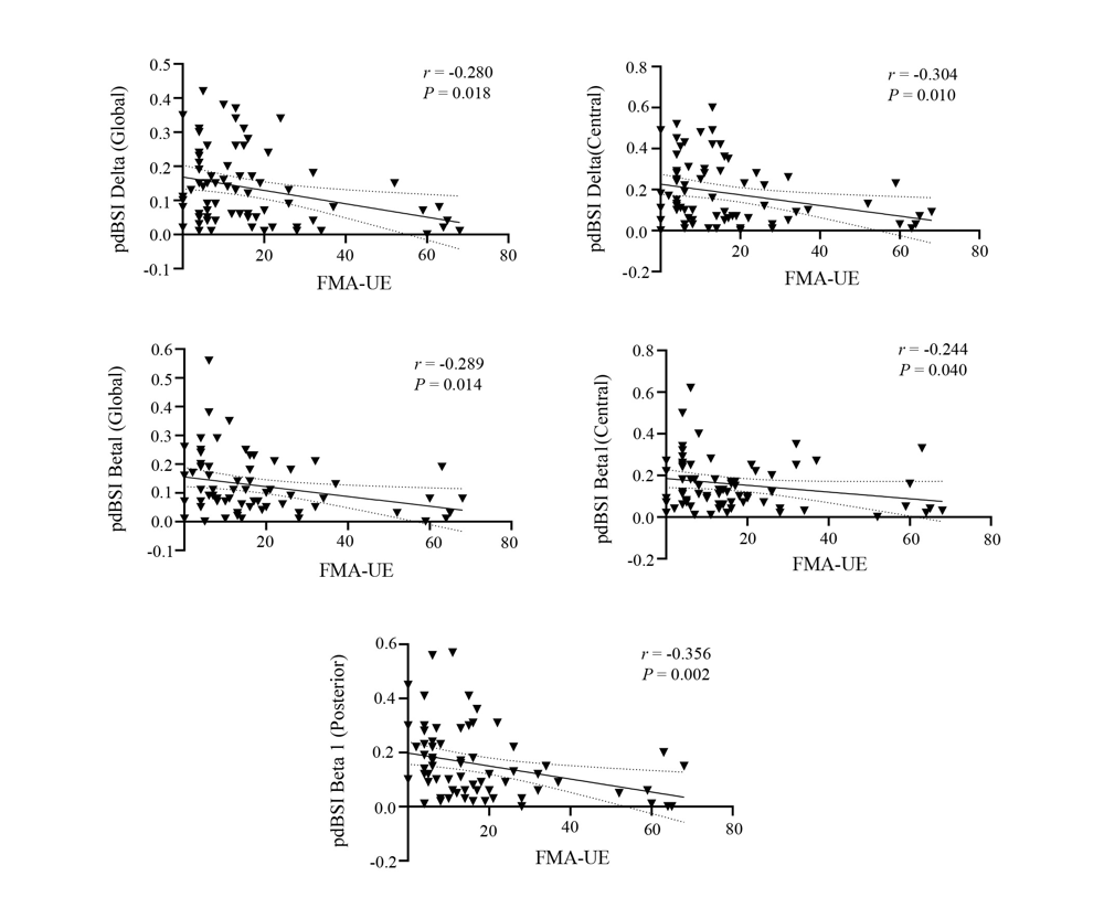

Figure 2
Correlation analysis between DAR and FMA-UE in the stroke group"
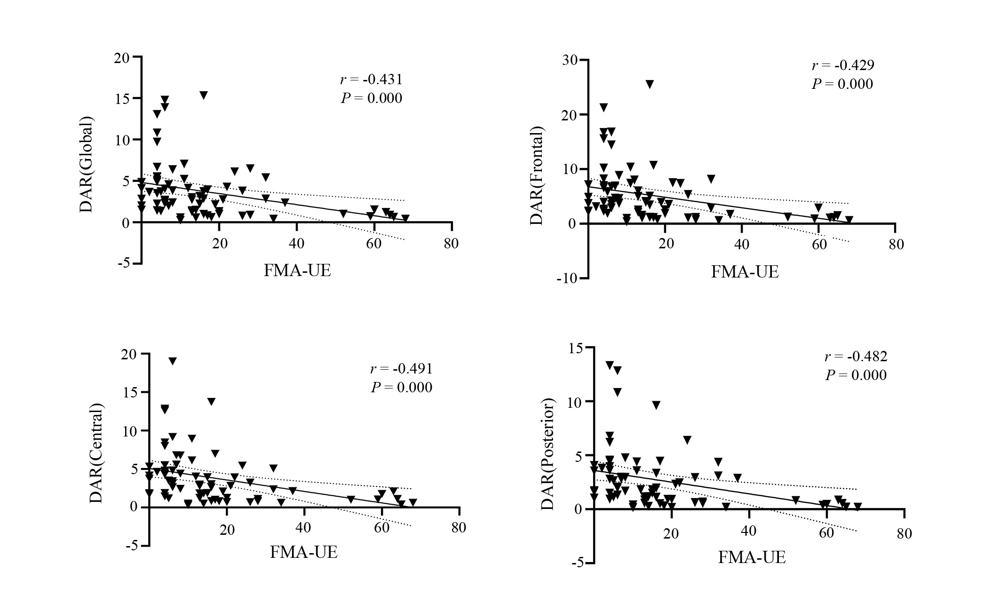
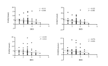
Figure 3
Correlation analysis between DAR and the Brunnstrom stages in the stroke group"

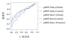
Figure 4
Results from ROC analysis to distinguish the stroke group from the control group with pdBSI"
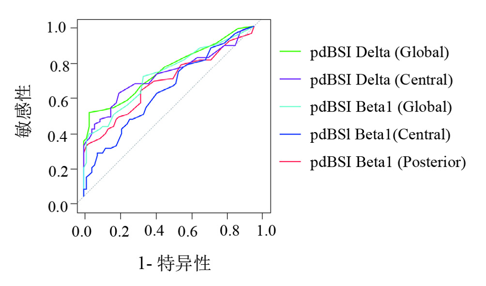
Table 3
Results from ROC analysis to distinguish the stroke group from the control group"
| rsEEG指标 | AUC | SE | 敏感性 | 特异性 | 95% CI | P值 |
|---|---|---|---|---|---|---|
| pdBSI Delta(Global) | 0.740 | 0.042 | 0.493 | 0.968 | 0.657~0.823 | < 0.001 |
| pdBSI Delta(Central) | 0.714 | 0.045 | 0.606 | 0.794 | 0.626~0.801 | < 0.001 |
| pdBSI Beta1(Global) | 0.714 | 0.044 | 0.704 | 0.651 | 0.627~0.800 | < 0.001 |
| pdBSI Beta1(Central) | 0.622 | 0.048 | 0.451 | 0.730 | 0.527~0.716 | 0.015 |
| pdBSI Beta1(Posterior) | 0.668 | 0.047 | 0.620 | 0.667 | 0.577~0.759 | 0.001 |
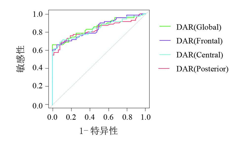
| DAR(Global) | 0.852 | 0.033 | 0.662 | 1.000 | 0.787~0.917 | < 0.001 |
| DAR(Frontal) | 0.842 | 0.034 | 0.662 | 0.952 | 0.776~0.909 | < 0.001 |
| DAR(Central) | 0.839 | 0.035 | 0.690 | 0.921 | 0.770~0.907 | < 0.001 |
| DAR(Psterior) | 0.829 | 0.037 | 0.676 | 0.921 | 0.758~0.901 | < 0.001 |
Figure 5
Results from ROC analysis to distinguish the stroke group from the control group with DAR"
| [1] |
ROTH G A, MENSAH G A, JOHNSON C O, et al. Global burden of cardiovascular diseases and risk factors, 1990-2019: Update from the GBD 2019 study[J]. J Am Coll Cardiol, 2020, 76(25): 2982-3021.
doi: 10.1016/j.jacc.2020.11.010 pmid: 33309175 |
| [2] |
STINEAR C M, LANG C E, ZEILER S, et al. Advances and challenges in stroke rehabilitation[J]. Lancet Neurol, 2020, 19(4): 348-360.
doi: S1474-4422(19)30415-6 pmid: 32004440 |
| [3] |
KWAH L K, HARVEY L A, DIONG J, et al. Models containing age and NIHSS predict recovery of ambulation and upper limb function six months after stroke: an observational study[J]. J Physiother, 2013, 59(3): 189-197.
doi: 10.1016/S1836-9553(13)70183-8 pmid: 23896334 |
| [4] | EWING A C, LI Y X, CHEN X X, et al. Stroke and activity limitation in Chinese adults 65 years or older[J]. Disabil Health J, 2023, 16(3): 101452. |
| [5] |
REINKENSMEYER D J, FARRENS A J, KAMPER D G. Facilitating limb movement after stroke[J]. Nat Med, 2023, 29(3): 535-536.
doi: 10.1038/s41591-023-02233-7 pmid: 36882528 |
| [6] | SHIN S, LEE Y, CHANG W H, et al. Multifaceted assessment of functional outcomes in survivors of first-time stroke[J]. JAMA Netw Open, 2022, 5(9): e2233094. |
| [7] | SEBASTIÁN-ROMAGOSA M, UDINA E, ORTNER R, et al. EEG biomarkers related with the functional state of stroke patients[J]. Front Neurosci, 2020, 14: 582. |
| [8] | 王冲, 周静, 李文豪, 等. 脑电在脑卒中运动功能评估中的应用研究进展[J]. 中国医学物理学杂志, 2023, 40(10): 1309-1315. |
| WANG C, ZHOU J, LI W H, et al. Application of electroencephalography in assessment of motor function in stroke patients: a review[J]. Chin J Med Phys, 2023, 40(10): 1309-1315. | |
| [9] |
DELCAMP C, SRINIVASAN R, CRAMER S C. EEG provides insights into motor control and neuroplasticity during stroke recovery[J]. Stroke, 2024, 55(10): 2579-2583.
doi: 10.1161/STROKEAHA.124.048458 pmid: 39171399 |
| [10] | RAY A M, FIGUEIREDO T D C, LÓPEZ-LARRAZ E, et al. Brain oscillatory activity as a biomarker of motor recovery in chronic stroke[J]. Hum Brain Mapp, 2019, 41(5): 1296-1308. |
| [11] | MILANI G, ANTONIONI A, BARONI A, et al. Relation between EEG measures and upper limb motor recovery in stroke patients: a scoping review[J]. Brain Topogr, 2022, 35(5/6): 651-666. |
| [12] |
LANZONE J, MOTOLESE F, RICCI L, et al. Quantitative measures of the resting EEG in stroke: a systematic review on clinical correlation and prognostic value[J]. Neurol Sci, 2023, 44(12): 4247-4261.
doi: 10.1007/s10072-023-06981-9 pmid: 37542545 |
| [13] |
WILKINSON C M, BURRELL J I, KUZIEK J W P, et al. Predicting stroke severity with a 3-min recording from the Muse portable EEG system for rapid diagnosis of stroke[J]. Sci Rep, 2020, 10(1): 18465.
doi: 10.1038/s41598-020-75379-w pmid: 33116187 |
| [14] |
SAES M, MESKERS C G M, DAFFERTSHOFER A, et al. Are early measured resting-state EEG parameters predictive for upper limb motor impairment six months poststroke?[J]. Clin Neurophysiol, 2021, 132(1): 56-62.
doi: 10.1016/j.clinph.2020.09.031 pmid: 33248434 |
| [15] | SATO Y, SCHMITT O, IP Z, et al. Pathological changes of brain oscillations following ischemic stroke[J]. J Cereb Blood Flow Metab, 2022, 42(10): 1753-1776. |
| [16] |
SAES M, MESKERS C G M, DAFFERTSHOFER A, et al. How does upper extremity Fugl-Meyer motor score relate to resting-state EEG in chronic stroke? A power spectral density analysis[J]. Clin Neurophysiol, 2019, 130(5): 856-862.
doi: S1388-2457(19)30026-4 pmid: 30902439 |
| [17] |
SAES M, ZANDVLIET S B, ANDRINGA A S, et al. Is resting-state EEG longitudinally associated with recovery of clinical neurological impairments early poststroke? A prospective cohort study[J]. Neurorehabil Neural Repair, 2020, 34(5): 389-402.
doi: 10.1177/1545968320905797 pmid: 32249674 |
| [18] | LEONARDI G, CIURLEO R, CUCINOTTA F, et al. The role of brain oscillations in post-stroke motor recovery: an overview[J]. Front Syst Neurosci, 2022, 16: 947421. |
| [19] | KANCHEVA I, VAN DER SALM S M A, RAMSEY N F, et al. Association between lesion location and sensorimotor rhythms in stroke: a systematic review with narrative synthesis[J]. Neurol Sci, 2023, 44(12): 4263-4289. |
| [20] | 李岑博, 陈龙, 顾斌, 等. 定量脑电图用于慢性缺血性卒中患者康复状态评估[J]. 中国生物医学工程学报, 2022, 41(5): 547-557. |
| LI C B, CHEN L, GU B, et al. Evaluation of rehabilitation status of patients with chronic ischemic stroke based on quantitative EEG[J]. Chin J Biomed Eng, 2022, 41(5): 547-557. | |
| [21] | AMINOV A, ROGERS J M, JOHNSTONE S J, et al. Acute single channel EEG predictors of cognitive function after stroke[J]. PLoS One, 2017, 12(10): e0185841. |
| [22] | WANG A X, TIAN X, JIANG D, et al. Rehabilitation with brain-computer interface and upper limb motor function in ischemic stroke: a randomized controlled trial[J]. Medcine, 2024, 5(6): 559-569.e4. |
| [23] |
SINGH N, SAINI M, KUMAR N, et al. Evidence of neuroplasticity with robotic hand exoskeleton for post-stroke rehabilitation: a randomized controlled trial[J]. J Neuroeng Rehabil, 2021, 18(1): 76.
doi: 10.1186/s12984-021-00867-7 pmid: 33957937 |
| [24] |
CASULA E P, PELLICCIARI M C, BONNì S, et al. Evidence for interhemispheric imbalance in stroke patients as revealed by combining transcranial magnetic stimulation and electroencephalography[J]. Hum Brain Mapp, 2021, 42(5): 1343-1358.
doi: 10.1002/hbm.25297 pmid: 33439537 |
| [25] | FINGELKURTS A A, FINGELKURTS A A. Altered structure of dynamic electroencephalogram oscillatory pattern in major depression[J]. Biol Psychiatry, 2015, 77(12): 1050-1060. |
| [26] | ZHANG Y C, YE L L, CAO L, et al. Resting-state electroencephalography changes in poststroke patients with visuospatial neglect[J]. Front Neurosci, 2022, 16: 974712. |
| [27] | TAKEUCHI N, IZUMI S I. Motor learning based on oscillatory brain activity using transcranial alternating current stimulation: a review[J]. Brain Sci, 2021, 11(8): 1095. |
| [28] | ESPENHAHN S, ROSSITER H E, VAN WIJK B C M, et al. Sensorimotor cortex beta oscillations reflect motor skill learning ability after stroke[J]. Brain Commun, 2020, 2(2): fcaa161. |
| [29] |
CASSIDY J M, WODEYAR A, WU J, et al. Low-frequency oscillations are a biomarker of injury and recovery after stroke[J]. Stroke, 2020, 51(5): 1442-1450.
doi: 10.1161/STROKEAHA.120.028932 pmid: 32299324 |
| [30] | HU Y X, WANG Y F, ZHANG R, et al. Assessing stroke rehabilitation degree based on quantitative EEG index and nonlinear parameters[J]. Cogn Neurodyn, 2022, 17(3): 661-669. |
| [31] |
ZHANG J J, BAI Z F, FONG K N K. Resting-state cortical electroencephalogram rhythms and network in patients after chronic stroke[J]. J Neuroeng Rehabil, 2024, 21(1): 32.
doi: 10.1186/s12984-024-01328-7 pmid: 38424592 |
| [32] | ZHANG J J, SÁNCHEZ VIDAÑA D I, CHAN J N M, et al. Biomarkers for prognostic functional recovery poststroke: a narrative review[J]. Front Cell Dev Biol, 2023, 10: 1062807. |
| [33] |
BARTUR G, PRATT H, SOROKER N. Changes in mu and beta amplitude of the EEG during upper limb movement correlate with motor impairment and structural damage in subacute stroke[J]. Clin Neurophysiol, 2019, 130(9): 1644-1651.
doi: S1388-2457(19)30916-2 pmid: 31326646 |
| [1] | CHEN Mengye, QU Qingming, ZHU Jie, CHEN Xianggui, JIA Jie. Characteristics of cardiorespiratory fitness in patients with post-stroke hemiplegia based on cardiopulmonary exercise testing [J]. Chinese Journal of Rehabilitation Theory and Practice, 2025, 31(4): 441-447. |
| [2] | LIU Pengcheng, QU Mengjian, LONG Liping, WANG Yalin, YANG Mingzhu, LIU Peiyong, ZHOU Jun, LIU Jing. Effect of pneumatic and electric hand training system with multiple sensory stimulation modalities combined with low-frequency repetitive transcranial magnetic stimulation on hand movement and tactile pressure sensation in patients with stroke [J]. Chinese Journal of Rehabilitation Theory and Practice, 2025, 31(4): 458-465. |
| [3] | SU Panpan, YE Peng, LU Qian, HE Chuan, LU Xiao. Effect of visual deprivation training combined with proprioceptive training on balance in hemiplegic patients after stroke [J]. Chinese Journal of Rehabilitation Theory and Practice, 2025, 31(3): 254-263. |
| [4] | LIN Changsheng, CAO Yu, WANG Tong, DAI Wenjun, HOU Hong, HU Cuiqin, BAO Shilei, PANG Sufang. Effect of closed-chain exercise training on hemiplegic shoulder pain and shoulder joint stability in stroke patients: a study with ultrasound [J]. Chinese Journal of Rehabilitation Theory and Practice, 2025, 31(3): 264-273. |
| [5] | WANG Xiaojun, WANG Hani, YU Hong, LI Yuanmei, ZHOU Yuda. Effect of high-definition transcranial direct current stimulation combined with upper limb robot on upper limb dysfunction after ischemic stroke [J]. Chinese Journal of Rehabilitation Theory and Practice, 2025, 31(2): 218-224. |
| [6] | MA Wenwen, WEN Yanzheng, Manripati ROZI, CUI Boya, Suyinqimei . Effect of healthy side tilt training on balance function in patients with Pusher syndrome after stroke [J]. Chinese Journal of Rehabilitation Theory and Practice, 2025, 31(2): 225-230. |
| [7] | QIN Qing, LIU Ye, YE Haiyan, LI Chen, CHEN Di. Robot-assisted therapy for upper limb of stoke: a bibliometrics analysis [J]. Chinese Journal of Rehabilitation Theory and Practice, 2025, 31(1): 85-98. |
| [8] | ZHANG Lu, MA Jiangping, YANG Erli, CHEN Qiuhua, DONG Lijun, YIN Xiaobing. Application of cognitive-motor dual-task training in stroke: a bibliometrics analysis [J]. Chinese Journal of Rehabilitation Theory and Practice, 2024, 30(9): 1034-1042. |
| [9] | LUO Hong, XU Li. Effect of repetitive transcranial magnetic stimulation combined with repetitive peripheral magnetic stimulation on upper extremities motor function in patients with cerebral hemorrhage: a randomized controlled trial based on resting state-functional magenetic resonance imaging [J]. Chinese Journal of Rehabilitation Theory and Practice, 2024, 30(9): 1060-1068. |
| [10] | WANG Min, FANG Lantian, HUANG Chenyi. Effect of modified graded motor imagery on upper limb motor function for stroke patients: a randomized controlled trial [J]. Chinese Journal of Rehabilitation Theory and Practice, 2024, 30(9): 1069-1073. |
| [11] | XIE Dandan, CHEN Shanjia, LEI Lei, YU Guo, YU Jiahui, ZHAO Jiapei, HE Xiaokuo. Characteristics of brain activation during treadmill walking with visual feedback in healthy subjects and hemiplegic patients: a functional near infrared spectroscopy study [J]. Chinese Journal of Rehabilitation Theory and Practice, 2024, 30(9): 1074-1081. |
| [12] | LI Dong, ZHANG Hao, LIU Nan, WANG Xinyue, XU Miao. Effect of cognitive-motor dual-task training on balance function and gait in convalescent stroke patients: a randomized contolled trial [J]. Chinese Journal of Rehabilitation Theory and Practice, 2024, 30(9): 1082-1091. |
| [13] | YU Tingting, CAI Fuliang, MIAO Guihua, GU Chen, PENG Yuan. Effect of structured therapy and education based on personal strength on ischemic stroke: a randomized controlled trial [J]. Chinese Journal of Rehabilitation Theory and Practice, 2024, 30(8): 965-971. |
| [14] | LIANG Tianjia, LONG Yaobin, LU Liyan, ZHOU Jinying, HUANG Fucai, HUANG Linpeng, WU Yingchao, LONG Yaoxiang, WEI Xiaocui, LIU Zhong. Effect of rope-assisted proprioceptive neuromuscular facilitation combined with rope-assisted brain-computer interface training on upper limb function in stroke patients with hemiplegia: a randomized controlled trial [J]. Chinese Journal of Rehabilitation Theory and Practice, 2024, 30(8): 972-978. |
| [15] | WANG Zhe, WAN Qin, HUANG Zhaoming, WANG Yongli, QIAN Hong. Characteristics of speech prosody function in adults with non-fluent aphasia after stroke [J]. Chinese Journal of Rehabilitation Theory and Practice, 2024, 30(8): 979-992. |
| Viewed | ||||||
|
Full text |
|
|||||
|
Abstract |
|
|||||
|
||