


《Chinese Journal of Rehabilitation Theory and Practice》 ›› 2020, Vol. 26 ›› Issue (1): 77-84.doi: 10.3969/j.issn.1006-9771.2020.01.014
Previous Articles Next Articles
WU Qiong1a,REN Shi-yuan2,YUE Zan2,GE Yun-xiang3,MA Di1a,ZHAO Hong-liang1b,LIU Gang2,WANG Jing2,PAN Yu1a( ),DOU Wei-bei3,4()
Received:2019-10-09
Revised:2019-11-04
Published:2020-01-25
Online:2020-02-07
Contact:
PAN Yu,DOU Wei-bei
E-mail:py10335@163.com;douwb@tsinghua.edu.cn
Supported by:CLC Number:
WU Qiong,REN Shi-yuan,YUE Zan,GE Yun-xiang,MA Di,ZHAO Hong-liang,LIU Gang,WANG Jing,PAN Yu,DOU Wei-bei. Brain-computer Interface and Comprehensive Training for Stroke: A Resting State Functional Magnetic Resonance Imaging Study[J]. 《Chinese Journal of Rehabilitation Theory and Practice》, 2020, 26(1): 77-84.
"
| 项目 | 统计量 | |
|---|---|---|
| 性别(n) | 男 | 9 |
| 女 | 5 | |
| 年龄(岁) | 54.501±3.550 | |
| 卒中类型(n) | 脑梗死 | 12 |
| 脑出血 | 2 | |
| 偏瘫侧(n) | 左 | 8 |
| 右 | 6 | |
| 病程(月) | 1.500±0.711 | |
"
| 时间 | FMA-UE | ARAT | WMFT |
|---|---|---|---|
| 训练前 | 20.14±13.21 | 9.50(3.00,23.25) | 30.07±12.66 |
| 训练后 | 35.36±15.92 | 24.00(10.00,45.25) | 47.79±18.68 |
| 训练前后差值 | 14.71±7.80 | 8.50(3.75,24.00) | 18.14±12.03 |
| t/Z值a | -6.612 | -3.297 | -5.298 |
| P值a | < 0.001 | 0.001 | < 0.001 |
"
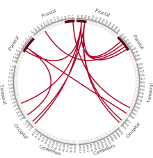
| 种子点 | 连接点 | 训练前 | 训练后 | t值 | P值 |
|---|---|---|---|---|---|
| BA4L | BA41R | -0.008±0.212 | 0.135±0.227 | 2.317 | 0.038 |
| BA6R | BA7L | -0.177±0.252 | 0.049±0.214 | 3.723 | 0.007 |
| BA6R | BA2R | 0.489±0.133 | 0.590±0.141 | 2.542 | 0.025 |
| BA6R | BA3R | 0.586±0.232 | 0.697±0.085 | 2.321 | 0.037 |
| BA6R | BA37L | -0.214±0.183 | -0.057±0.213 | 3.092 | 0.009 |
| BA6R | BA7R | 0.045±0.228 | 0.198±0.270 | 2.774 | 0.016 |
| BA6R | BA19L | -0.207±0.227 | -0.0132±0.287 | 2.181 | 0.048 |
| BA5L | BA48R | -0.128±0.174 | -0.006±0.224 | 2.204 | 0.046 |
| BA5L | BA48L | -0.022±0.224 | 0.088±0.196 | 2.185 | 0.048 |
| BA5R | BA44L | -0.309±0.1312 | -0.200±0.152 | 2.682 | 0.019 |
| BA8R | BA7R | 0.187±0.292 | 0.051±0.276 | 2.251 | 0.042 |
| BA8R | BA18R | -0.337±0.140 | -0.146±0.175 | 3.371 | 0.005 |
| BA8R | BA18L | -0.340±0.128 | -0.192±0.161 | 3.978 | 0.002 |
| BA8R | BA19R | -0.382±0.142 | -0.208±0.191 | 4.764 | 0.000 |

"

"

"
| FC | 临床评分 | r值 | P值 |
|---|---|---|---|
| BA4L-BA41R | FMA-UE | 0.485 | 0.009 |
| ARAT | 0.416 | 0.028 | |
| WMFT | 0.505 | 0.006 | |
| BA5L-BA48R | FMA-UE | 0.522 | 0.004 |
| ARAT | 0.544 | 0.003 | |
| WMFT | 0.696 | < 0.001 |
| [1] | 王陇德, 刘建民, 杨弋, 等. 我国脑卒中防治仍面临巨大挑战——《中国脑卒中防治报告2018》概要[J]. 中国循环杂志, 2019, 34(2):105-119. |
| [2] |
Tedesco Triccas L, Kennedy N, Smith T, et al. Predictors of upper limb spasticity after stroke? A systematic review and meta-analysis[J]. Physiotherapy, 2019, 105(2):163-173.
doi: S0031-9406(19)30009-4 pmid: 30745061 |
| [3] |
Allison R, Shenton L, Bamforth K, et al. Incidence, time course and predictors of impairments relating to caring for the profoundly affected arm after stroke: a systematic review[J]. Physiother Res Int, 2016, 21(4):210-227.
doi: 10.1002/pri.v21.4 |
| [4] |
Harvey R L. Predictors of functional outcome following stroke[J]. Phys Med Rehabil Clin N Am, 2015, 26(4):583-598.
doi: 10.1016/j.pmr.2015.07.002 |
| [5] |
Mazrooyisebdani M, Nair V A, Loh P L, et al. Evaluation of changes in the motor network following BCI therapy based on graph theory analysis[J]. Front Neurosci, 2018, 12:861.
doi: 10.3389/fnins.2018.00861 |
| [6] |
Biasiucci A, Leeb R, Iturrate I, et al. Brain-actuated functional electrical stimulation elicits lasting arm motor recovery after stroke[J]. Nat Commun, 2018, 9(1):2421.
doi: 10.1038/s41467-018-04673-z pmid: 29925890 |
| [7] |
Chowdhury A, Meena Y K, Raza H, et al. Active physical practice followed by mental practice using BCI-driven hand exoskeleton: a pilot trial for clinical effectiveness and usability[J]. IEEE J Biomed Health Inform, 2018, 22(6):1786-1795.
doi: 10.1109/JBHI.2018.2863212 |
| [8] |
Ramos-Murguialday A, Broetz D, Rea M, et al. Brain-machine interface in chronic stroke rehabilitation: a controlled study[J]. Ann Neurol, 2013, 74(1):100-108.
doi: 10.1002/ana.23879 pmid: 23494615 |
| [9] |
Bundy D T, Souders L, Baranyai K, et al. Contralesional brain-computer interface control of a powered exoskeleton for motor recovery in chronic stroke survivors[J]. Stroke, 2017, 48(7):1908-1915.
doi: 10.1161/STROKEAHA.116.016304 |
| [10] |
Pichiorri F, Morone G, Petti M, et al. Brain-computer interface boosts motor imagery practice during stroke recovery[J]. Ann Neurol, 2015, 77(5):851-865.
doi: 10.1002/ana.24390 pmid: 25712802 |
| [11] | 中华医学会神经病学分会, 中华医学会神经病学分会脑血管病学组. 中国急性脑卒中临床研究规范共识2018[J]. 中华神经科杂志, 2018, 51(4):247-255. |
| [12] | 刘华, 程钰琦, 李洋, 等. 中文版运动觉-视觉想象问卷的结构效度[J]. 中国康复理论与实践, 2017, 23(5):580-583. |
| [13] | Li Y C, Liao W W, Hsieh Y W, et al. Predictors of clinically important changes in actual and perceived functional arm use of the affected upper limb after rehabilitative therapy in chronic stroke[J]. Arch Phys Med Rehabil, 2019. DOI: 10.1016/j.apmr.2019.08.483.[Epub ahead of print]. |
| [14] | Maenza C, Good D C, Winstein C J, et al. Functional deficits in the less-impaired arm of stroke survivors depend on hemisphere of damage and extent of paretic arm impairment[J]. Neurorehabil Neural Repair, 2019. DOI: 10.1177/1545968319875951.[Epub ahead of print]. |
| [15] |
Page S J H E, Persch A. Psychometrics of the wrist stability and hand mobility subscales of the Fugl-Meyer Assessment in moderately impaired stroke[J]. Phys Ther, 2015, 95(1):103-108.
doi: 10.2522/ptj.20130235 |
| [16] |
Simpson L A, Eng J J. Functional recovery following stroke: capturing changes in upper-extremity function[J]. Neurorehabil Neural Repair, 2013, 27(3):240-250.
doi: 10.1177/1545968312461719 pmid: 23077144 |
| [17] |
Pan Y, Dou W B, Wang Y H, et al. Non-concomitant cortical structural and functional alterations in sensorimotor areas following incomplete spinal cord injury[J]. Neural Regen Res, 2017, 12(12):2059-2066.
doi: 10.4103/1673-5374.221165 pmid: 29323046 |
| [18] |
Horn A, Blankenburg F. Toward a standardized structural-functional group connectome in MNI space[J]. Neuroimage, 2016, 124(Pt A):310-322.
doi: 10.1016/j.neuroimage.2015.08.048 |
| [19] |
Paxinos G. Human brainnetome atlas: a new chapter of brain cartography[J]. Sci China Life Sci, 2016, 59(9):965-967.
doi: 10.1007/s11427-016-5110-x pmid: 27473861 |
| [20] |
Fan L, Li H, Zhuo J, et al. The human brainnetome atlas: a new brain atlas based on connectional architecture[J]. Cereb Cortex, 2016, 26(8):3508-3526.
doi: 10.1093/cercor/bhw157 |
| [21] |
Arya K N, Verma R, Garg R K. Estimating the minimal clinically important difference of an upper extremity recovery measure in subacute stroke patients[J]. Top Stroke Rehabil, 2011, 18(Suppl 1):599-610.
doi: 10.1310/tsr18s01-599 |
| [22] |
Jaeschke R, Singer J, Guyatt G H. Measurement of health status. Ascertaining the minimal clinically important difference[J]. Control Clin Trials, 1989, 10(4):407-415.
pmid: 2691207 |
| [23] |
Page S J H E, Persch A. Predictors of motor outcomes in severe sub-acute stroke patients after upper limb intensive combined (robot-mediated + usual care) training[J]. Phys Ther, 2015, 95(1):103-108.
doi: 10.2522/ptj.20130235 |
| [24] | 杜鹃. 基于多模态功能磁共振的重复经颅磁刺激促进脑卒中运动功能恢复的作用机制研究[D]. 上海:第二军医大学, 2017. |
| [25] | 田强. 基于多模态MRI影像组学策略的胶质瘤术前分级预测研究[D]. 西安:第四军医大学, 2017. |
| [26] |
Huda R, Goard M J, Pho G N, et al. Neural mechanisms of sensorimotor transformation and action selection[J]. Eur J Neurosci, 2019, 49(8):1055-1060.
doi: 10.1111/ejn.2019.49.issue-8 |
| [27] | Luz María Alonso-Valerdi V R M. Enrichment of human-computer interaction in brain-computer interfaces via virtual environments[J]. Comput Intell Neurosci, 2017, 2017:6076913. |
| [28] | 陈树耿, 束小康, 贾杰. 基于闭环脑机接口的脑卒中患者的手功能康复研究[J]. 中国康复医学杂志, 2016, 31(11):1189-1194. |
| [29] |
Murata A, Wen W, Asama H. The body and objects represented in the ventral stream of the parieto-premotor network[J]. Neurosci Res, 2016, 104:4-15.
doi: 10.1016/j.neures.2015.10.010 pmid: 26562332 |
| [30] |
Park C H, Chang W H, Lee M, et al. Which motor cortical region best predicts imagined movement?[J]. Neuroimage, 2015, 113:101-110.
doi: 10.1016/j.neuroimage.2015.03.033 pmid: 25800212 |
| [31] |
McGregor H R, Gribble P L. Functional connectivity between somatosensory and motor brain areas predicts individual differences in motor learning by observing[J]. J Neurophysiol, 2017, 118(2):1235-1243.
doi: 10.1152/jn.00275.2017 pmid: 28566463 |
| [32] |
Bracci S, Caramazza A, Peelen M V. View-invariant representation of hand postures in the human lateral occipitotemporal cortex[J]. Neuroimage, 2018, 181:446-452.
doi: 10.1016/j.neuroimage.2018.07.001 |
| [33] |
Borich M R, Brodie S M, Gray W A, et al. Understanding the role of the primary somatosensory cortex: opportunities for rehabilitation[J]. Neuropsychologia, 2015, 79(Pt B):246-255.
doi: 10.1016/j.neuropsychologia.2015.07.007 pmid: 26164474 |
| [34] | Bruni S, Gerbella M, Bonini L, et al. Cortical and subcortical connections of parietal and premotor nodes of the monkey hand mirror neuron network[J]. Brain Struct Funct, 2018, 223(4):1713-1729. |
| [35] |
Berneiser J, Jahn G, Grothe M, et al. From visual to motor strategies: training in mental rotation of hands[J]. Neuroimage, 2018, 167:247-255.
doi: S1053-8119(16)30247-6 pmid: 27321046 |
| [36] |
Makary M M, Eun S, Soliman R S, et al. Functional topography of the primary motor cortex during motor execution and motor imagery as revealed by functional MRI[J]. Neuroreport, 2017, 28(12):731-738.
doi: 10.1097/WNR.0000000000000825 pmid: 28617759 |
| [37] |
Kruschwitz J D, Waller L, List D, et al. Anticipating the good and the bad: a study on the neural correlates of bivalent emotion anticipation and their malleability via attentional deployment[J]. Neuroimage, 2018, 183:553-564.
doi: S1053-8119(18)30746-8 pmid: 30145207 |
| [1] | LIN Na, GAO Hanlu, LU Huiping, CHEN Yanqing, ZHENG Junfan, CHEN Shurong. Effect of virtual reality on upper limb function after stroke: a study of diffusion tensor imaging [J]. 《Chinese Journal of Rehabilitation Theory and Practice》, 2024, 30(1): 61-67. |
| [2] | WANG Haoyi, SHI Yawei, LU Jun, XU Guangxu. Impact of subjective vertical perception impairment on function in stroke patients: a retrospective study [J]. 《Chinese Journal of Rehabilitation Theory and Practice》, 2024, 30(1): 68-73. |
| [3] | CHEN Junwen, CHEN Qian, CHEN Cheng, LI Shuyue, LIU Lingling, WU Cunshu, GONG Xiang, LU Jun, XU Guangxu. Effect of modified Baduanjin exercise on cardiopulmonary function, motor function and activities of daily living for stroke patients [J]. 《Chinese Journal of Rehabilitation Theory and Practice》, 2024, 30(1): 74-80. |
| [4] | HU Yonglin, MA Ying, DOU Chao, LU Anmin, JIANG Xiaoge, SONG Xinjian, XIAO Yuhua. Effect of neural mobilization based on shoulder control training on shoulder pain and upper limb function in stroke patients with hemiplegia [J]. 《Chinese Journal of Rehabilitation Theory and Practice》, 2024, 30(1): 81-86. |
| [5] | WANG He, HAN Liang, KAN Mengfan, YU Shaohong. Efficacy of electrical stimulation on shoulder-hand syndrome after stroke: a systematic review and meta-analysis [J]. 《Chinese Journal of Rehabilitation Theory and Practice》, 2023, 29(9): 1048-1056. |
| [6] | SUN Tengfang, REN Mengting, YANG Lin, WANG Yaoting, WANG Hongyu, YAN Xingzhou. Effect of hyperbaric oxygen therapy combined with repetitive peripheral magnetic stimulation on ankle motor function and balance of stroke patients [J]. 《Chinese Journal of Rehabilitation Theory and Practice》, 2023, 29(8): 875-881. |
| [7] | WANG Ya'nan, LIU Xihua. Correlation and predictive effect of subjective and objective balance function measurements in stroke patients with hemiplegia [J]. 《Chinese Journal of Rehabilitation Theory and Practice》, 2023, 29(8): 890-895. |
| [8] | WANG Haiyun, WANG Yin, ZHOU Xinjie, HE Aiqun. Effect of transcranial direct current stimulation combined with acupuncture on central and upper limb function in stroke patients based on central-peripheral-central theory [J]. 《Chinese Journal of Rehabilitation Theory and Practice》, 2023, 29(8): 919-925. |
| [9] | CHEN Yiting, WANG Qian, CUI Shenhong, LI Yingcai, ZHANG Siyu, WEI Yanxu, REN Hui, LENG Jun, CHEN Bin. Effect of bilateral sequential repetitive transcranial magnetic stimulation on motor function of upper limbs in stroke patients [J]. 《Chinese Journal of Rehabilitation Theory and Practice》, 2023, 29(8): 926-932. |
| [10] | LI Zhenya, SUN Jie, GUO Pengfei, WANG Guangming. Correlation between changes of swallowing function in oral and pharyngeal phases, and aspiration in stroke patients based on videofluroscopic swallowing study [J]. 《Chinese Journal of Rehabilitation Theory and Practice》, 2023, 29(8): 933-939. |
| [11] | LI Ziyi, SONG Weiqun, DU Jubao, CAO Guanglei, ZHANG Yanming, LI Ran. Effect of motor imagery on knee function after unicompartmental knee arthroplasty [J]. 《Chinese Journal of Rehabilitation Theory and Practice》, 2023, 29(7): 745-749. |
| [12] | HUA Ling, ZHANG Yi'nan, ZHENG Yu, SUN Qiaoyi, FANG Hui, SONG Da. Effect of hand controlled rhythm music therapy on unilateral spatial neglect after stroke [J]. 《Chinese Journal of Rehabilitation Theory and Practice》, 2023, 29(7): 833-838. |
| [13] | JIANG Xiaocui, LIU Zhen, SU Qinglun, ZHAO Qin, XIA Xiaomei, LU Fei. Effect of intermittent theta burst transcranial magnetic stimulation on non-fluent aphasia after stroke [J]. 《Chinese Journal of Rehabilitation Theory and Practice》, 2023, 29(7): 839-843. |
| [14] | XU Miaomiao, LI Nan, YING Ying, YANG Kaixiang, YANG Jingrui, LI Jie, QIU Yanqun. Effect of repetitive peripheral magnetic stimulation on upper limb motor function of stroke patients after contralateral seventh cervical nerve transfer [J]. 《Chinese Journal of Rehabilitation Theory and Practice》, 2023, 29(6): 686-690. |
| [15] | ZHENG Li, BAO Zhicheng, ZHANG Qi, REN Xuyan, SU Min. Effect of transcutaneous auricular vagus nerve stimulation combined with robot-assisted therapy on upper limb function of stroke patients [J]. 《Chinese Journal of Rehabilitation Theory and Practice》, 2023, 29(6): 691-696. |
| Viewed | ||||||
|
Full text |
|
|||||
|
Abstract |
|
|||||
|
||