



Chinese Journal of Rehabilitation Theory and Practice ›› 2026, Vol. 32 ›› Issue (5): 522-533.doi: 10.3969/j.issn.1006-9771.2026.05.003
Previous Articles Next Articles
HAN Tianxing1, LIU Haotian1, XU Ziqi1, PENG Xiaohan1, WANG Yang2, CAO Danna2( )
Received:2025-11-19
Published:2026-05-25
Online:2026-05-26
Contact:
CAO Danna
E-mail:hljanna@126.com
Supported by:CLC Number:
HAN Tianxing, LIU Haotian, XU Ziqi, PENG Xiaohan, WANG Yang, CAO Danna. Functional connectivity in default mode network and medial temporal lobe in mild cognitive impairment stage of Alzheimer's disease based on resting-state functional magnetic resonance imaging: a meta-analysis[J]. Chinese Journal of Rehabilitation Theory and Practice, 2026, 32(5): 522-533.
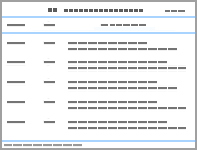
Table 1
PICO framework for rs-fMRI examination of AD patients at MCI stage"
| 人群(Population) | 干预(Intervention) | 比较(Comparison) | 结局(Outcome) |
|---|---|---|---|
| 临床诊断为AD源性MCI患者 | 基于种子点rs-fMRI分析所获得的DMN区域影像指标:BOLD、FC、fALFF、ReHo 基于种子点rs-fMRI分析所获得的MTL区域影像指标:BOLD、FC、fALFF、ReHo | HC同期同设备完成的rs-fMRI检测结果 rs-fMRI对DMN和MTL的形态学检测结果 | 诊断效能指标:敏感性、特异性、LR+、LR-、DOR、SROC-AUC 神经影像特征指标:DMN与MTL核心脑区的BOLD 信号变化趋势、rsFC强度值、ReHo/fALFF量化值,异常脑区的MNI坐标、团块大小及t值 |
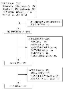
Figure 1
Flowchart of literature screening"
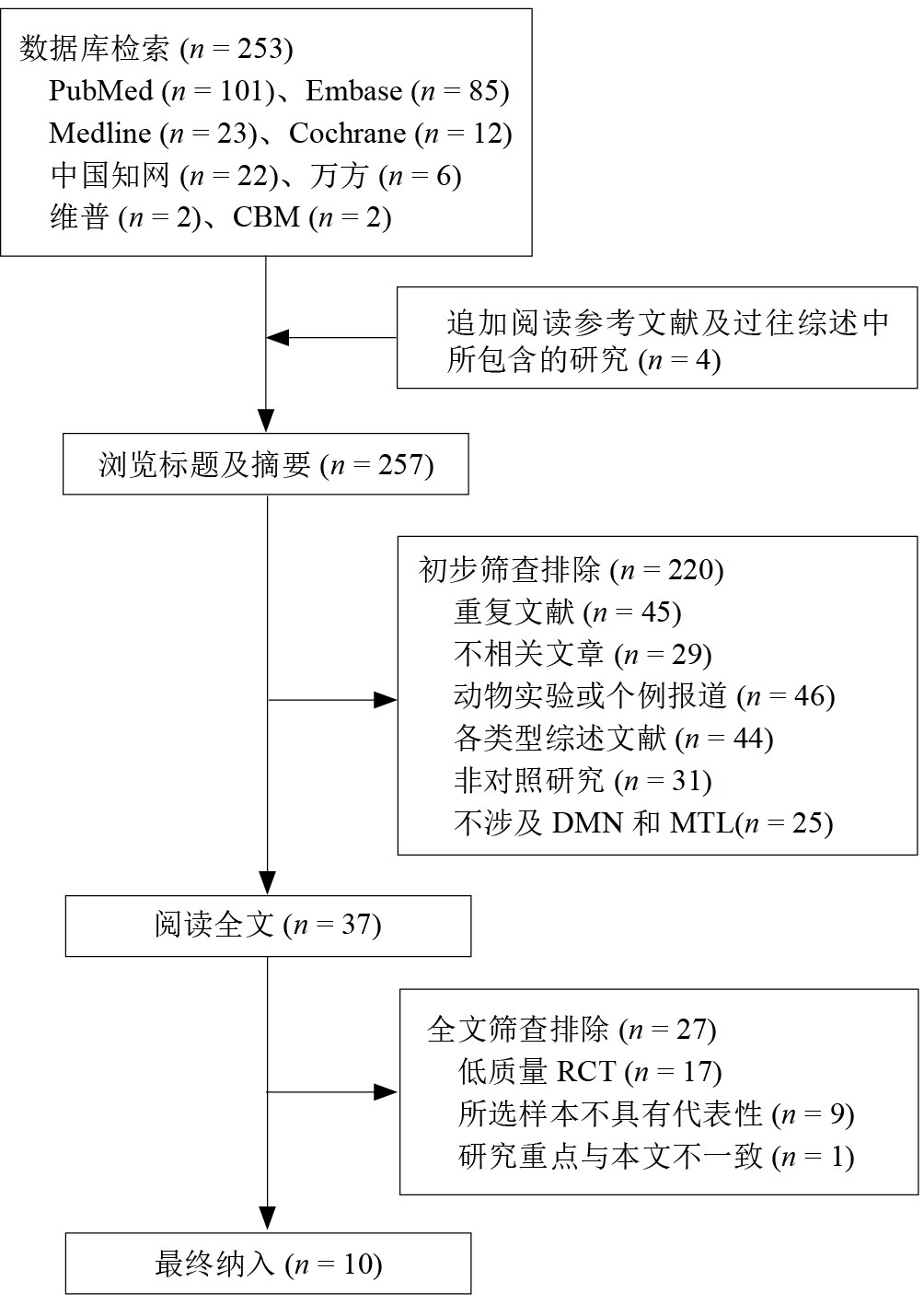
Table 2
QUADAS-2 quality evaluation"
| 条目 | 是a | 否b | 不能回答c | 不适用d |
|---|---|---|---|---|
| 1.研究对象是否为连续或随机样本? | √ | |||
| 2.研究对象的选择是否避免了不恰当的选择标准? | √ | |||
| 3.待评价试验的测量方法是否有效和可靠? | √ | |||
| 4.待评价试验的结果判读是否是在不知晓结局的情况下进行的? | √ | |||
| 5.测量结局的方法是否有效和可靠? | √ | |||
| 6.衡量所有研究对象结局的方法是否相同? | √ | |||
| 7.结果判读是否使用了盲法? | √ | |||
| 8.所有进行评价试验后的研究对象是否都避免了治疗? | √ | |||
| 9.是否所有的研究对象均纳入统计分析? | √ | |||
| 10.是否采用了恰当的方法来说明删失? | √ |
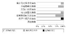
Figure 2
Bia risk assessment chart of methodological quality"
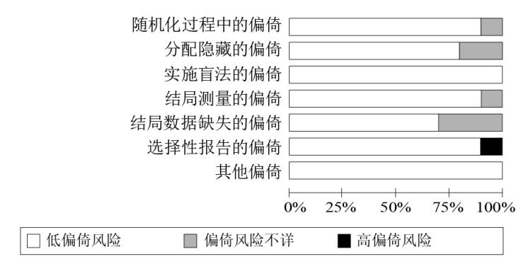
Table 3
Basic characteristics of included studies"
| 纳入文献 | 种子点 | 样本量(男/女)/n | 平均年龄(C/T)/岁 | 诊断标准 | |
|---|---|---|---|---|---|
| C | T | ||||
| Cao等[ | PCC、HIP、MTL | 40/29 | 25/13 | (69.16±4.96)/(68.46±4.97) | AD临床评估、ADAS-Cog |
| Liao等[ | PCC、右HIP、mPFC、PFcm | 37/36 | 76/64 | (68.28±9.64)/(66.30±9.52) | MMSE、MoCA |
| Xiong等[ | PCC、PF | 6/10 | 17/15 | (68.28±9.64)/(66.30±9.52) | ADNI、MMSE、GDS、CDR、FAQ、NPI-Q |
| Sendi等[ | PCC | 498/530 | 166/191 | (75.1±7.85)/(69.83±8.64) | 影像数据分析、CDR |
| Soman等[ | PCC、MTL | 12/19 | 26/34 | (65.7±6.0)/(65.1±6.7) | ACE-M |
| Ghahremani等[ (2023) | PFcm、Pfop、PFt、PFm、 Pga、PGp | 35/28 | 18/14 | (73.9±8.1)/(70.6±6.7) | MBIC、MoCA |
| Hojjati等[ | PCC、HIT、MTL | 176/185 | 88/80 | (68.41±6.15)/(64.96±3.17) | MMSE、MoCA |
| Takao等[ | mPFC、PCC、HIP | 18/13 | 38/42 | (71.9±7.0)/(76.0±7.2) | MMSE |
| Saberian等[ | PCC、MTL | 13/15 | 23/51 | (71.74±6.51)/(72.26±5.95) | CDR、MMSE |
| Mohammadian等[ | PCC、HIT、MTL | 4/10 | 12/17 | (72.87±4.34)/(71.17±2.59) | MMSE |
Table 4
Image characteristic and literature quality evaluation of the included studies"
| 纳入文献 | 敏感性/% | 特异性/% | 影像特征 | 异常脑区MNI坐标与团块大小 | NOS评分 | |||
|---|---|---|---|---|---|---|---|---|
| x | y | z | 体积/mm3 | |||||
| Cao等[ | 95.30 | 74.49 | DMN中双侧内侧前额皮质、后扣带楔前叶、腹侧前扣带回BOLD活动降低;mPFC与PCC之间FC下降;楔前叶与PCC fALFF下降;前额叶皮质ReHo降低 | 3 | -47 | 22 | 442 | 8 |
| Liao等[ | 88.43 | 78.21 | 右中额回、右扣带后回BOLD活动降低;PCC和MTL FC下降;内侧前颞叶fALFF升高不显著,楔前叶与PCC fALFF下降;后部DMN、海马区、前额叶皮质ReHo下降 | 6 | -55 | 30 | 376 | 8 |
| Xiong等[ | 96.90 | 81.20 | 尾状核与小脑亚区、尾状核与额下回、尾状核、内侧前额叶皮质BOLD活动降低;DMN内部FC广泛降低;双侧内侧前额皮质和PCC之间fALFF下降,后扣带皮质ReHo降低 | -4 | -47 | 24 | 209 | 9 |
| Sendi等[ | 95.00 | 72.00 | 视交叉上核、运动神经元存活基因、自动决策支持系统、细胞间基质蛋白族BOLD活动降低;后扣带楔前页和腹侧前扣带回存在FC下降情况,内侧前颞叶ReHo变化不一致,楔前叶ReHo下降 | ±47 | -57 | 33 | 588 | 9 |
| Soman等[ | 89.30 | 73.50 | DMN、运动神经元存活基因、顶网络、背侧注意网络BOLD活动降低;PCC与海马体之间FC下降;尾状和前额叶皮质之间fALFF下降,DMN内ReHo普遍性降低 | ±38 | -66 | 28 | 381 | 7 |
| Ghahremani等[ | 95.35 | 80.15 | DMN、脑皮质区域BOLD活动降低;MTL内FC下降;右扣带后回fALFF下降;海马体边缘与楔前叶ReHo下降 | 2 | -46 | 24 | 761 | 8 |
| Hojjati等[ | 91.40 | 77.30 | 视觉网络、运动网络、背侧注意网络、凸显网络、控制网络、边缘网络和DMN中tau蛋白和Aβ蛋白沉积,BOLD活动降低;楔前叶ReHo下降 | -5 | 46 | -13 | 447 | 8 |
| Takao等[ | 91.90 | 74.30 | 感觉运动网络、执行控制网络、视觉网络、DMN、背侧注意网络、凸显网络、双侧内侧前额皮质BOLD活动降低;MTL及海马区fALFF下降 | 4 | 47 | -9 | 736 | 8 |
| Saberian等[ | 92.40 | 80.70 | 神经丝轻链、DMN BOLD活动降低,右扣带后回fALFF下降,PCC与海马体之间FC下降,多基因风险评估显示AD风险增加 | ±66 | -60 | 28 | 337 | 9 |
| Mohammadian等[ | 87.80 | 83.60 | DMN、凸显网络、背侧注意网络、视觉网络、前额顶叶、语言中枢和小脑皮质神经元BOLD活动降低;PCC、内侧前额叶皮质和颞顶联合区存在不同程度FC下降;MTL特别是海马旁回和海马体之间ReHo降低、fALFF下降 | 3 | 55 | -11 | 509 | 7 |
Table 5
Diagnostic efficacy results"
| 结局指标 | 结果 | 异质性检验 | SMD(95%CI) | 效应模型 | |
|---|---|---|---|---|---|
| I2值/% | P值 | ||||
| 合并敏感性 | 0.92 | 46.66% | <0.001 | 0.88~0.94 | 随机效应 |
| 合并特异性 | 0.79 | 52.33% | <0.001 | 0.75~0.83 | 随机效应 |
| LR+ | 0.89 | 39.73% | <0.001 | 0.82~0.93 | 随机效应 |
| LR- | 0.31 | 41.49% | <0.001 | 0.25~0.38 | 随机效应 |
| DOR | 0.66 | 55.13% | <0.001 | 0.58~0.75 | 随机效应 |
| SROC-AUC | 0.91 | — | — | 0.88~0.93 | 固定效应 |
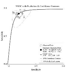
Figure 3
SROC curve"
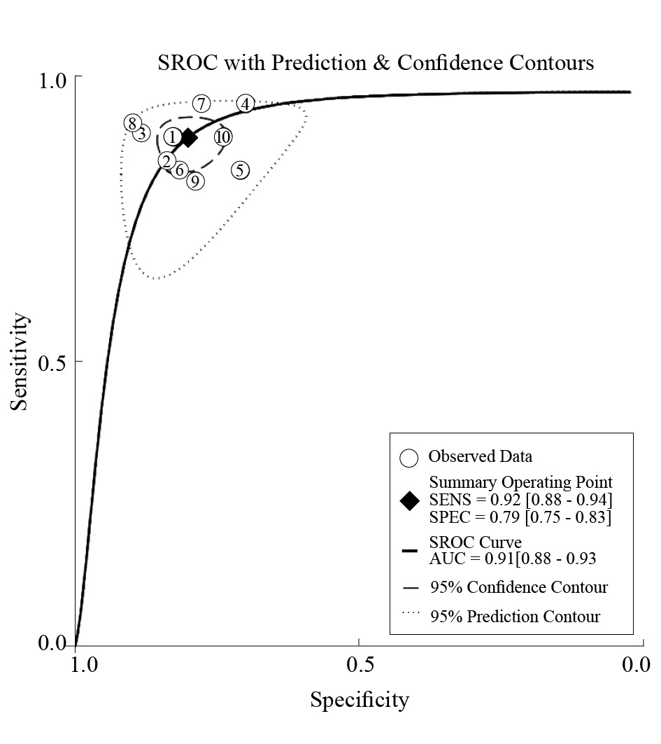
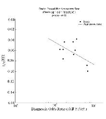
Figure 4
Deeks funnel plot assessed publication bias"
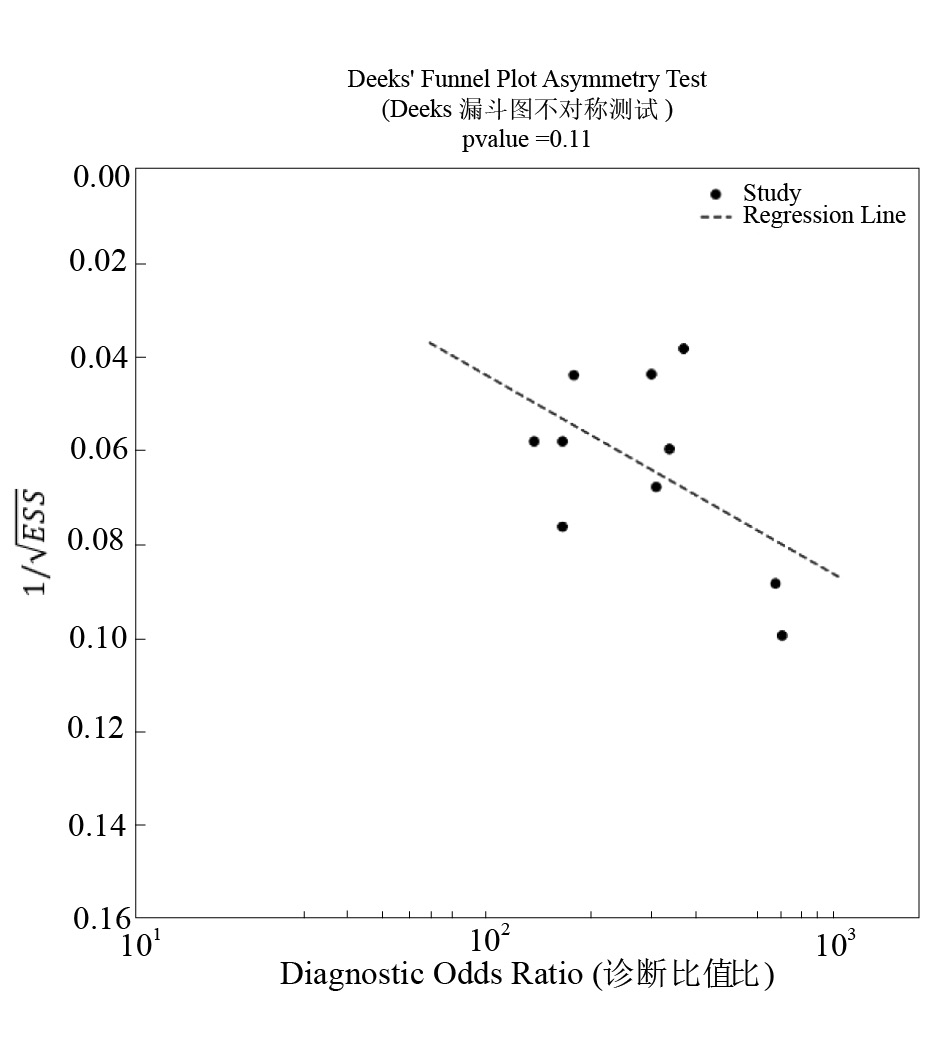
Table 6
Subgroup analysis"
| 亚组变量 | 纳入分析数量 | 合并敏感性(95%CI) | 合并特异性(95%CI) | Meta回归联合P值 |
|---|---|---|---|---|
| 病例来源 ADNI数据库 非ADNI数据库 | 566 789 | 0.88 (0.82, 0.89) 0.84 (0.81, 0.92) | 0.94 (0.92, 0.96) 0.93 (0.87, 0.95) | 0.67 |
| 场强 1.5 T 3.0 T | 842 204 | 0.85 (0.82, 0.88) 0.90 (0.88, 0.91) | 0.91 (0.88, 0.95) 0.95 (0.91, 0.99) | 0.03 |
| 线圈类型 PA ER | 649 703 | 0.86 (0.82, 0.88) 0.89 (0.86, 0.90) | 0.90 (0.88, 0.93) 0.90 (0.86, 0.92) | 0.86 |
| [1] | 戴西件, 骆嵩. 阿尔茨海默病源性认知障碍早期诊疗专家共识(2025)[J]. 中国医学影像学杂志, 2025, 33(4): 337-346. |
| DAI X J, LUO S. Expert Consensus on Early Diagnosis and Treatment of Alzheimer's Disease-Associated Cognitive Impairment(2025)[J]. Chin J Med Imag, 2025, 33(4): 337-346. | |
| [2] |
JACOBS T, BRIEN C O, FIGUEREDO L, et al. Body mass index and blood volume influence plasma biomarkers and positron emission tomography classification in preclinical Alzheimer's disease[J]. Alzheimers Dement, 2025, 21(10): e70765.
doi: 10.1002/alz.v21.10 |
| [3] |
YUAN S, MA W, YANG R, et al. Sleep duration, genetic susceptibility, and Alzheimer's disease: a longitudinal UK Biobank-based study[J]. BMC Geriatr, 2022, 22(1): 638.
doi: 10.1186/s12877-022-03298-8 pmid: 35918656 |
| [4] |
蒋嘉蕊, 牛振东. 深度学习在阿尔茨海默病诊断中应用近5年文献的可视化分析[J]. 中国康复理论与实践, 2022, 28(11): 1318-1324.
doi: 10.3969/j.issn.1006-9771.2022.11.011 |
| JIANG J R, NIU Z D. Deep learning for diagnosis of Alzheimer's disease in the past five years: a visuallized analysis[J]. Chin J Rehabil Theory Pract, 2022, 28(11): 1318-1324. | |
| [5] | 胡晓飞, 桂莉, 陈晓, 等. 2024版NIA-AA阿尔茨海默病诊断与分期标准更新解读:多模态影像的诊疗意义[J]. 中国医学影像学杂志, 2025, 33(5): 454-460. |
| HU X F, GUI L, CHEN X, et al. Interpretation of updates for diagnosis and staging criterion of NIA-AA Alzheimer's Disease (2024 Edition): diagnostic and therapeutic significance of multimodal imaging[J]. Chin J Med Imag, 2025, 33(5): 454-460. | |
| [6] |
GREICIUS M D, SRIVASTAVA G, REISS A L, et al. Default-mode network activity distinguishes Alzheimer's disease from healthy aging: evidence from functional MRI[J]. Proc Natl Acad Sci U S A, 2004, 101(13): 4637-4642.
doi: 10.1073/pnas.0308627101 |
| [7] |
SOHN W S, YOO K, NA D L, et al. Progressive changes in hippocampal resting-state connectivity across cognitive impairment: a cross-sectional study from normal to Alzheimer disease[J]. Alzheimer Dis Assoc Disord, 2014, 28(3): 239-246.
doi: 10.1097/WAD.0000000000000027 |
| [8] |
SULLIVAN M D, ANDERSON J A, TURNER G R, et al. Alzheimer's Disease Neuroimaging Initiative. Intrinsic neurocognitive network connectivity differences between normal aging and mild cognitive impairment are associated with cognitive status and age[J]. Neurobiol Aging, 2019, 73: 219-228.
doi: S0197-4580(18)30357-9 pmid: 30391818 |
| [9] |
THERRIAULT J, WANG S, MATHOTAARACHCHI S, et al. Rostral-caudal hippocampal functional convergence is reduced across the Alzheimer's disease spectrum[J]. Mol Neurobiol, 2019, 56(12): 8336-8344.
doi: 10.1007/s12035-019-01671-0 pmid: 31230260 |
| [10] |
WANG S M, KANG D W, UM Y H, et al. Cognitive normal older adults with APOE-2 allele show a distinctive functional connectivity pattern in response to cerebral Aβ deposition[J]. Int J Mol Sci, 2023, 24(14): 11250.
doi: 10.3390/ijms241411250 |
| [11] |
IBRAHIM K M, AGCAOGLU O, DUDA M, et al. Parallel multilink joint ICA for multimodal fusion of gray matter and multiple resting fMRI networks[J]. Annu Int Conf IEEE Eng Med Biol Soc, 2024, 2024: 1-4.
doi: 10.1109/EMBC53108.2024.10781919 pmid: 40039205 |
| [12] |
JALILIANHASANPOUR R, BEHESHTIAN E, SHERBAF G, et al. Functional connectivity in neurodegenerative disorders: Alzheimer's disease and frontotemporal dementia[J]. Top Magn Reson Imaging, 2019, 28(6): 317-324.
doi: 10.1097/RMR.0000000000000223 pmid: 31794504 |
| [13] |
RAO Y L, GANARAJA B, MURLIMANJU B V, et al. Hippocampus and its involvement in Alzheimer's disease: a review[J]. 3 Biotech, 2022, 12(2): 55.
doi: 10.1007/s13205-022-03123-4 pmid: 35116217 |
| [14] |
BECKER M, REPANTIS D, DRESLER M, et al. Cognitive enhancement: Effects of methylphenidate, modafinil, and caffeine on latent memory and resting state functional connectivity in healthy adults[J]. Hum Brain Mapp, 2022, 43(14): 4225-4238.
doi: 10.1002/hbm.25949 pmid: 35670369 |
| [15] |
YANG A, ZHUANG H, DU L, et al. Evaluation of whole-brain oxygen metabolism in Alzheimer's disease using QSM and quantitative BOLD[J]. Neuroimage, 2023, 282: 120381.
doi: 10.1016/j.neuroimage.2023.120381 |
| [16] |
ALMEIDA V N, RADANOVIC M. Semantic processing and neurobiology in Alzheimer's disease and mild cognitive impairment[J]. Neuropsychologia, 2022, 174: 108337.
doi: 10.1016/j.neuropsychologia.2022.108337 |
| [17] | ABDELAZIZ M, WANG T, ANWAAR W, et al. Multi-scale multimodal deep learning framework for Alzheimer's disease diagnosis[J]. Comput BiolMed, 2025, 184: 109438. |
| [18] |
LENZI D, SERRA L, PERRI R, et al. Single domain amnestic MCI: a multiple cognitive domains fMRI investigation[J]. Neurobiol Aging, 2011, 32(9): 1542-1557.
doi: 10.1016/j.neurobiolaging.2009.09.006 pmid: 19880216 |
| [19] | 厍映霞, 王帅文, 马文玲, 等. 阿尔茨海默病患者静息态局部一致性与体素镜像同伦连接研究[J]. 中国医学影像学杂志, 2018, 26(10): 721-725. |
| SHE Y X, WANG S W, MA W L, et al. Regional homogeneity of Alzheimer's disease at resting-state and voxel-mirrored homotopic connectivity[J]. Chin J Med Imag, 2018, 26(10): 721-725. | |
| [20] |
DING S, LI X, HUANG Z, et al. Alterations of brain structural and functional connectivity networks topology and decoupling in pediatric myelin oligodendrocyte glycoprotein antibody-associated disease[J]. Mult Scler Relat Disord, 2024, 87: 105699.
doi: 10.1016/j.msard.2024.105699 |
| [21] |
IBRAHIM B, SUPPIAH S, IBRAHIM N, et al. Diagnostic power of resting-state fMRI for detection of network connectivity in Alzheimer's disease and mild cognitive impairment: a systematic review[J]. Hum Brain Mapp, 2021, 42(9): 2941-2968.
doi: 10.1002/hbm.25369 pmid: 33942449 |
| [22] |
JACKSON D, TURNER R. Power analysis for random-effects meta-analysis[J]. Res Synth Methods, 2017, 8(3): 290-302.
doi: 10.1002/jrsm.1240 pmid: 28378395 |
| [23] | SALAMEH J P, BOSSUYT P M, MCGRATH T A, et al. Preferred Reporting Items for Systematic Review and Meta-Analysis of Diagnostic Test Accuracy Studies (PRISMA-DTA): explanation, elaboration, and checklist[J]. BMJ, 2020, 370: m2632. |
| [24] |
DE J F, SCHAEFFERS A, KOOREMAN Z E, et al. Botulinum toxin A treatment in facial palsy synkinesis: a systematic review and meta-analysis[J]. Eur Arch Otorhinolaryngol, 2023, 280(4): 1581-1592.
doi: 10.1007/s00405-022-07796-8 |
| [25] |
ISO-MARKKU P, KUJALA U M, KNITTLE K, et al. Physical activity as a protective factor for dementia and Alzheimer's disease: systematic review, meta-analysis and quality assessment of cohort and case-control studies[J]. Br J Sports Med, 2022, 56(12): 701-709.
doi: 10.1136/bjsports-2021-104981 |
| [26] |
LIU S, ZHAO M, YANG X, et al. Exosomal microRNA biomarkers in Alzheimer's disease: systematic review and meta-analysis[J]. Clin Chim Acta, 2026, 578: 120542.
doi: 10.1016/j.cca.2025.120542 |
| [27] |
SEITIDIS G, NIKOLAKOPOULOS S, HENNESSY E A, et al. Network meta-analysis techniques for synthesizing prevention science evidence[J]. Prev Sci, 2022, 23(3): 415-424.
doi: 10.1007/s11121-021-01289-6 |
| [28] |
PULJAK L, BALA M M, MATHES T, et al. AMSTAR 2 is only partially applicable to systematic reviews of non-intervention studies: a meta-research study[J]. J Clin Epidemiol, 2023, 163: 11-20.
doi: 10.1016/j.jclinepi.2023.08.021 pmid: 37659582 |
| [29] |
CAO Y, YANG H, ZHOU Z, et al. Abnormal default-mode network homogeneity in patients with mild cognitive impairment in Chinese communities[J]. Front Neurol, 2021, 11: 569806.
doi: 10.3389/fneur.2020.569806 |
| [30] |
LIAO Z, SUN W, LIU X, et al. Altered dynamic intrinsic brain activity of the default mode network in Alzheimer's disease: a resting-state fMRI study[J]. Front Hum Neurosci, 2022, 16: 951114.
doi: 10.3389/fnhum.2022.951114 |
| [31] |
XIONG Y, YE C, CHEN Y, et al. Altered functional connectivity of basal ganglia in mild cognitive impairment and Alzheimer's disease[J]. Brain Sci, 2022, 12(11): 1555.
doi: 10.3390/brainsci12111555 |
| [32] |
SENDI M S, ZENDEHROUH E, MILLER R L, et al. Alzheimer's disease projection from normal to mild dementia reflected in functional network connectivity: a longitudinal study[J]. Front Neural Circuits, 2021, 14: 593263.
doi: 10.3389/fncir.2020.593263 |
| [33] |
SOMAN S M, RAGHAVAN S, RAJESH P G, et al. Does resting state functional connectivity differ between mild cognitive impairment and early Alzheimer's dementia?[J]. J Neurol Sci, 2020, 418: 117093.
doi: 10.1016/j.jns.2020.117093 |
| [34] | GHAHREMANI M, NATHAN S, SMITH E E, et al. Functional connectivity and mild behavioral impairment in dementia-free elderly[J]. Alzheimers Dement (N Y), 2023, 9(1): e12371. |
| [35] |
HOJJATI S H, BUTLER T A, DE LEON M, et al. Inter-network functional connectivity increases by beta-amyloid and may facilitate the early stage of tau accumulation[J]. Neurobiol Aging, 2025, 148: 16-26.
doi: 10.1016/j.neurobiolaging.2025.01.005 pmid: 39879839 |
| [36] |
TAKAO H, AMEMIYA S, ABE O. Longitudinal stability of resting-state networks in normal aging, mild cognitive impairment, and Alzheimer's disease[J]. Magn Reson Imaging, 2021, 82: 55-73.
doi: 10.1016/j.mri.2021.06.020 pmid: 34153437 |
| [37] |
SABERIAN P, NASIRI H, KAFFASHIAN F, et al. Potential mediating role of polygenic hazard score in the association between neurofilament light chain and default mode network connectivity across the Alzheimer's disease continuum[J]. IBRO Neurosci Rep, 2025, 18: 732-738.
doi: 10.1016/j.ibneur.2025.04.018 pmid: 40476223 |
| [38] |
MOHAMMADIAN F, ZARE SADEGHI A, NOROOZIAN M, et al. Quantitative assessment of resting-state functional connectivity MRI to differentiate amnestic mild cognitive impairment, late-onset Alzheimer's disease from normal subjects[J]. J Magn Reson Imaging, 2023, 57(6): 1702-1712.
doi: 10.1002/jmri.v57.6 |
| [39] |
WU J, HE Y, LIANG S, et al. Effects of computerized cognitive training on structure-function coupling and topology of multiple brain networks in people with mild cognitive impairment: a randomized controlled trial[J]. Alzheimers Res Ther, 2023, 15(1): 158.
doi: 10.1186/s13195-023-01292-9 |
| [40] |
PAN T, QU H, PAN Y, et al. Effective connectivity of default mode network subsystems in Parkinson's disease with mild cognitive impairment based on spectral dynamic causal modeling[J]. J Integr Neurosci, 2024, 23(6): 110.
doi: 10.31083/j.jin2306110 pmid: 38940086 |
| [41] |
WANG C, JI D, SU X, et al. Cerebral perfusion correlates with amyloid deposition in patients with mild cognitive impairment due to Alzheimer's disease[J]. J Prev Alzheimers Dis, 2025, 12(2): 100031.
doi: 10.1016/j.tjpad.2024.100031 |
| [42] |
COHEN J S, PHILLIPS J, DAS S R, et al. Posterior hippocampal sparing in Lewy body disorders with Alzheimer's copathology: an in vivo MRI study[J]. Neuroimage Clin, 2025, 45: 103714.
doi: 10.1016/j.nicl.2024.103714 |
| [43] |
CHAO L L, LEE J A, MARTINEZ S, et al. Preventing loss of independence through exercise (PLIÉ): a pilot trial in older adults with subjective memory decline and mild cognitive impairment[J]. J Alzheimers Dis, 2021, 82(4): 1543-1557.
doi: 10.3233/JAD-210159 |
| [44] |
MCGETTIGAN C, WARREN J E, EISNER F, et al. Neural correlates of sublexical processing in phonological working memory[J]. J Cogn Neurosci, 2011, 23(4): 961-977.
doi: 10.1162/jocn.2010.21491 |
| [45] |
KIM M, SEKIYA H, YAO G, et al. Diagnosis of Alzheimer disease and tauopathies on whole-slide histopathology images using a weakly supervised deep learning algorithm[J]. Lab Invest, 2023, 103(6): 100127.
doi: 10.1016/j.labinv.2023.100127 |
| [46] |
RABINOVICI G D, KNOPMAN D S, ARBIZU J, et al. Updated appropriate use criteria for amyloid and tau PET: a report from the Alzheimer's Association and Society for Nuclear Medicine and Molecular Imaging Workgroup[J]. Alzheimers Dement. 2025, 21(1): e14338.
doi: 10.1002/alz.v21.1 |
| [47] |
AHMED N, NGADI A B, SHARIF J M, et al. Network threat detection using machine/deep learning in SDN-Based platforms: a comprehensive analysis of state-of-the-art solutions, discussion, challenges, and future research direction[J]. Sensors (Basel), 2022, 22(20): 7896.
doi: 10.3390/s22207896 |
| [48] |
STEINBERG S N, KING T Z. Within-individual BOLD signal variability and its implications for task-based cognition: a systematic review[J]. Neuropsychol Rev, 2024, 34(4): 1115-1164.
doi: 10.1007/s11065-023-09619-x |
| [1] | CHE Ruixin, TANG Yongtian, PAN Shiqin, TONG Yaqian. Effect of transcutaneous electrical acupoint stimulation on chronic obstructive pulmonary disease: a meta-analysis [J]. Chinese Journal of Rehabilitation Theory and Practice, 2026, 32(4): 426-436. |
| [2] | DU Wenqian, ZHANG Xu, CHEN Jiwei, WANG Xing, ZHU Kun. Effect of exercise on freezing of gait in Parkinson's disease: a meta-analysis [J]. Chinese Journal of Rehabilitation Theory and Practice, 2025, 31(7): 781-789. |
| [3] | YANG Yicheng, WANG Dandan, SHEN Qunce, ZHANG Lei, WU Xueping. Effect of exercise on cancer patients with anxiety and depression during chemotherapy: a meta-analysis [J]. Chinese Journal of Rehabilitation Theory and Practice, 2025, 31(2): 184-193. |
| [4] | ZHU Airong, LIU Peihao, GUO Jing, GUAN Tingyu, CHEN Xizhu, CAI Shining, HU Yan, ZHANG Yuxia. Motor-cognitive dual-task training for cognitive function in patients with Parkinson's disease: a meta-analysis [J]. Chinese Journal of Rehabilitation Theory and Practice, 2025, 31(12): 1376-1384. |
| [5] | LIU Dongfei, YAN Huiyi, YANG Fan, ZHANG Shuai. Effect of exercise interventions on multidimensional health outcomes in children and adolescents with Down syndrome: a meta-analysis [J]. Chinese Journal of Rehabilitation Theory and Practice, 2025, 31(12): 1398-1413. |
| [6] | ZHANG Hao, XU Chuanlei, WEI Zhenxing, MA Lihong. Comparison of different virtual reality technologies on motor function in Parkinson's disease: a network meta-analysis [J]. Chinese Journal of Rehabilitation Theory and Practice, 2025, 31(11): 1290-1302. |
| [7] | MAO Tao, YOU Maolin. Effect of exercise on respiratory function in Parkinson's disease: a meta-analysis [J]. Chinese Journal of Rehabilitation Theory and Practice, 2025, 31(11): 1303-1313. |
| [8] | CHEN Yuhang, YANG Yu'ai, LU Xuanyu, WANG Yuhang, ZHANG Mengyuan, ZHANG Zihan, WANG Huiying, CHANG Jingling. Spontaneous brain activity changes in post-stroke aphasia and post-stroke depression: a meta-analysis of resting state functional magnetic resonance imaging [J]. Chinese Journal of Rehabilitation Theory and Practice, 2025, 31(10): 1143-1155. |
| [9] | YANG Sanxia, LIU Qingluan, WAN Jiamin. Regional differences in brain activation after music intervention for Alzheimer's disease: a meta-analysis of functional magnetic resonance imaging [J]. Chinese Journal of Rehabilitation Theory and Practice, 2025, 31(10): 1156-1163. |
| [10] | ZHAO Yingnan, ZHENG Xintong, LIU Junling, WANG Zidan, WU Hongyue, LI Bing, LI Yan. Effect of threshold inspiratory muscle training on pulmonary rehabilitation in postoperative lung cancer patients: a meta-analysis [J]. Chinese Journal of Rehabilitation Theory and Practice, 2025, 31(10): 1164-1171. |
| [11] | WEN Yanfei, YANG Lu, BAN Yue, Ykabaru Daniela BERBESI NORIEGA, ZHANG Haoqi, WANG Li, LIU Hua. Effect of exercise therapy on chronic nonspecific neck pain: a meta-analysis based on ICF [J]. Chinese Journal of Rehabilitation Theory and Practice, 2024, 30(7): 778-788. |
| [12] | LÜ Meiling, WANG Jie, ZENG Weisi, WEN Xiaoting, CHU Xin. Effect of virtual reality on cognitive function and quality of life in patients with Parkinson's disease: a meta-analysis [J]. Chinese Journal of Rehabilitation Theory and Practice, 2024, 30(6): 648-656. |
| [13] | CUI Tiantian, YANG Yulin, CUI Tengteng, MA Lihong. Effect of different intensive training on upper limb motor function in children with cerebral palsy: a network meta-analysis [J]. Chinese Journal of Rehabilitation Theory and Practice, 2024, 30(4): 437-448. |
| [14] | WEI Chen, WANG Zixian, LI Shufan, WANG Peng, JIA Shuqi, TIAN Ying. Effect of mirror therapy on upper extremity motor function and activities of daily living in stroke patients: a meta-analysis [J]. Chinese Journal of Rehabilitation Theory and Practice, 2024, 30(3): 281-291. |
| [15] | WANG Mingchen, ZHANG Wenyu, ZHANG Xianzuo, ZANG Wanli. Effect of transcranial direct current stimulation on cognitive function and quality of life in patients with Parkinson's disease: a meta-analysis [J]. Chinese Journal of Rehabilitation Theory and Practice, 2024, 30(2): 183-188. |
| Viewed | ||||||
|
Full text |
|
|||||
|
Abstract |
|
|||||
|
||
